

Differential phenotype and behavior in culture of CD34 positive cells from peripheral blood and adipose tissue
- PMID: 34458617
- PMCID: PMC8377488
- DOI: 10.1016/j.heliyon.2021.e07779
Differential phenotype and behavior in culture of CD34 positive cells from peripheral blood and adipose tissue
Abstract
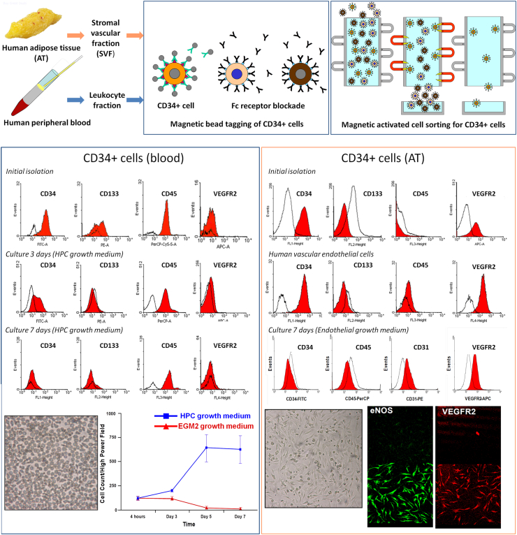
The localization and quantification of endothelial progenitor cells (EPCs) are controversial. Circulating CD34 + cells in blood have been identified as EPCs and as biomarkers of cardiovascular disease. We discuss in this paper the current data describing differential phenotype and behavior in vitro of CD34 positive cells from the circulation and adipose tissue (AT). We also describe in brief our own findings from CD34 + cells isolated from leukopheresis cones derived from healthy platelet donors and from patients undergoing bariatric surgery. We conclude that CD34 + cells in blood and in AT are different in antigenic profile and behavior in culture. The findings described assert that CD34 + cells detected in blood previously identified as biomarkers of cardiovascular disease are predominantly HPCs rather than EPCs, and that true CD34 + EPCs can be readily identified and extracted from AT, supportive of the current evidence which suggests EPCs are resident in the tissue vasculature.
Keywords: Adipose tissue; Cell culture; Endothelial progenitor cells; Flow cytometry; Hematopoietic progenitor cells; Peripheral blood.
© 2021 The Author(s).
Conflict of interest statement
The authors declare no conflict of interest.
Figures
Similar articles
-
Human CD34+AC133+VEGFR-2+ cells are not endothelial progenitor cells but distinct, primitive hematopoietic progenitors.Exp Hematol. 2007 Jul;35(7):1109-18. doi: 10.1016/j.exphem.2007.04.002. Exp Hematol. 2007. PMID: 17588480
-
Circulating CD34+, CD133+, and vascular endothelial growth factor receptor 2-positive endothelial progenitor cells in myelofibrosis with myeloid metaplasia.J Clin Oncol. 2005 Aug 20;23(24):5688-95. doi: 10.1200/JCO.2005.09.021. J Clin Oncol. 2005. PMID: 16110028
-
Analysis of origin and optimization of expansion and transduction of circulating peripheral blood endothelial progenitor cells in the rhesus macaque model.Hum Gene Ther. 2002 Nov 20;13(17):2041-50. doi: 10.1089/10430340260395893. Hum Gene Ther. 2002. PMID: 12489999
-
Endothelial progenitor cells: isolation and characterization.Trends Cardiovasc Med. 2003 Jul;13(5):201-6. doi: 10.1016/s1050-1738(03)00077-x. Trends Cardiovasc Med. 2003. PMID: 12837583 Review.
-
Dysfunction and Therapeutic Potential of Endothelial Progenitor Cells in Diabetes Mellitus.J Clin Med Res. 2018 Oct;10(10):752-757. doi: 10.14740/jocmr3581w. Epub 2018 Sep 10. J Clin Med Res. 2018. PMID: 30214646 Free PMC article. Review.
Cited by
-
Endothelial Progenitor Cells as Biomarkers of Cardiovascular Pathologies: A Narrative Review.Cells. 2022 May 18;11(10):1678. doi: 10.3390/cells11101678. Cells. 2022. PMID: 35626716 Free PMC article. Review.
-
Vascular tissue engineering from human adipose tissue: fundamental phenotype of its resident microvascular endothelial cells and stromal/stem cells.Biomater Biosyst. 2022 Apr 11;6:100049. doi: 10.1016/j.bbiosy.2022.100049. eCollection 2022 Jun. Biomater Biosyst. 2022. PMID: 36824164 Free PMC article.
-
Adipose Tissue Development Relies on Coordinated Extracellular Matrix Remodeling, Angiogenesis, and Adipogenesis.Biomedicines. 2022 Sep 8;10(9):2227. doi: 10.3390/biomedicines10092227. Biomedicines. 2022. PMID: 36140327 Free PMC article. Review.
References
-
- Asahara T. Isolation of putative progenitor endothelial cells for angiogenesis. Science. 1997;275(5302):964–967. - PubMed
-
- Friedrich E.B. CD34-/CD133+/VEGFR-2+ endothelial progenitor cell subpopulation with potent vasoregenerative capacities. Circ. Res. 2006;98(3):e20–e25. - PubMed
-
- Heiss C. Impaired progenitor cell activity in age-related endothelial dysfunction. J. Am. Coll. Cardiol. 2005;45(9):1441–1448. - PubMed
-
- Schmidt-Lucke C. Reduced number of circulating endothelial progenitor cells predicts future cardiovascular events: proof of concept for the clinical importance of endogenous vascular repair. Circulation. 2005;111(22):2981–2987. - PubMed
-
- Vasa M. Number and migratory activity of circulating endothelial progenitor cells inversely correlate with risk factors for coronary artery disease. Circ. Res. 2001;89(1):E1–7. - PubMed
Publication types
LinkOut - more resources
Full Text Sources
