

Looking beneath the surface: the importance of subcortical structures in frontotemporal dementia
- PMID: 34458729
- PMCID: PMC8390477
- DOI: 10.1093/braincomms/fcab158
Looking beneath the surface: the importance of subcortical structures in frontotemporal dementia
Abstract
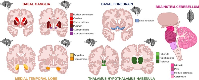
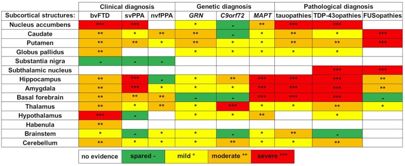
Whilst initial anatomical studies of frontotemporal dementia focussed on cortical involvement, the relevance of subcortical structures to the pathophysiology of frontotemporal dementia has been increasingly recognized over recent years. Key structures affected include the caudate, putamen, nucleus accumbens, and globus pallidus within the basal ganglia, the hippocampus and amygdala within the medial temporal lobe, the basal forebrain, and the diencephalon structures of the thalamus, hypothalamus and habenula. At the most posterior aspect of the brain, focal involvement of brainstem and cerebellum has recently also been shown in certain subtypes of frontotemporal dementia. Many of the neuroimaging studies on subcortical structures in frontotemporal dementia have been performed in clinically defined sporadic cases. However, investigations of genetically- and pathologically-confirmed forms of frontotemporal dementia are increasingly common and provide molecular specificity to the changes observed. Furthermore, detailed analyses of sub-nuclei and subregions within each subcortical structure are being added to the literature, allowing refinement of the patterns of subcortical involvement. This review focuses on the existing literature on structural imaging and neuropathological studies of subcortical anatomy across the spectrum of frontotemporal dementia, along with investigations of brain-behaviour correlates that examine the cognitive sequelae of specific subcortical involvement: it aims to 'look beneath the surface' and summarize the patterns of subcortical involvement have been described in frontotemporal dementia.
Keywords: MR imaging; frontotemporal dementia; subcortical structures.
© The Author(s) (2021). Published by Oxford University Press on behalf of the Guarantors of Brain.
Figures

References
-
- Woollacott IO, Rohrer JD. The clinical spectrum of sporadic and familial forms of frontotemporal dementia. J Neurochem. 2016;138 (Suppl 1):6–31. - PubMed
-
- Mackenzie IR, Neumann M. Molecular neuropathology of frontotemporal dementia: Insights into disease mechanisms from postmortem studies. J Neurochem. 2016;138 (Suppl 1):54–70. - PubMed
Publication types
Grants and funding
LinkOut - more resources
Full Text Sources