. 2020 May 19;1(2):56-59.
doi: 10.1039/d0cb00029a. eCollection 2020 Jun 1.

Synthetic hyperacetylation of nucleosomal histones

Affiliations

Synthetic hyperacetylation of nucleosomal histones

Hidetoshi Kajino et al. RSC Chem Biol. .

Abstract

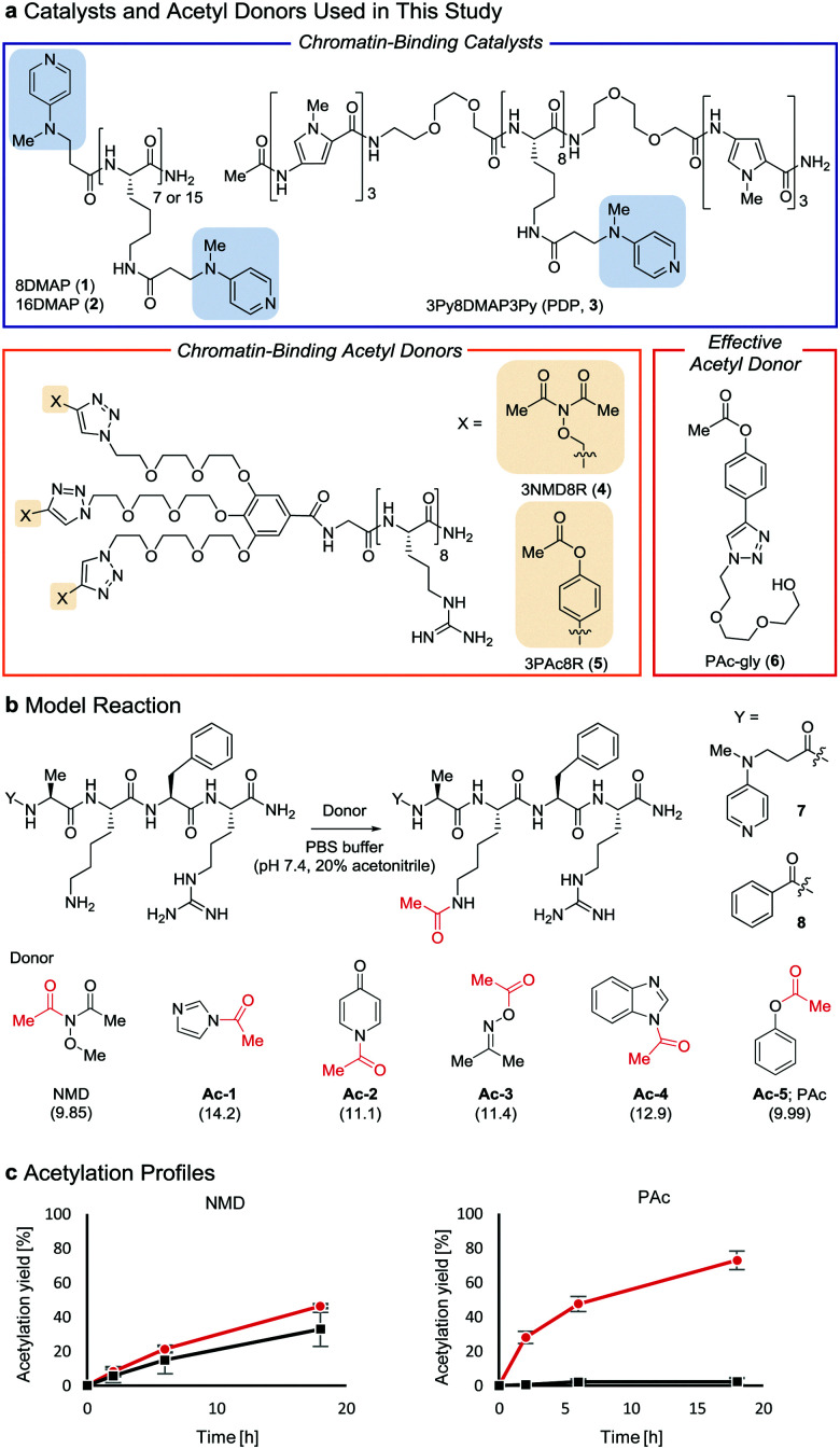
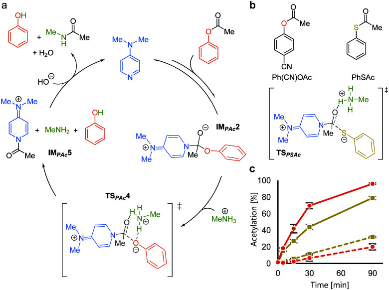
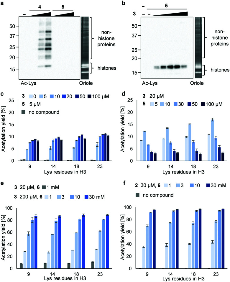
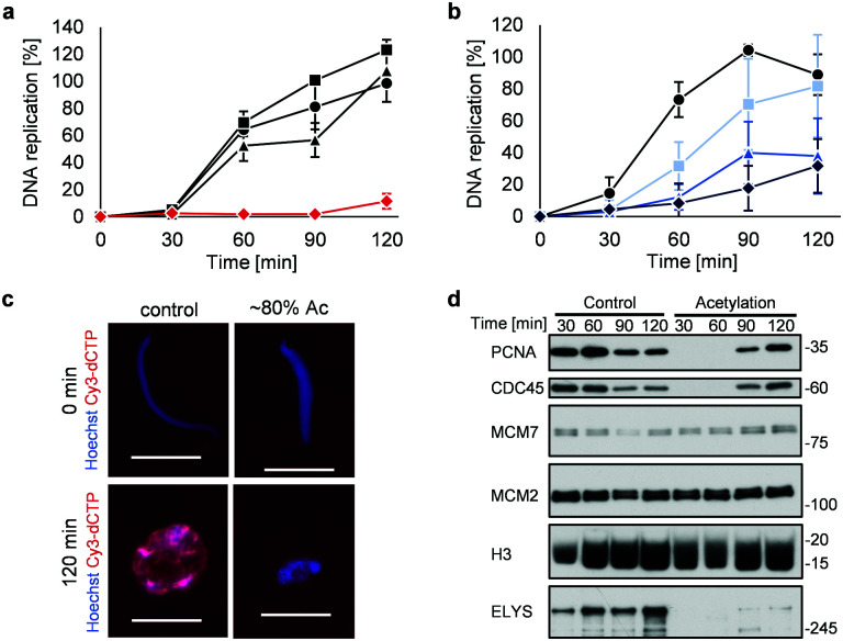
We report combinations of a DMAP-based catalyst and phenyl acetate with optimal electron density as a new chemical system for high-yield, selective synthetic acetylation of histone lysine residues. The utility of this chemical system as a unique biologic tool is demonstrated by applying it to Xenopus laevis sperm chromatin.


Conflict of interest statement

There are no conflicts to declare.

Figures

Fig. 1
Fig. 1. Properties of acetyl donors. (a) Chemical structures of the catalysts and acetyl donors. (b) Screening of acetyl donors. The pKa value of each leaving group conjugate acid is shown in parentheses. (c) Comparison of NMD and Ac-5 using 7 () and 8 (■) at 30 °C. The average and SD (bars) are indicated (n = 3 independent experiments).
Fig. 2
Fig. 2. Mechanistic analyses of DMAP-catalyzed acetylation with PAc. (a) A plausible catalytic cycle based on DFT calculations. (b) Comparison between phenyl acetate and phenyl thioacetate. (c) Reaction profiles of the acetylation of 7 (solid lines) and 8 (dotted lines) using Ph(CN)OAc () and PhSAc () at 30 °C.
Fig. 3
Fig. 3. Acetylation of a mixture of recombinant nucleosomes (0.2 μM as DNA concentration) and HeLa cell extract (non-histone proteins) at 25 °C for 3 h. (a) Background histone acetylation by 4 (2, 5, and 10 μM) or 5 (2, 5, and 10 μM). (b) Catalyzed histone acetylation by 3 (5, 10, 20, 50, and 100 μM) and 5 (5 μM). In (a and b), acetylated lysines were detected by immunoblotting using an anti-Ac-Lys antibody. Proteins were visualized by Oriole staining. (c) Yield of histone acetylation by 3 (5, 10, 20, 50, and 100 μM) and 5 (5 μM). (d) Yield of histone acetylation by 3 (20 μM) and 5 (5, 10, 30, 50, and 100 μM). (e) Yield of histone acetylation by 3 (20 μM) and 6 (1 mM) or 3 (200 μM) and 6 (1, 3, 10, and 30 mM). (f) Yield of histone acetylation by 2 (30 μM) and 6 (1, 3, 10, and 30 mM). In (c–f), the yield of acetylated lysines of the H3 tail was determined by LC-MS/MS analysis. The average and error range (bars) are indicated (n = 2 independent experiments).
Fig. 4
Fig. 4. Synthetic histone acetylation inhibits DNA replication. (a and b) XSC (20 000 sperm per μL) was treated with 2 (30 μM) and 6 (30 mM) for (a) 7 h or (b) the indicated time at 25 °C. After the reaction, control or acetylated XSC was incubated with interphase egg extracts containing radiolabeled [α-32P]-dCTP. The DNA replication kinetics in egg extracts was assessed by the incorporation of radioactivity, determined using a scintillation counter. Data shown are the average and SD (bars) from (a) three or (b) five independent experiments. (a) ●: control; ■: 2 only; ▲: 6 only; : 2 + 6. (b) ●: control. Reaction time = : 0.5 h; : 1 h; : 3 h. (c) and (d) XSC (20 000 sperm per μL) was treated with 2 (30 μM) and 6 (30 mM) for 7 h. (c) Control or acetylated XSC was incubated with interphase egg extracts containing Cy3-dCTP. After incubation, the samples were fixed and DNA was visualized with Hoechst 33258 (blue image). DNA replication was monitored as the incorporation of Cy3-dCTP (red image) into DNA. The scale bar represents 20 μm. (d) Control or acetylated XSC was incubated with interphase egg extracts. Chromatin was isolated from the egg extracts at the indicated times and the chromatin samples were analyzed by immunoblotting using the indicated antibodies.

References

    1. Shikama N. Ivon J. La Thangue N. Trends Cell Biol. 1997;7:230–236. doi: 10.1016/S0962-8924(97)01048-9. - DOI - PubMed
    1. Dawson M. A. Kouzarides T. Cell. 2012;150:12–27. doi: 10.1016/j.cell.2012.06.013. - DOI - PubMed
    1. Elsässer S. J. Ernst R. J. Walker O. S. Chin J. W. Nat. Methods. 2016;13:158–164. doi: 10.1038/nmeth.3701. - DOI - PMC - PubMed
    1. David Y. Vila-Perelló M. Verma S. Muir T. W. Nat. Chem. 2015;7:394–402. doi: 10.1038/nchem.2224. - DOI - PMC - PubMed
    1. Pelton J. G. Wemmer D. E. Proc. Natl. Acad. Sci. U. S. A. 1989;86:5723–5727. doi: 10.1073/pnas.86.15.5723. - DOI - PMC - PubMed