Review
. 2020 Jul 16;1(3):110-127.
doi: 10.1039/d0cb00073f. eCollection 2020 Aug 1.

Matters of class: coming of age of class III and IV lanthipeptides

Affiliations
Review

Matters of class: coming of age of class III and IV lanthipeptides

Julian D Hegemann et al. RSC Chem Biol. .

Abstract

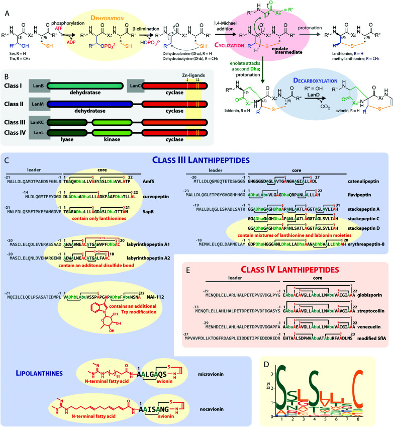
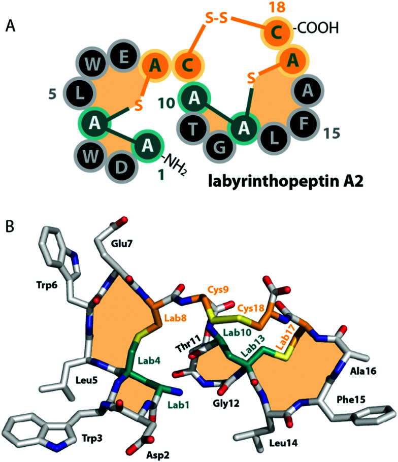
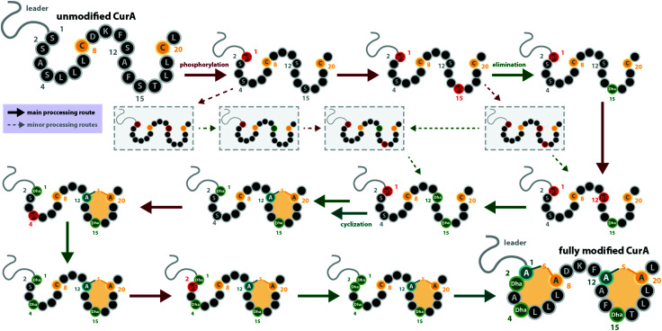
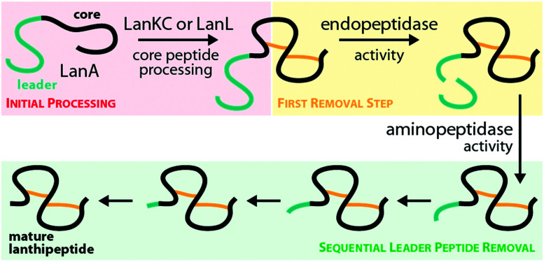
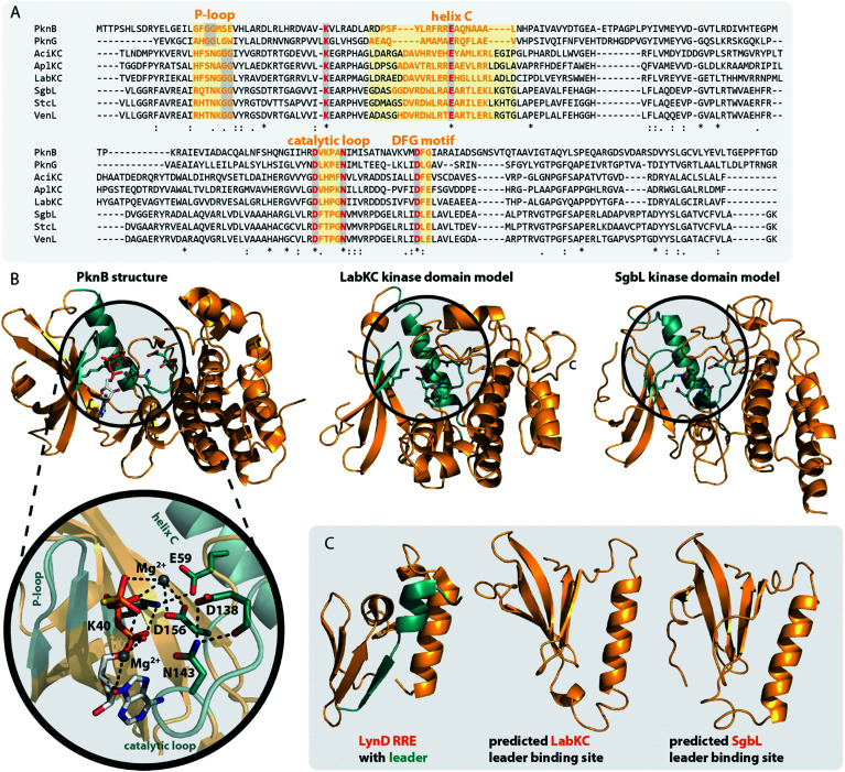
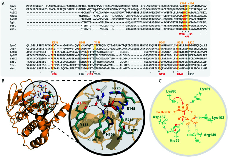
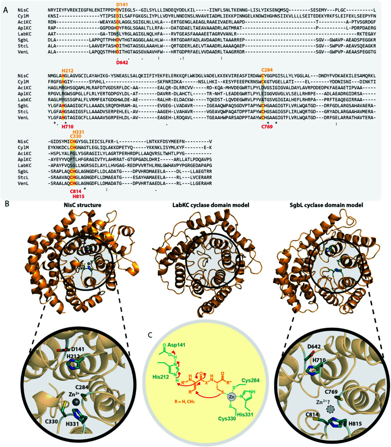
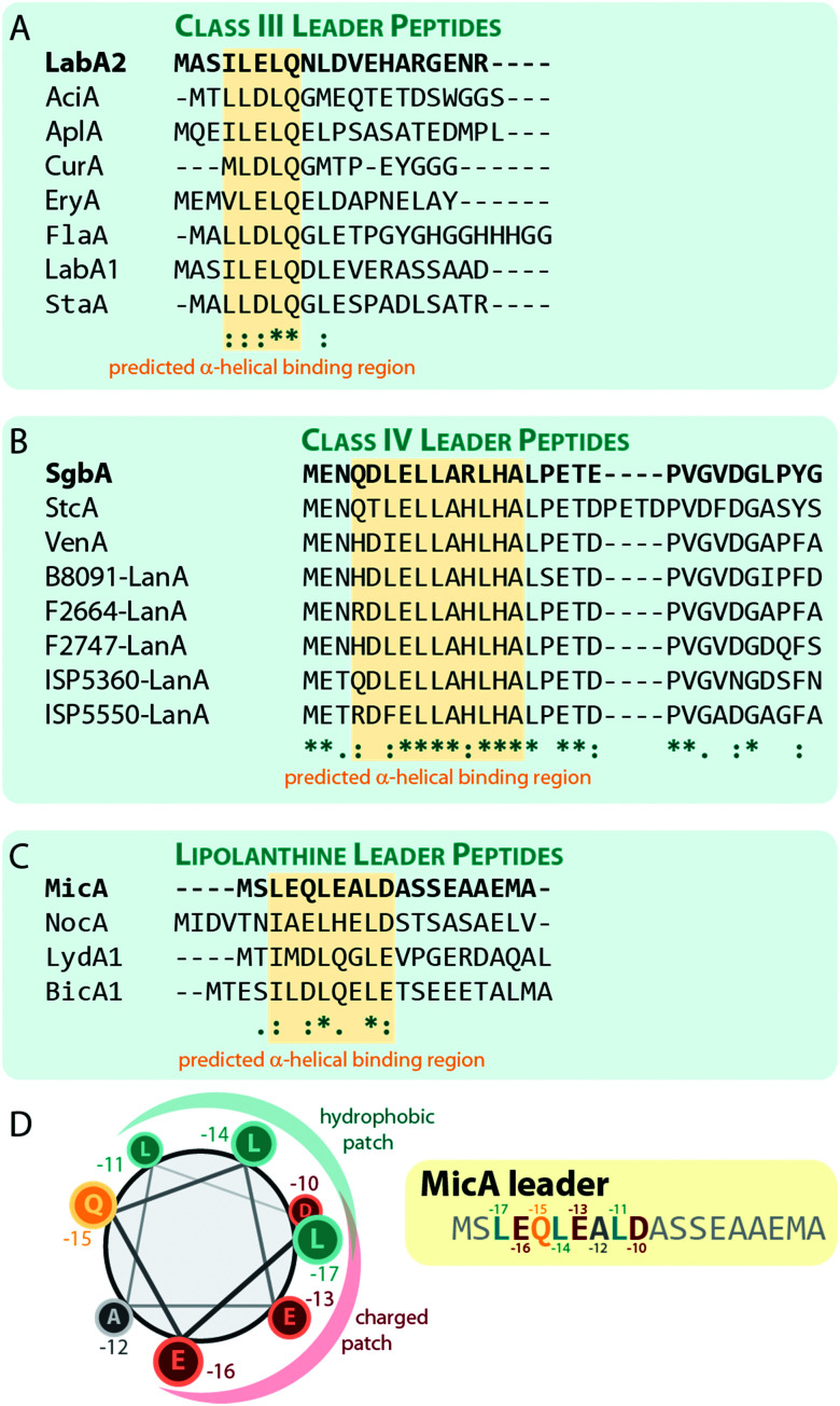
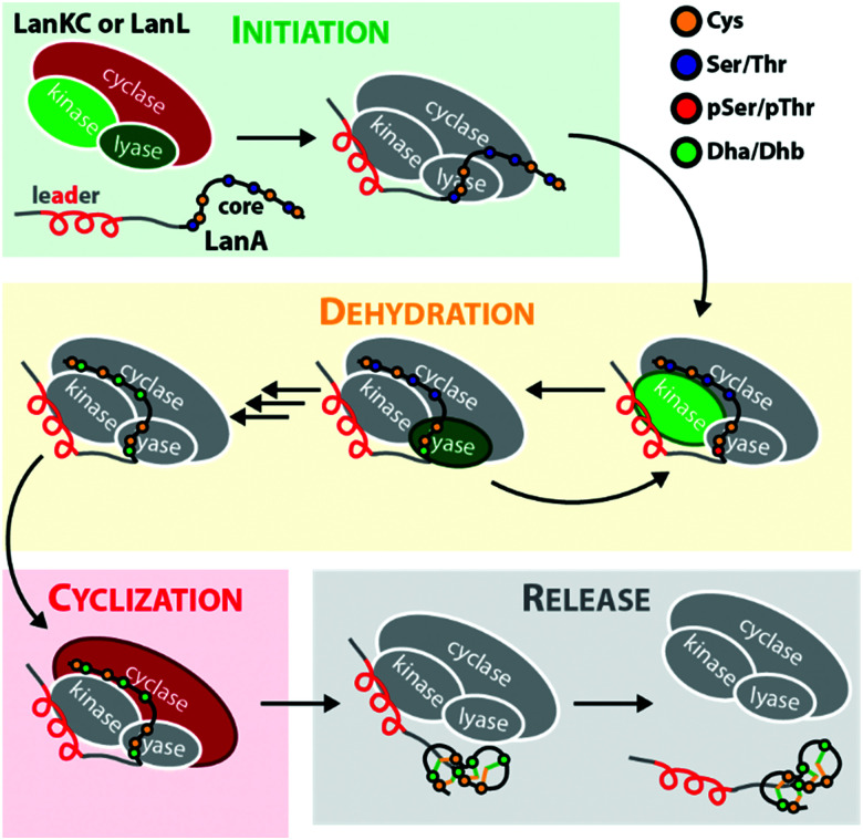
Lanthipeptides belong to the superfamily of ribosomally-synthesized and posttranslationally-modified peptides (RiPPs). Despite the fact that they represent one of the longest known RiPP subfamilies, their youngest members, classes III and IV, have only been described more recently. Since then, a plethora of studies furthered the understanding of their biosynthesis. While there are commonalities between classes III and IV due to the similar domain architectures of their processing enzymes, there are also striking differences that allow their discrimination. In this concise review article, we summarize what is known about the underlying biosynthetic principles of these lanthipeptides and discuss open questions for future research.


Conflict of interest statement

There are no conflicts to declare.

Figures

Fig. 1
Fig. 1. (A) Underlying principles of class III and IV lanthipeptide biosynthesis. (B) Overview of the four known lanthipeptide classes. Schematic showing the primary structures of important (C) class III and (D) class IV lanthipeptides. The color coding is as follows: leader peptide residues are grey, unmodified core peptide residues are black, cysteines involved in (methyl)lanthionines and labionins are orange, other residues in (methyl)lanthionines and labionins are teal, Dha/Dhb residues are green, additional posttranslational modifications are red. (E) MEME motif predicted on basis of all Sx2Sx2–5C motifs from the peptides in (C) highlighting the most common residues and arrangements of these motifs in studied class III lanthipeptides. Dha = Dehydroalanine. Dhb = Dehydrobutyrine. Abu = Aminobutyric acid.
Fig. 2
Fig. 2. (A) Schematic representation and (B) crystal structure (CCDC number 721326) of labyrinthopeptin A2. The labionin macrocycles are highlighted by orange shading in the background.
Fig. 3
Fig. 3. Processing order of the CurA precursor peptide by the CurKC processing enzyme, which was investigated in-depth using the deuterium-labeling approach as described in the main text. This case exemplifies that while a general C-to-N-terminal directionality is observed, there can be exceptions to this order (here, Ser1 is phosphorylated first) and that besides the major processing route, minor routes can be detected with sufficient scrutiny (note that only a few exemplary early minor processing routes are shown and not all that were described in the original publication). This example also emphasizes that while usually the same residue becomes phosphorylated and eliminated before the next residue is processed, there are also exceptions to this rule. pS = phosphoserine.
Fig. 4
Fig. 4. Hypothesized general route for leader peptide removal in class III and IV lanthipeptide systems. It should be noted that the endopeptidase step might not be required in all cases.
Fig. 5
Fig. 5. (A) Excerpt of an alignment of the mycobacterial protein kinases PknB and PknG with the kinase domains of exemplary class III (AciKC, AplKC, LabKC) and class IV (SgbL, StcL, VenL) lanthipeptide synthetases. Conserved catalytic residues are shown in red and structural features important for activity are highlighted. (B) Comparison of the crystal structure of PknB binding the ATP-mimic adenosine 5′-[γ-thio]triphosphate (PDB code 1MRU) with the Phyre2 models of the LabKC and SgbL kinase domains. A close up of the active site of PknB is shown. Catalytic residues and regions of importance are highlighted in teal. (C) Structure of the RRE domain of the cyanobactin heterocyclase LynD from Lyngbya aestuarii with bound leader (PDB code 4V1T) compared with the segments of the LabKC and SgbL structure models that are implied in leader binding.
Fig. 6
Fig. 6. (A) Excerpt of an alignment of OspF from Shigella dysenteriae and SpvC from Salmonella paratyphi with the lyase domains of exemplary class III (AciKC, AplKC, LabKC) and class IV (SgbL, StcL, VenL) lanthipeptide synthetases. Catalytic residues of the OspF protein family pThr lyases are highlighted in orange, catalytic residues of class III/IV lyase domains in red. Above the alignment, numbering of the catalytic residues of SpvC are shown. Below the alignment, numbering of the catalytic residues of VenL are shown. (B) Crystal structure of a K136A variant of SpvC (PDB code 2Q8Y) with bound substrate zoomed in on the active site. For clarity, only a fragment of the pThr-containing peptide substrate is shown. The K136 residue of WT SpvC acts as catalytic base during the β-elimination reaction. (C) Schematic showing the catalyzed phosphate elimination reaction with the hypothesized functions for the catalytically important residues in VenL.
Fig. 7
Fig. 7. (A) Excerpt of an alignment of the NisC cyclase (involved in the biosynthesis of the class I lanthipeptide nisin) and the CylM cyclase domain (involved in the biosynthesis of the class II cytolysin lanthipeptides) with the cyclase domains of exemplary class III (AciKC, AplKC, LabKC) and class IV (SgbL, StcL, VenL) lanthipeptide synthetases. Catalytic residues of class I and II cyclases are highlighted in orange, catalytic residues of the class IV cyclase domains in red. Above the alignment, numbering of the catalytic residues of NisC are shown. Below the alignment, numbering of the predicted catalytic residues of SgbL are shown. (B) Comparison of the crystal structure of NisC (PDB code 2G0D) with the Phyre2 models of the LabKC and SgbL cyclase domains. Close ups of the active site of NisC and the predicted active site of the SgbL cyclase domain are shown. Catalytic residues are highlighted in teal. (C) Schematic showing the catalyzed cyclization reaction with the hypothesized functions for the catalytically important residues in NisC.
Fig. 8
Fig. 8. Alignments of a selection of (A) class III, (B) class IV, and (C) lipolanthine leader peptides highlighting the predicted α-helical regions that are suggested to be involved in substrate recognition by the respective processing enzymes. The peptides LabA2, SgbA, and MicA, which were used for the experimental validation of the importance of these regions, are shown on the top of each alignment and are highlighted in bold. (D) Helical-wheel representation of the MicA leader peptide highlighting the hydrophobic and charged patches on the helix surface.
Fig. 9
Fig. 9. Simplified mechanism of the precursor processing through LanKC and LanL enzymes. Domains active in the respective steps are highlighted. Note that the directionality of the dehydration reactions was chosen arbitrarily to demonstrate that a specific Ser/Thr residue often undergoes first phosphorylation and then β-elimination, before the next residue is modified in the peptide. As discussed earlier, cyclization can sometimes be initiated before the complete dehydration of the core peptide is accomplished. In some systems, the formation of a thioether crosslink is even a prerequisite for further dehydrations to occur, while in other instances, complete dehydration in absence of any macrocycles is possible.
None
Julian D. Hegemann
None
Roderich D. Süssmuth

References

    1. Arnison P. G. Bibb M. J. Bierbaum G. Bowers A. A. Bugni T. S. Bulaj G. Camarero J. A. Campopiano D. J. Challis G. L. Clardy J. Cotter P. D. Craik D. J. Dawson M. Dittmann E. Donadio S. Dorrestein P. C. Entian K. D. Fischbach M. A. Garavelli J. S. Goransson U. Gruber C. W. Haft D. H. Hemscheidt T. K. Hertweck C. Hill C. Horswill A. R. Jaspars M. Kelly W. L. Klinman J. P. Kuipers O. P. Link A. J. Liu W. Marahiel M. A. Mitchell D. A. Moll G. N. Moore B. S. Muller R. Nair S. K. Nes I. F. Norris G. E. Olivera B. M. Onaka H. Patchett M. L. Piel J. Reaney M. J. Rebuffat S. Ross R. P. Sahl H. G. Schmidt E. W. Selsted M. E. Severinov K. Shen B. Sivonen K. Smith L. Stein T. Süssmuth R. D. Tagg J. R. Tang G. L. Truman A. W. Vederas J. C. Walsh C. T. Walton J. D. Wenzel S. C. Willey J. M. van der Donk W. A. Ribosomally synthesized and post-translationally modified peptide natural products: overview and recommendations for a universal nomenclature. Nat. Prod. Rep. 2012;30(1):108. doi: 10.1039/C2NP20085F. - DOI - PMC - PubMed
    1. Repka L. M. Chekan J. R. Nair S. K. van der Donk W. A. Mechanistic Understanding of Lanthipeptide Biosynthetic Enzymes. Chem. Rev. 2017;117(8):5457. doi: 10.1021/acs.chemrev.6b00591. - DOI - PMC - PubMed
    1. Ortega M. A. Hao Y. Zhang Q. Walker M. C. van der Donk W. A. Nair S. K. Structure and mechanism of the tRNA-dependent lantibiotic dehydratase NisB. Nature. 2015;517(7535):509. doi: 10.1038/nature13888. - DOI - PMC - PubMed
    1. van der Donk W. A. Nair S. K. Structure and mechanism of lanthipeptide biosynthetic enzymes. Curr. Opin. Struct. Biol. 2014;29:58. doi: 10.1016/j.sbi.2014.09.006. - DOI - PMC - PubMed
    1. Li B. Yu J. P. Brunzelle J. S. Moll G. N. van der Donk W. A. Nair S. K. Structure and mechanism of the lantibiotic cyclase involved in nisin biosynthesis. Science. 2006;311(5766):1464. doi: 10.1126/science.1121422. - DOI - PubMed