Review
. 2020 Dec 23;2(1):30-46.
doi: 10.1039/d0cb00197j. eCollection 2021 Feb 1.

The chemical biology of coronavirus host-cell interactions

Affiliations
Review

The chemical biology of coronavirus host-cell interactions

Suprama Datta et al. RSC Chem Biol. .

Abstract

Severe acute respiratory syndrome coronavirus 2 (SARS-CoV-2) is responsible for the current coronavirus disease 2019 (COVID-19) pandemic that has led to a global economic disruption and collapse. With several ongoing efforts to develop vaccines and treatments for COVID-19, understanding the molecular interaction between the coronavirus, host cells, and the immune system is critical for effective therapeutic interventions. Greater insight into these mechanisms will require the contribution and combination of multiple scientific disciplines including the techniques and strategies that have been successfully deployed by chemical biology to tease apart complex biological pathways. We highlight in this review well-established strategies and methods to study coronavirus-host biophysical interactions and discuss the impact chemical biology will have on understanding these interactions at the molecular level.


Conflict of interest statement

E. C. H., K. A. V., D. J. H., R. C. O., and O. O. F. are employees of Merck Sharp & Dohme Corp., a subsidiary of Merck & Co., Inc., Kenilworth, NJ, USA.

Figures
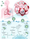
Fig. 1
Fig. 1. SARS-CoV-2 infection lifecycle. Following entry into host cells by engagement of the severe acute respiratory syndrome coronavirus 2 (SARS-CoV-2) viral envelope glycoprotein spike (S) with receptors on the surface of epithelial cells located in distal lung, e.g. angiotensin-converting enzyme 2 (ACE2) on alveolar cells, and establishment of upper respiratory tract infection via nose, eyes and mouth, the viral lifecycle proceeds with the fusion of viral envelope with cell membrane and subsequent release of viral RNA into the cell. The RNA is then translated to produce viral pre-proteins that are processed by its own proteases to release over two dozen effector proteins which eventually participate in viral replication, hijacking of host pathways, and assembly of virions followed by release.
Fig. 2
Fig. 2. Host signaling and metabolic responses to SARS-CoV-2 infection predicted based on strong similarities with two previous highly pathogenic human β-coronaviruses SARS-CoV and MERS-CoV. After viral entry, the single-stranded RNA genome (gRNA) of SARS-CoV-2 is released in the host cell cytoplasm. ORF1a and ORF1ab mRNAs are translated to produce two large polyproteins, pp1a and pp1ab, which are proteolytically cleaved into 16 non-structural proteins (nsps), including papain-like protease (PLpro), 3C-like protease (3CLpro), RNA-dependent RNA polymerase (RdRp), helicase (Hel), exonuclease (ExoN), and 4 structural proteins. The envelope glycoprotein spike (S) forms a layer of glycoproteins that protrude from the envelope. Two additional transmembrane glycoproteins are incorporated in the virion: envelope (E) and membrane (M). Inside the viral envelope resides the helical nucleocapsid, which consists of the viral positive-sense RNA ((+)RNA) genome encapsidated by protein nucleocapsid (N). An additional 9–12 ORFs are encoded through the transcription of a nested set of sub-genomic RNAs. Functions of these truncated or untruncated sub-genomic RNAs are still unknown. The viral elements (ssRNA or sub-genomic RNAs) are recognized by pattern recognition receptors such as Toll-like receptors (TLRs), retinoic-acid-inducible protein I (RIG-I), RIG-I like receptor, and melanoma differentiation-associated protein 5 (MDA-5) that recruit adapter mitochondrial antiviral-signaling protein (MAVS) and leads to activation of interferon regulatory factor 3 (IRF3) and NF-κB and the subsequent production of type I Interferons (IFNs) and pro-inflammatory cytokines (e.g. IL-1β and IL-6), respectively. Itaconate, an anti-inflammatory metabolite transported by TCA cycle intermediates (e.g. citrate), results in activation of pathways involved in proinflammatory cytokines. Proinflammatory cytokines activate the mammalian target of rapamycin (mTOR) signaling in addition to the increased energy metabolism through aerobic glycolysis. The increased production of tricarboxylic acid cycle (TCA) intermediates e.g. succinate and citrate activate NF-κB downstream signaling resulting in the production of proinflammatory cytokines. The antiviral activity of type I IFNs limit viral replication and modulate the innate and adaptive immune responses. They bind to interferon-α/β receptors (IFNARs), which are expressed on a number of different cells including macrophages and activate the JAK/STAT signaling pathway. This signaling leads to the formation of the STAT1/2/IRF9 complex and the induction of a plethora of IFN-stimulated genes (ISGs). Cytokines released by infected cells modulate the adaptive immune response by recruiting and activating immune cells such as macrophages, B-cells, and T-cells to orchestrate the elimination of the virus. However, an unbalanced immune response has been reported to cause hyper-inflammation, a condition termed as ‘cytokine storm’ in cases of severe clinical symptoms of COVID-19. Many, but not all, aspects of this model have been directly verified in the rapidly emerging SARS-CoV-2 experimental literature.
Fig. 3
Fig. 3. Imaging-based interaction analysis using viral reporter strains for monitoring virus–host interactions. Workflow illustrates fluorescence imaging using immunolabeling of fixed cells or tissues to stain a protein of interest, or reporter viruses stained with antibodies labeled with fluorophores.
Fig. 4
Fig. 4. Computational approaches for virus–host interaction analysis. Bioinformatics pipeline for identifying virus–host interactions by data parsing from high quality virus–host protein–protein interactomes (PPIs) databases and mapped to form putative domain–domain interaction networks (DDIs) for conserved domains of both virus and host proteins that can be assessed for their network distribution properties, such as node topology, as well as enrichment for gene ontology (GO) functional annotations and disease associations.
Fig. 5
Fig. 5. Molecular biology-based investigations of coronavirus–host interactions. (A) The yeast 2-hybrid methods of screening PPIs involve mating two haploid complementary yeast strains each expressing a distinct expression plasmid. The first strain expresses a protein bait fused to a DNA-binding domain (DBD) that binds to its cognate binding site, usually upstream of a reporter gene. The second strain expresses a protein prey fused to a transcription activation domain (AD). If there is interaction between bait and prey, the AD is brought into the proximity of the DBD which causes transcriptional activation of the reporter gene leading to selection. (B) Engineered CRISPR systems contain two components, a single guide RNA (sgRNA) and a CRISPR-associated endonuclease (Cas9 protein) to edit (insert or delete) DNA sequences by generating double stranded breaks (DSB) and introducing indels (insertions/deletions) for generation of gene knock-out libraries. In CRISPR–Cas9-pooled screen methods, single-guide RNA (sgRNA) libraries are initially synthesized as lentiviral vectors and amplified and packaged into lentiviruses. Lentivirus pools are then used to transduce Cas9 positive target cells at low multiplicity of infection (MOI). The genomic DNA is extracted from input versus selected cell pools, and the integrated sgRNA sequences are PCR-amplified. The sgRNA abundance is then determined by next-generation DNA sequencing. (C) Workflow showing the application of the CRISPR–Cas screening platform in detection of SARS-CoV-2 infection. A SARS-CoV-2 gene specific CRISPR–Cas sgRNA library is created from identifying the conserved gene sequences of pan-coronaviral genome and validated for detection of SARS-CoV-2 RNAs (PAC-MAN method). This library is then used to generate lateral flow strip-based assays to detect the viral RNAs in nasopharyngeal swabs from COVID-19 patients.
Fig. 6
Fig. 6. Mass spectrometry-based techniques used to identify virus–host interaction networks. (A) Overview of proteomic tools utilized in the of study host–pathogen interactions and their integration with omics approaches. (B) Quantitative multi-omic analysis workflow to define the host response to an infection. The resulting datasets are mapped to known metabolic pathways to measure the up- or downregulation upon infection and integrated with phenotype data to construct correlation networks. (C) Schematic of immunoaffinity or epitope tag-based affinity purification coupled to mass spectrometry (IP-MS/AP-MS) workflow. Components include immunoaffinity purification of protein complexes, enzymatic digestion of proteins, nano-liquid chromatography coupled to mass spectrometry (nLC-MS/MS), and bioinformatic analysis to identify proteins. Label-free protein quantification may be performed by MS/MS spectral counting or precursor ion intensity (MS1) integration. (D) Global proteomic approaches can be used to study alterations throughout infection in protein abundance. These changes can be quantified at the MS level, by pulse labeling via Stable Isotope Labeling in Cell Culture (SILAC) and/or isobaric tagging (such as tandem mass tagging, TMT) of samples after proteolysis and comparing the ion intensities to define proteome alterations at multiple time points of infection. In this workflow, cells are harvested at different infection times after pulse labeling and digested peptides from each sample are labeled with isobaric tags consisting of unique reporter masses. The samples are mixed together for MS analysis, and peptide quantification is assessed at the MS/MS level using the reporter ion intensities. Peptide quantitative values derived from sequences assigned to the same protein are used to calculate the overall relative protein abundance. (E) Workflow showing differential analysis of global proteomic and metabolomic profiles of COVID-19 patient cohorts vs. healthy individuals via an untargeted LC-MS/MS platform. (A–D) adapted from ref. 95.
Fig. 7
Fig. 7. Chemo-proteomic analysis of drug or cellular metabolites in coronavirus host interactions. (A) Schematic illustrating small molecule ligand pull down workflow. A SARS-CoV-2 protein of interest or bait fused to an epitope tag is expressed in cells either by transfection of bait plasmid or transduction of SARS-CoV-2 typed lentivirus and immobilized on an affinity matrix specific for the epitope tag. The protein bound matrix is then exposed to metabolites or synthetic compounds to identify high-affinity binders which are then recovered and eluted from the bait protein and identified by untargeted metabolomics. (B) The principle of cellular thermal shift assay coupled to mass spectrometry (CETSA-MS) for the identification of the endogenous target(s) of a bioactive compound is based on measuring changes in the enthalpy of SARS-CoV-2 protein structures upon binding with its ligand, which is reflected in a shift in its melting temperature (Tm). The assay involves treatment of cells or cell free extracts with desired compound/s and subsequent sample heating followed by quantitative proteomic analysis.
Fig. 8
Fig. 8. Virus–host protein microenvironment mapping. Protein environments associated with coronavirus–host cell interactions can be assessed using proximity labeling technologies that rely on proximal protein labeling via an enzyme or small molecule catalyst. The catalyst can be used to probe cell surface and cytosolic environments through attachment to viral components such as the spike protein that engages host cell surfaces or viral replication and transcription proteins responsible for production of RNA. Catalytic proximity labeling can be induced enzymatically or through visible light activation of small molecule photocatalysts to covalently capture host protein interaction environments. Labeled proteins can then be affinity enriched and identified through LC-MS based proteomics.

Similar articles

Cited by

References

    1. Wu F. Zhao S. Yu B. Chen Y. M. Wang W. Song Z. G. Hu Y. Tao Z. W. Tian J. H. Pei Y. Y. Yuan M. L. Zhang Y. L. Dai F. H. Liu Y. Wang Q. M. Zheng J. J. Xu L. Holmes E. C. Zhang Y. Z. Nature. 2020;579:265–269. doi: 10.1038/s41586-020-2008-3. - DOI - PMC - PubMed
    1. Krammer F. Nature. 2020;586:516–527. doi: 10.1038/s41586-020-2798-3. - DOI - PubMed
    1. Corum J., Wu K. J. and Zimmer C., Coronavirus Drug and Treatment Tracker, nytimes.com/interactive/2020/science/coronavirus-drugs-treatments.html
    1. Sheahan T. P. Sims A. C. Leist S. R. Schafer A. Won J. Brown A. J. Montgomery S. A. Hogg A. Babusis D. Clarke M. O. Spahn J. E. Bauer L. Sellers S. Porter D. Feng J. Y. Cihlar T. Jordan R. Denison M. R. Baric R. S. Nat. Commun. 2020;11:222. doi: 10.1038/s41467-019-13940-6. - DOI - PMC - PubMed
    1. Yurkovetskiy L. Wang X. Pascal K. E. Tomkins-Tinch C. Nyalile T. P. Wang Y. Baum A. Diehl W. E. Dauphin A. Carbone C. Veinotte K. Egri S. B. Schaffner S. F. Lemieux J. E. Munro J. B. Rafique A. Barve A. Sabeti P. C. Kyratsous C. A. Dudkina N. V. Shen K. Luban J. Cell. 2020;183:739–751. doi: 10.1016/j.cell.2020.09.032. - DOI - PMC - PubMed