Review
. 2021 Jan 26;2(2):368-386.
doi: 10.1039/d0cb00218f. eCollection 2021 Apr 1.

Brief survey on organometalated antibacterial drugs and metal-based materials with antibacterial activity

Affiliations
Review

Brief survey on organometalated antibacterial drugs and metal-based materials with antibacterial activity

Przemysław Biegański et al. RSC Chem Biol. .

Abstract

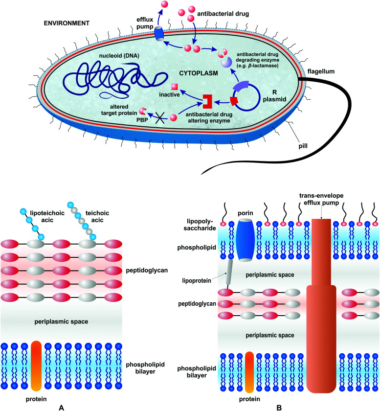
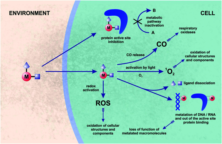
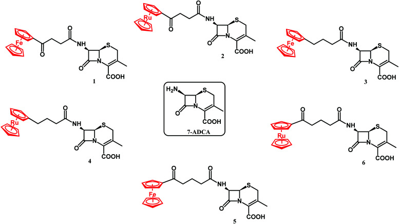
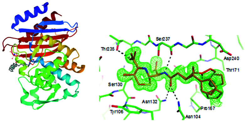
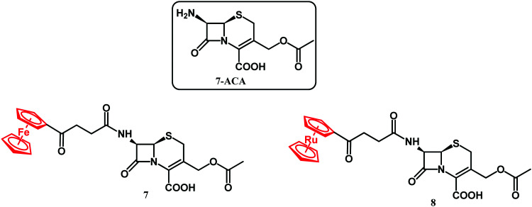
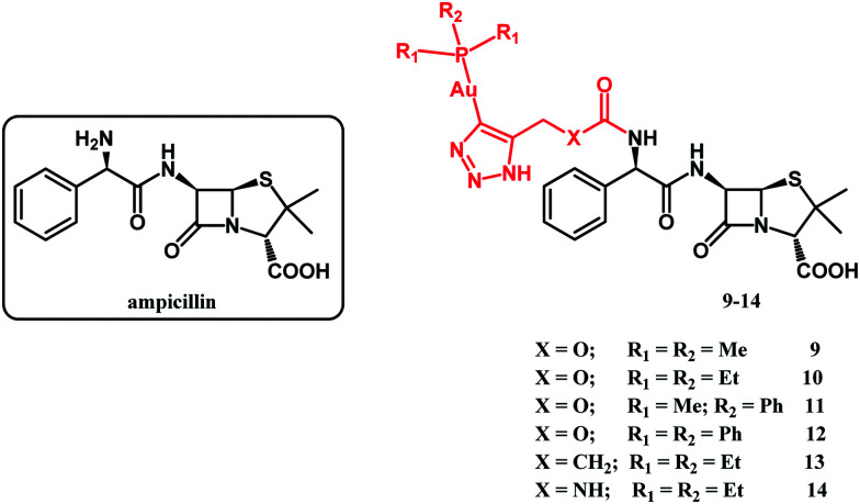
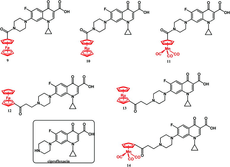
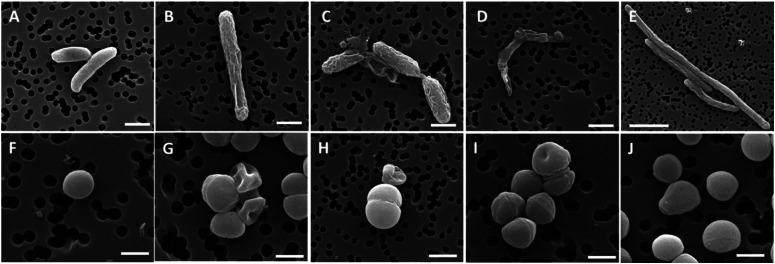
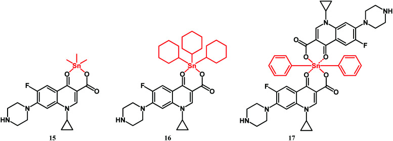
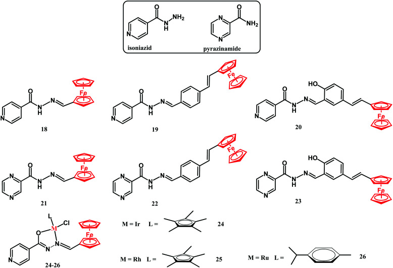
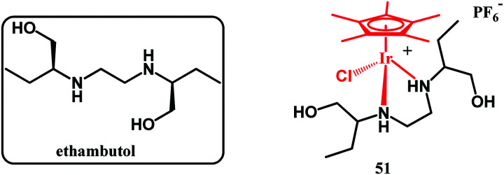
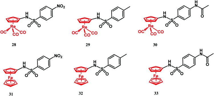
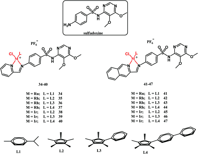
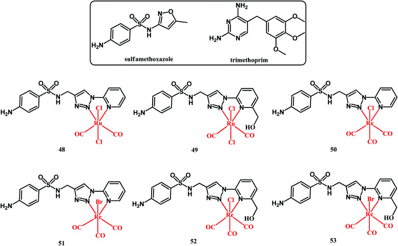
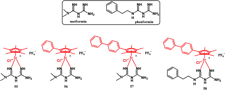
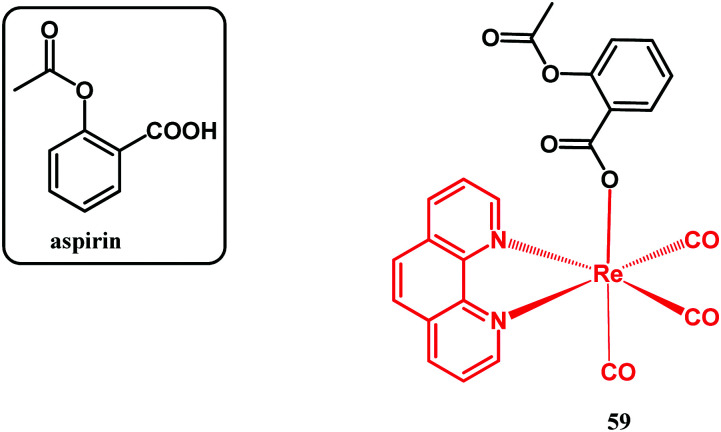
Rising bacterial antibiotic resistance is a global threat. To deal with it, new antibacterial agents and antiseptic materials need to be developed. One alternative in this quest is the organometallic derivatization of well-established antibacterial drugs and also the fabrication of advanced metal-based materials having antibacterial properties. Metal-based agents and materials often show new modes of antimicrobial action which enable them to overcome drug resistance in pathogenic bacterial strains. This review summarizes recent (2017-2020) progress in the field of organometallic-derived antibacterial drugs and metal-based materials having antibacterial activity. Specifically, it covers organometallic derivatives of antibacterial drugs including β-lactams, ciprofloxacin, isoniazid, trimethoprim, sulfadoxine, sulfamethoxazole, and ethambutol as well as non-antibacterial drugs like metformin, phenformin and aspirin. Recent advances and reported clinical trials in the use of metal-based nanomaterials as antibiofouling coatings on medical devices, as photocatalytic agents in indoor air pollutant control, and also as photodynamic/photothermal antimicrobial agents are also summarized.


Conflict of interest statement

There are no conflicts to declare.

Figures

Fig. 1
Fig. 1. Different mechanisms of drug resistance in bacteria and a schematic representation of the Gram-positive (A) and Gram-negative (B) bacterial cell envelope.
Fig. 2
Fig. 2. General view on possible mechanisms of action of organometallic compounds in bacteria.
Fig. 3
Fig. 3. Structures of 7-ADCA and compounds 1–6.
Fig. 4
Fig. 4. Left: Structure of the acyl–enzyme complex of CTX-M E166A with compound 2 (compound 2 depicted in grey; adopts two alternative conformations). Image from the RCSB PDB (rcsb.org) of PDB ID 5UJO (E. M. Lewandowski, Ł. Szczupak, S. Wong, J. Skiba, A. Guśpiel, J. Solecka, V. Vrček, K. Kowalski and Y. Chen, Organometallics, 2017, 36, 1673–1676). Right: Enlarged view of the acyl–enzyme complex nested in the protein binding site. The protein and compound are shown in green and gold, respectively. The unbiased FoFc density map is shown in green at 2σ. The figure was reproduced from ref. 107 with permission from the American Chemical Society.
Fig. 5
Fig. 5. Structures of 7-ACA and compounds 7 and 8.
Fig. 6
Fig. 6. Structures of ampicillin and compounds 9–14.
Fig. 7
Fig. 7. Structures of ciprofloxacin and compounds 9–14.
Fig. 8
Fig. 8. SEM micrographs of the bacterial cell morphology after exposure to compounds 12–14 and ciprofloxacin. Top panels, E. coli cells; bottom panels, S. aureus cells. (A and F) control, non-treated samples; and bacteria treated with compounds 12 (B and G), 13 (C and H), 14 (D and I), and ciprofloxacin (E and J). Scale bars: 500 nm in F, G, I, and J; 1 μm in A–D and H; and 3 μm in E. Reproduced from ref. 114 with permission from the Royal Society of Chemistry.
Fig. 9
Fig. 9. Structures of compounds 15–17.
Fig. 10
Fig. 10. Structures of isoniazid, pyrazinamide and compounds 18–26.
Fig. 11
Fig. 11. Structures of ethambutol and compound 27.
Fig. 12
Fig. 12. Structures of compounds 28–33.
Fig. 13
Fig. 13. Structures of sulfadoxine and compounds 34–47.
Fig. 14
Fig. 14. Structures of sulfamethoxazole, trimethoprim and compounds 48–53.
Fig. 15
Fig. 15. Structure of compound 54.
Fig. 16
Fig. 16. Structures of metformin, phenformin and compounds 55–58.
Fig. 17
Fig. 17. Structure of aspirin and compound 59.
None
Przemysław Biegański
None
Łukasz Szczupak
None
Manuel Arruebo
None
Konrad Kowalski

References

    1. Planavsky N. J. Asael D. Hofmann A. Reinhard C. T. Lalonde S. V. Knudsen A. Wang X. Ossa F. O. Pecoits E. Smith A. J. B. Beukes N. J. Bekker A. Johnson T. M. Konhauser K. O. Lyons T. W. Rouxel O. J. Nat. Geosci. 2014;7:283–286. doi: 10.1038/ngeo2122. - DOI
    1. Thomazo C. Ader M. Philippot P. Geobiology. 2011;9:107–120. doi: 10.1111/j.1472-4669.2011.00271.x. - DOI - PubMed
    1. Lepot K. Earth-Sci. Rev. 2020;209:103296. doi: 10.1016/j.earscirev.2020.103296. - DOI
    1. Lepot K. Addad A. Knoll A. H. Wang J. Troadec D. Béché A. Javaux E. J. Nat. Commun. 2017;8:14890–14900. doi: 10.1038/ncomms14890. - DOI - PMC - PubMed
    1. Jannasch H. W. Mottl M. J. Science. 1985;229:717–725. doi: 10.1126/science.229.4715.717. - DOI - PubMed

LinkOut - more resources