. 2021 Jan 19;2(2):577-591.
doi: 10.1039/d0cb00200c. eCollection 2021 Apr 1.

Identifying cysteine residues susceptible to oxidation by photoactivatable atomic oxygen precursors using a proteome-wide analysis

Affiliations

Identifying cysteine residues susceptible to oxidation by photoactivatable atomic oxygen precursors using a proteome-wide analysis

Ankita Isor et al. RSC Chem Biol. .

Abstract

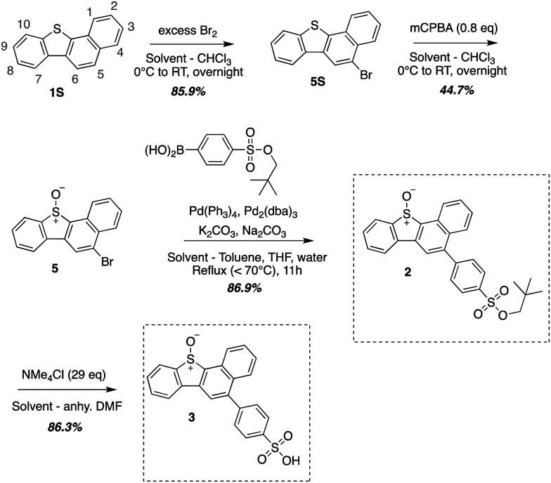
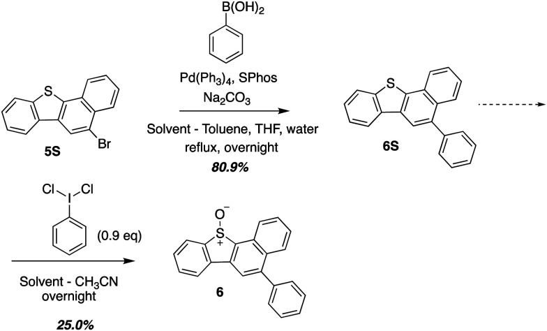
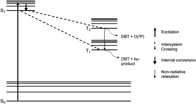
The reactivity profile of atomic oxygen [O(3P)] in the condensed phase has shown a preference for the thiol group of cysteines. In this work, water-soluble O(3P)-precursors were synthesized by adding aromatic burdens and water-soluble sulphonic acid groups to the core structure of dibenzothiophene-S-oxide (DBTO) to study O(3P) reactivity in cell lysates and live cells. The photodeoxygenation of these compounds was investigated using common intermediates, which revealed that an increase in aromatic burdens to the DBTO core structure decreases the total oxidation yield due to competitive photodeoxygenation mechanisms. These derivatives were then tested in cell lysates and live cells to profile changes in cysteine reactivity using the isoTOP-ABPP chemoproteomics platform. The results from this analysis indicated that O(3P) significantly affects cysteine reactivity in the cell. Additionally, O(3P) was found to oxidize cysteines within peptide sequences with leucine and serine conserved at the sites surrounding the oxidized cysteine. O(3P) was also found to least likely oxidize cysteines among membrane proteins.


Conflict of interest statement

There are no conflicts to declare.

Figures

Fig. 1
Fig. 1. Benzannulated DBTO derivative: benzonaphthothiophene-S-oxide.
Fig. 2
Fig. 2. Oxidation pathways for thiols using DBTO as an O(3P) pre-cursor outlined in Omlid et al., 2017.
Fig. 3
Fig. 3. Synthesized water-soluble and red-shifted DBTO derivatives to study cysteine oxidation in cell lysates.
Scheme 1
Scheme 1. Synthetic outline for 1 (Zheng et al., 2016).
Scheme 2
Scheme 2. Synthetic outline for 2 and 3.
Scheme 3
Scheme 3. Synthesis of 6 which represents the core structure of 2 and 3.
Fig. 4
Fig. 4. UV-Vis absorption spectra for DBTO, 1–3, and 6.
Fig. 5
Fig. 5. Common intermediate experiment with toluene.
Fig. 6
Fig. 6. Modified Jablonski diagram for photodeoxygenation of DBTO.
Fig. 7
Fig. 7. Cysteine reactivity surveyed by Iodoacetamide labeling (A) iodoacetamide labeling in lysate treated with DBTO derivatives 1–3 ± UV irradiation; (B) iodoacetamide labeling in lysates from live HeLa cells ± treatment with 1 ± UV irradiation; (C) cysteine reactivity from HeLa cells treated with 1 ± UV irradiation; (D) cysteine reactivity from HeLa cells treated with 1 – UV irradiation.
Fig. 8
Fig. 8. Cellular location of peptides that showed two-fold reduction in labelling. The inner circle represents the peptide count for cellular components and the outer circle represents the specific location within those components.
Fig. 9
Fig. 9. (Generated using WebLogo): (A) sequence Logos for peptides with a single modified cysteine exhibiting two-fold reduction in labeling segmented by “C” in the sequence (B) sequence Logos for peptides with a single modified cysteine that showed an increase in labeling segmented by “C” in the sequen.
Fig. 10
Fig. 10. Solvent accessible surface area of sulphur atoms in cysteines with highest reduction in labeling of peptides with resolved crystal structures.

References

    1. Fleury C. Mignotte B. Vayssière J. L. Biochimie. 2002;84:131–141. doi: 10.1016/S0300-9084(02)01369-X. - DOI - PubMed
    1. Kalyanaraman B. Cheng G. Hardy M. Ouari O. Bennett B. Zielonka J. Redox Biol. 2018;15:347–362. doi: 10.1016/j.redox.2017.12.012. - DOI - PMC - PubMed
    1. Schieber M. Chandel N. S. Curr. Biol. 2014;24:R453–R462. doi: 10.1016/j.cub.2014.03.034. - DOI - PMC - PubMed
    1. Nietzel T. Mostertz J. Hochgräfe F. Schwarzländer M. Mitochondrion. 2017;33:72–83. doi: 10.1016/j.mito.2016.07.010. - DOI - PubMed
    1. Zadok E. Amar D. Mazur Y. J. Am. Chem. Soc. 1980;102:6369–6370. doi: 10.1021/ja00540a046. - DOI