. 2021 Nov;24(5):746.
doi: 10.3892/mmr.2021.12386. Epub 2021 Aug 30.

Involvement of autophagy in diosgenin‑induced megakaryocyte differentiation in human erythroleukemia cells

Affiliations

Involvement of autophagy in diosgenin‑induced megakaryocyte differentiation in human erythroleukemia cells

Dima Diab et al. Mol Med Rep. 2021 Nov.

Abstract

Natural agents have been used to restart the process of differentiation that is inhibited during leukemic transformation of hematopoietic stem or progenitor cells. Autophagy is a housekeeping pathway that maintains cell homeostasis against stress by recycling macromolecules and organelles and plays an important role in cell differentiation. In the present study, an experimental model was established to investigate the involvement of autophagy in the megakaryocyte differentiation of human erythroleukemia (HEL) cells induced by diosgenin [also known as (25R)‑Spirosten‑5‑en‑3b‑ol]. It was demonstrated that Atg7 expression was upregulated from day 1 of diosgenin‑induced differentiation and was accompanied by a significant elevation in the conversion of light chain 3 A/B (LC3‑A/B)‑I to LC3‑A/B‑II. Autophagy was modulated before or after the induction of megakaryocyte differentiation using 3‑methyladenine (3‑MA, autophagy inhibitor) and metformin (Met, autophagy initiation activator). 3‑MA induced a significant accumulation of the LC3 A/B‑II form at day 8 of differentiation. It was revealed that 3‑MA had a significant repressive effect on the nuclear (polyploidization) and membrane glycoprotein V [(GpV) expression] maturation. On the other hand, autophagy activation increased GpV genomic expression, but did not change the nuclear maturation profile after HEL cells treatment with Met. It was concluded that autophagy inhibition had a more prominent effect on the diosgenin‑differentiated cells than autophagy activation.

Keywords: 3‑methyladenine; autophagy; diosgenin; human erythroleukemia cells; megakaryocyte differentiation.


Conflict of interest statement

The authors declare that they have no competing interests.

Figures
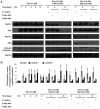
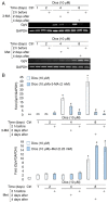
Figure 1.
Figure 1.
Time-dependent expression of autophagy-related protein expression during diosgenin-induced megakaryocytic differentiation in human erythroleukemia cells and effect of autophagy inhibitor or activator. (A) Beclin1, Atg7, Atg12-5, Atg3 and LC3A/B expression levels were evaluated in the total cellular pool using western blot analysis (GAPDH was used as a loading control and the blot shown is representative of three separate experiments). (B) The fold increase values are expressed as the mean ± SEM (n=3). *P<0.05 vs. ctrl group. Dios, diosgenin; LC3A/B, light chain 3 A/B; Ctrl, control; 3-MA, 3-methyladenine; Met, metformin.
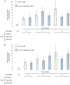
Figure 2.
Figure 2.
Effect of autophagy inhibitor or activator on cell ploidy during diosgenin-induced megakaryocyte differentiation in human erythroleukemia cells. Relative percentage of nuclear ploidy classes were determined by flow cytometry and values are expressed as the mean ± SEM (n=3). *P<0.05 vs. dios alone group (>4N). Dios, diosgenin; Ctrl, control; 3-MA, 3-methyladenine; Met, metformin.
Figure 3.
Figure 3.
Effect of autophagy inhibitor or activator on GpV mRNA expression during diosgenin-induced megakaryocyte differentiation in human erythroleukemia cells. (A) Reverse transcription-semi-quantitative PCR analysis was performed after total RNA extraction and semi-quantification of GpV transcripts using GAPDH as an internal control. Agarose gels shown are representative of three separate experiments. (B) The fold ratios are expressed as the mean ± SEM (n=5). *P<0.05 vs. ctrl group; #P<0.05 vs. dios group. Dios, diosgenin; Ctrl, control; 3-MA, 3-methyladenine; Met, metformin; GpV, glycoprotein V.
Figure 4.
Figure 4.
Effect of autophagy (A) inhibitor or (B) activator on DNA fragmentation during diosgenin-induced megakaryocytic differentiation in human erythroleukemia cells. DNA fragmentation was quantified from cytosol extracts by ELISA. Results are reported as n-fold compared with the control. Data are shown as the mean ± SEM (n=3). Dios, diosgenin; Ctrl, control; 3-MA, 3-methyladenine; Met, metformin.

References

    1. Sell S. Leukemia: Stem cells, maturation arrest, and differentiation therapy. Stem Cell Rev. 2005;1:197–205. doi: 10.1385/SCR:1:3:197. - DOI - PubMed
    1. Nowak D, Stewart D, Koeffler HP. Differentiation therapy of leukemia: 3 decades of development. Blood. 2009;113:3655–3665. doi: 10.1182/blood-2009-01-198911. - DOI - PMC - PubMed
    1. Gocek E, Marcinkowska E. Differentiation therapy of acute myeloid leukemia. Cancers (Basel) 2011;3:2402–2420. doi: 10.3390/cancers3022402. - DOI - PMC - PubMed
    1. Yang J, Qiu J, Hu Y, Zhang Y, Chen L, Long Q, Chen J, Song J, Rao Q, Li Y, et al. A natural small molecule induces megakaryocytic differentiation and suppresses leukemogenesis through activation of PKCδ/ERK1/2 signaling pathway in erythroleukemia cells. Biomed Pharmacother. 2019;118:109265. doi: 10.1016/j.biopha.2019.109265. - DOI - PubMed
    1. Gao X, Wu J, Zou W, Dai Y. Two ellagic acids isolated from roots of Sanguisorba officinalis L. promote hematopoietic progenitor cell proliferation and megakaryocyte differentiation. Molecules. 2014;19:5448–5458. doi: 10.3390/molecules19045448. - DOI - PMC - PubMed