. 2021 Nov;30(11):2338-2345.
doi: 10.1002/pro.4176. Epub 2021 Sep 13.

Quantitative analyses for effects of neddylation on CRL2VHL substrate ubiquitination and degradation

Affiliations

Quantitative analyses for effects of neddylation on CRL2VHL substrate ubiquitination and degradation

Kankan Wang et al. Protein Sci. 2021 Nov.

Abstract

Through catalyzing the ubiquitination of key regulatory proteins, cullin-RING ubiquitin ligases (CRLs) play essential biological roles and their activities are controlled by multiple mechanisms including neddylation, the conjugation of NEDD8 to cullins. Upon neddylation, a CRL, such as the CUL1-based CRL1, undergoes conformational changes that accelerate substrate ubiquitination. Given the structural diversity across subfamilies of CRLs and their substrates, to what extent neddylation modulates the activity of individual CRLs remains to be evaluated. Here, through reconstituting the CRL2 ubiquitination reaction in vitro, we showed that neddylation promotes CRL2VHL -dependent degradation of both full-length HIF1α and the degron peptide of HIF1α, resulting in more than 10-fold increase in the rate of substrate ubiquitination. Consistently, pevonedistat (also known as MLN4924), an inhibitor of neddylation, inhibits the degradation of HIF1α in RCC4 cells stably expressing VHL in cycloheximide chase assays. However, such inhibitory effect of pevonedistat on HIF1α degradation was not observed in HEK293 cells, which was further found to be due to CRL2VHL -independent degradation that was active in HEK293 but not RCC4 cells. After truncating HIF1α to its Carboxy-terminal Oxygen-Dependent Degradation (CODD) domain, we showed that pevonedistat inhibited the degradation of CODD and increased its half-life by six-fold in HEK293 cells. Our results demonstrate that neddylation plays a significant role in activating CRL2, and the cellular activity of CRL2VHL is better reflected by the degradation of CODD than that of HIF1α, especially under conditions where CRL2-independent degradation of HIF1α is active.

Keywords: CODD; CRL2 ubiquitin ligase; neddylation; protein degradation; ubiquitination.


Conflict of interest statement

The authors declare no conflict of interest.

Figures

FIGURE 1
FIGURE 1
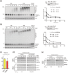
Eliminating cullin neddylation failed to inhibit the degradation of HIF1α in HEK293 cells. (a) HEK293 cells were pretreated with DFX for 5 h and pevonedistat or equivalent volume of DMSO for 1 h. After washout of inhibitors, cells were treated by CHX with or without pevonedistat for indicated time and were collected for WB analyses. (b) same as in (a) except that HEK293 cells containing tetracycline induced FLAGHIF1α were used
FIGURE 2
FIGURE 2
Neddylation is required for efficient CRL2VHL‐dependent degradation of HIF1α in vitro and in RCC4 (+VHL) cells. (a,b) Full‐length FLAGHIF1α purified from RCC4 (VHL null) cells stably expressing FLAGHIF1α (a) or HIF1α degron peptide (b) were ubiquitinated in vitro for indicated time with or without CUL2 neddylation. Samples without CUL2 served as negative controls. Intensities of unmodified (u.m.) FLAGHIF1α or degron peptide bands were normalized to intensities of VHL bands for regression analyses. WB of CUL2 from each group and regression curves of remaining unmodified substrates were shown on the right. (c) Average relative changes of substrate t 1/2 by neddylation from experiments in (a,b). Error bars: range of values, n = 2. (D) RCC4 (VHL null) and RCC4 (+VHL) cells were pretreated with DFX for 3 h and pevonedistat or equivalent volume of DMSO for 1 h. After washout of inhibitors, cells were treated by CHX with or without pevonedistat for indicated time and were collected for WB analyses. (e) Same assays as performed in (d) with FLAGHIF1α stably expressed in RCC4 (+VHL) cells. In (d,e), relative t 1/2 was shown as average ± range of values (n = 2)
FIGURE 3
FIGURE 3
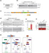
The degradation of CODD reports CRL2VHL activity in HEK293 cells in which VHL‐independent degradation of full‐length HIF1α is active. (a) HEK293 cells containing tetracycline induced FLAGHIF1α were treated by 100 μM VH‐298 for indicated time and were collected for WB analyses. (b) The same cells as in (a) were pretreated with DFX for 5 h and VH‐298 or equivalent volume of DMSO for 1 h. After washout of inhibitors, cells were treated by CHX with or without VH‐298 for indicated time and were collected for WB analyses. (c) Schematic illustration of the CODD truncation of HIF1α. Red vertical lines denote ubiquitination sites reported in GGBase. (d) Same assays as in Figure 1b with HEK293 cells containing tetracycline induced FLAGHIF1α or FLAGCODD. Relative t 1/2 of FLAGHIF1α or FLAGCODD elimination are shown below the corresponding blot. (e) Average changes in relative t 1/2 from experiments in (d). Error bars: SEM, n = 3, p < 0.01. (f) Unmodified (upper band) and hydroxylated (lower band) FLAGCODD separated on a 12.5% SDS‐PAGE gel. HEK293 cells with tetracycline induced expression of FLAGCODD were treated for 4 h with DMSO (Lane 1), or pevonedistat (Lane 2), or DFX (Lane 3), and were collected WB analyses. Equal volumes of samples in Lanes 2 and 3 were mixed and included in Lane 4. The bottom blot shows overlay of signals from FLAG and hydroxy‐HIF‐1α (Pro564) antibodies. (g) Summary of HIF1α degradation machineries. CRL2VHL binds the hydroxylated ODD domain of HIF1α or the hydroxylated CODD truncation to catalyze their ubiquitination and degradation. This CRL2VHL‐dependent degradation is accelerated by neddylation and can be inhibited by pevonedistat. CRL2VHL‐independent degradation acts through regions other than the HIF1α CODD domain and cannot be inhibited by pevonedistat

Similar articles

Cited by

References

    1. Hershko A. The ubiquitin system for protein degradation. Annu Rev Biochem. 1992;61:761–807. - PubMed
    1. Varshavsky A. The ubiquitin system. Trends Biochem Sci. 1997;22:383–387. - PubMed
    1. Petroski MD, Deshaies RJ. Function and regulation of cullin‐RING ubiquitin ligases. Nat Rev Mol Cell Biol. 2005;6:9–20. - PubMed
    1. Skaar JR, Pagan JK, Pagano M. SCF ubiquitin ligase‐targeted therapies. Nat Rev Drug Discov. 2014;13:889–903. - PMC - PubMed
    1. Zheng N, Schulman BA, Song L, et al. Structure of the Cul1‐Rbx1‐Skp1‐F boxSkp2 SCF ubiquitin ligase complex. Nature. 2002;416:703–709. - PubMed

Publication types

MeSH terms