. 2021 Oct 1;321(4):F505-F516.
doi: 10.1152/ajprenal.00041.2021. Epub 2021 Aug 30.

Natural antibody and complement activation characterize patients with idiopathic nephrotic syndrome

Affiliations

Natural antibody and complement activation characterize patients with idiopathic nephrotic syndrome

Howard Trachtman et al. Am J Physiol Renal Physiol. .

Abstract

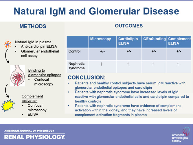
Focal segmental glomerulosclerosis (FSGS) and minimal change disease (MCD) are common forms of idiopathic nephrotic syndrome. The causes of these diseases are incompletely understood, but the response of patients to immunosuppressive therapies suggests that their pathogenesis is at least in part immune mediated. Preclinical and clinical research indicates that activation of the classical pathway of complement contributes to glomerular injury in FSGS. Glomerular IgM deposits are also prominent in some patients, raising the possibility that IgM is a trigger of classical pathway activation. In the present study, we examined the pattern of complement activation in the glomeruli and plasma of patients with nephrotic syndrome. We also tested whether patients with FSGS and MCD have elevated levels of natural IgM reactive with epitopes on glomerular endothelial cells and cardiolipin. We found evidence of classical pathway activation in patients with idiopathic nephrotic syndrome compared with healthy control subjects. We also detected higher levels of self-reactive IgM to both targets. Based on these results, IgM and classical pathway activation may contribute to disease pathogenesis in some patients with FSGS and MCD.NEW & NOTEWORTHY IgM is detected in biopsies from some patients with nephrotic syndrome, although this has been attributed to passive trapping of the protein. We found, however, that IgM colocalizes with complement activation fragments in some glomeruli. We also found that affected patients had higher levels of IgM reactive to glomerular endothelial cell epitopes. Thus, IgM activates the complement system in the glomeruli of some patients with nephrotic syndrome and may contribute to injury.

Keywords: IgM; complement; focal segmental glomerulosclerosis; minimal change disease; nephrotic syndrome.


Conflict of interest statement

J.M.T. and V.M.H. receive royalties from Alexion Pharmaceuticals, Inc., and are consultants for Q32 Bio, Inc., a company developing complement inhibitors. They also hold stock and will receive royalty income from Q32 Bio, Inc. S.L.K. and F.L. are employed by Q32 Bio, Inc. H.T. is a consultant to Otsuka (Chair, DSMB) and Chemocentryx (DSMB). He has consultancy agreements through NYU Grossman School of Medicine with Retrophin Inc. and Goldfinch Bio Inc. None of the other authors has any conflicts of interest, financial or otherwise, to disclose.

Figures

None
Graphical abstract
Figure 1.
Figure 1.
IgM and complement deposits in focal segmental glomerulosclerosis (FSGS). Biopsy samples from patients with FSGS or thin glomerular basement membrane disease (TBM) were stained for IgM, C4d, iC3b/C3d, and C9neo. A: representative images from a single primary FSGS biopsy containing three glomeruli. Individual glomeruli are indicated with a circle, rectangle, and hexagon. iC3b/C3d (red) and C9neo (shown in turquoise in the right image) were seen in only a subset of the glomeruli that were positive for IgM (blue) and C4d (green). Original magnification: ×200. Scale bars = 100 µm. B: high-powered views of a single glomerulus from a FSGS biopsy showing that IgM (blue), C4d (green), and iC3b/C3d (red) were deposited in a similar pattern within the glomerulus. Original magnification: ×600. Scale bars = 20 µm. C: high-powered views of a glomerulus from a TBM biopsy showing that all of the immune proteins could be detected, although to a lesser degree than in FSGS. Original magnification: ×600.
Figure 2.
Figure 2.
Complement activation fragments in plasma from patients with nephrotic syndrome. We measured C4a, Ba, and sC5b-9 in baseline plasma from patients with nephrotic syndrome and healthy controls. A: C4a levels were higher in both cohorts of nephrotic patients compared with healthy controls. B: sC5b-9 levels were higher in samples from the Nephrotic Syndrome Study Network Consortium (NEPTUNE) cohort of patients with nephrotic syndrome compared with healthy controls. C: Ba levels did not significantly differ between patients with nephrotic syndrome or control subjects, but they were significantly higher in patient with focal segmental glomerulosclerosis (FSGS) compared with patients with minimal change disease (MCD). Ba levels were also higher in those who did not achieve a complete or partial remission during follow-up (no PR) compared with those who did (CR/PR). n =29 for the healthy controls, 90 for the NEPTUNE samples, and 15 for the validation cohort. The red bars indicate medians for each group. **P < 0.01; ***P < 0.001.
Figure 3.
Figure 3.
Changes in complement activation fragments over time and after immunosuppression. We measured C4a, Ba, and sC5b-9 in plasma collected in 83 patients with nephrotic syndrome from the Nephrotic Syndrome Study Network Consortium cohort at baseline and during follow-up visits. No significant changes in any of the complement fragments were seen over time. Graphs show the medians for each time point; bars indicate interquartile ranges.
Figure 4.
Figure 4.
Complement activation fragments in urine from patients with nephrotic syndrome. We measured C4a, Ba, and sC5b-9 in baseline urine from patients with nephrotic syndrome and healthy controls. C4a levels were higher in both cohorts of patients compared with healthy controls. Ba levels were not significantly elevated in either of the patient cohorts. sC5b-9 levels were higher in samples from the Nephrotic Syndrome Study Network Consortium (NEPTUNE) cohort of patients with nephrotic syndrome compared with healthy controls. n =29 for the healthy controls, 90 for the NEPTUNE samples, and 15 for the validation cohort. The red bars indicate medians and interquartile ranges. ***P < 0.001.
Figure 5.
Figure 5.
IgM in the serum of patients with nephrotic syndrome binds to glomerular endothelial cells (GenCs). A: we tested binding of various natural IgM clones to human GenCs and confirmed that the natural antibody clone C2 reacted with human cells (dark black line). The isotype control is shown as a shaded gray peak. The other curves represent distinct natural antibody clones, illustrating the variable ability to bind GEnCs. B: serum from healthy controls (n =78) and from the Nephrotic Syndrome Study Network Consortium (NEPTUNE) cohort of patients (n =115) with nephrotic syndrome were incubated with GEnCs in vitro, and binding of IgM was analyzed by flow cytometry (GEnC IgM). Greater binding of IgM to GEnCs was seen in patients with nephrotic syndrome. *P < 0.05. C: GEnC IgM was inversely correlated with estimated glomerular filtration rate (eGFR). D: GEnC IgM levels were in patients who had a complete or partial remission (CR/PR) during follow-up compared with patients who did not have either a complete or partial remission (no PR). E: total serum IgM levels were lower in serum samples from the NEPTUNE cohort of patients (n =30) compared with healthy controls (n =10). **P < 0.01. RFU, relative fluorescence units.
Figure 6.
Figure 6.
IgM in the serum of patients with nephrotic syndrome binds to cardiolipin. We measured levels of Ig that reacted with cardiolipin in serum from healthy controls, patients with chronic kidney disease (CKD), the Nephrotic Syndrome Study Network Consortium (NEPTUNE) cohort of patients with nephrotic syndrome, and a second cohort of patients with nephrotic syndrome (validation cohort). A: anti-cardiolipin IgM (CL-M) levels were significantly higher in the NEPTUNE and validation cohorts than in healthy subjects or patients with CKD. *P < 0.05. B: anti-cardiolipin IgG (CL-G) levels were not significantly different among the groups. C: CL-M levels were measured in serially collected samples from the NEPTUNE cohort of patients with nephrotic syndrome (n = 94). Plotted data are means for each time point; error bars show SDs.
Figure 7.
Figure 7.
Effects of immunosuppression on levels of anti-cardiolipin IgM (CL-M) and complement fragments. A: in the subset of Nephrotic Syndrome Study Network Consortium (NEPTUNE) patients treated with glucocorticoids (n = 47), a significant interaction between treatment with glucocorticoids and CL-M levels was seen, and the level of CL-M decreased over time in this group. No interaction was seen between CL-M levels and age, sex, or race (not shown). In NEPTUNE patients started on prednisone during the study, CL-M (B) and plasma Ba (C) were lower at the subsequent visit. All of the patients in the validation cohort were treated with tacrolimus. D: the level of C4a was significantly decreased at the subsequent visit after tacrolimus was started. *P < 0.05.

References

    1. Rosenberg AZ, Kopp JB. Focal segmental glomerulosclerosis. Clin J Am Soc Nephrol 12: 502–517, 2017. [Erratum in Clin J Am Soc Nephrol 13: 1889, 2018]. doi:10.2215/CJN.05960616. - DOI - PMC - PubMed
    1. Andreoli SP. Racial and ethnic differences in the incidence and progression of focal segmental glomerulosclerosis in children. Adv Ren Replace Ther 11: 105–109, 2004. doi:10.1053/j.arrt.2003.10.015. - DOI - PubMed
    1. Waldman M, Crew RJ, Valeri A, Busch J, Stokes B, Markowitz G, D'Agati V, Appel G. Adult minimal-change disease: clinical characteristics, treatment, and outcomes. Clin J Am Soc Nephrol 2: 445–453, 2007. doi:10.2215/CJN.03531006. - DOI - PubMed
    1. Bose B, Cattran D; Toronto Glomerulonephritis Registry. Glomerular diseases: FSGS. Clin J Am Soc Nephrol 9: 626–632, 2014. doi:10.2215/CJN.05810513. - DOI - PMC - PubMed
    1. Habib R, Girardin E, Gagnadoux MF, Hinglais N, Levy M, Broyer M. Immunopathological findings in idiopathic nephrosis: clinical significance of glomerular “immune deposits.” Pediatr Nephrol 2: 402–408, 1988. doi:10.1007/BF00853431. - DOI - PubMed

Publication types

MeSH terms