Xylose-Configured Cyclophellitols as Selective Inhibitors for Glucocerebrosidase
- PMID: 34459538
- PMCID: PMC8596838
- DOI: 10.1002/cbic.202100396
Xylose-Configured Cyclophellitols as Selective Inhibitors for Glucocerebrosidase
Erratum in
-
Corrigendum: Xylose-Configured Cyclophellitols as Selective Inhibitors for Glucocerebrosidase.Chembiochem. 2023 Feb 1;24(3):e202300728. doi: 10.1002/cbic.202300728. Chembiochem. 2023. PMID: 36725348 Free PMC article. No abstract available.
Abstract
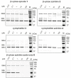
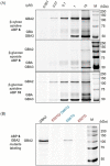
Glucocerebrosidase (GBA), a lysosomal retaining β-d-glucosidase, has recently been shown to hydrolyze β-d-xylosides and to transxylosylate cholesterol. Genetic defects in GBA cause the lysosomal storage disorder Gaucher disease (GD), and also constitute a risk factor for developing Parkinson's disease. GBA and other retaining glycosidases can be selectively visualized by activity-based protein profiling (ABPP) using fluorescent probes composed of a cyclophellitol scaffold having a configuration tailored to the targeted glycosidase family. GBA processes β-d-xylosides in addition to β-d-glucosides, this in contrast to the other two mammalian cellular retaining β-d-glucosidases, GBA2 and GBA3. Here we show that the xylopyranose preference also holds up for covalent inhibitors: xylose-configured cyclophellitol and cyclophellitol aziridines selectively react with GBA over GBA2 and GBA3 in vitro and in vivo, and that the xylose-configured cyclophellitol is more potent and more selective for GBA than the classical GBA inhibitor, conduritol B-epoxide (CBE). Both xylose-configured cyclophellitol and cyclophellitol aziridine cause accumulation of glucosylsphingosine in zebrafish embryo, a characteristic hallmark of GD, and we conclude that these compounds are well suited for creating such chemically induced GD models.
Keywords: Gaucher disease; activity-based probe; conduritol B-epoxide; cyclophellitol; glucocerebrosidase; xylose.
© 2021 The Authors. ChemBioChem published by Wiley-VCH GmbH.
Conflict of interest statement
The authors declare no conflict of interest.
Figures


Similar articles
-
In vivo inactivation of glycosidases by conduritol B epoxide and cyclophellitol as revealed by activity-based protein profiling.FEBS J. 2019 Feb;286(3):584-600. doi: 10.1111/febs.14744. Epub 2019 Feb 2. FEBS J. 2019. PMID: 30600575 Free PMC article.
-
Functionalized Cyclophellitols Are Selective Glucocerebrosidase Inhibitors and Induce a Bona Fide Neuropathic Gaucher Model in Zebrafish.J Am Chem Soc. 2019 Mar 13;141(10):4214-4218. doi: 10.1021/jacs.9b00056. Epub 2019 Mar 4. J Am Chem Soc. 2019. PMID: 30811188 Free PMC article.
-
Exploring functional cyclophellitol analogues as human retaining beta-glucosidase inhibitors.Org Biomol Chem. 2014 Oct 21;12(39):7786-91. doi: 10.1039/c4ob01611d. Epub 2014 Aug 26. Org Biomol Chem. 2014. PMID: 25156485
-
Mechanism-based inhibitors of glycosidases: design and applications.Adv Carbohydr Chem Biochem. 2014;71:297-338. doi: 10.1016/B978-0-12-800128-8.00004-2. Adv Carbohydr Chem Biochem. 2014. PMID: 25480507 Review.
-
Advances in GBA-associated Parkinson's disease--Pathology, presentation and therapies.Neurochem Int. 2016 Feb;93:6-25. doi: 10.1016/j.neuint.2015.12.004. Epub 2015 Dec 30. Neurochem Int. 2016. PMID: 26743617 Review.
Cited by
-
Synthesis and Biological Evaluation of Deoxycyclophellitols as Human Retaining β-Glucosidase Inhibitors.Chemistry. 2024 Dec 13;30(70):e202402988. doi: 10.1002/chem.202402988. Epub 2024 Nov 3. Chemistry. 2024. PMID: 39305182 Free PMC article.
-
Selective labelling of GBA2 in cells with fluorescent β-d-arabinofuranosyl cyclitol aziridines.Chem Sci. 2024 Sep 3;15(37):15212-20. doi: 10.1039/d3sc06146a. Online ahead of print. Chem Sci. 2024. PMID: 39246358 Free PMC article.
References
-
- Aerts J. M., Kuo C. L., Lelieveld L. T., Boer D. E. C., van der Lienden M. J. C., Overkleeft H. S., Artola M., Curr. Opin. Chem. Biol. 2019, 53, 204–215. - PubMed
-
- Ferraz M. J., Marques A. R., Appelman M. D., Verhoek M., Strijland A., Mirzaian M., Scheij S., Ouairy C. M., Lahav D., Wisse P., Overkleeft H. S., Boot R. G., Aerts J. M., FEBS Lett. 2016, 590, 716–725. - PubMed
Publication types
MeSH terms
Substances
Grants and funding
LinkOut - more resources
Full Text Sources
Molecular Biology Databases

