Review
. 2021 Oct 1;78(10):1262-1272.
doi: 10.1001/jamaneurol.2021.1312.

Emerging Neuroimaging Biomarkers Across Disease Stage in Parkinson Disease: A Review

Affiliations
Review

Emerging Neuroimaging Biomarkers Across Disease Stage in Parkinson Disease: A Review

Trina Mitchell et al. JAMA Neurol. .

Abstract

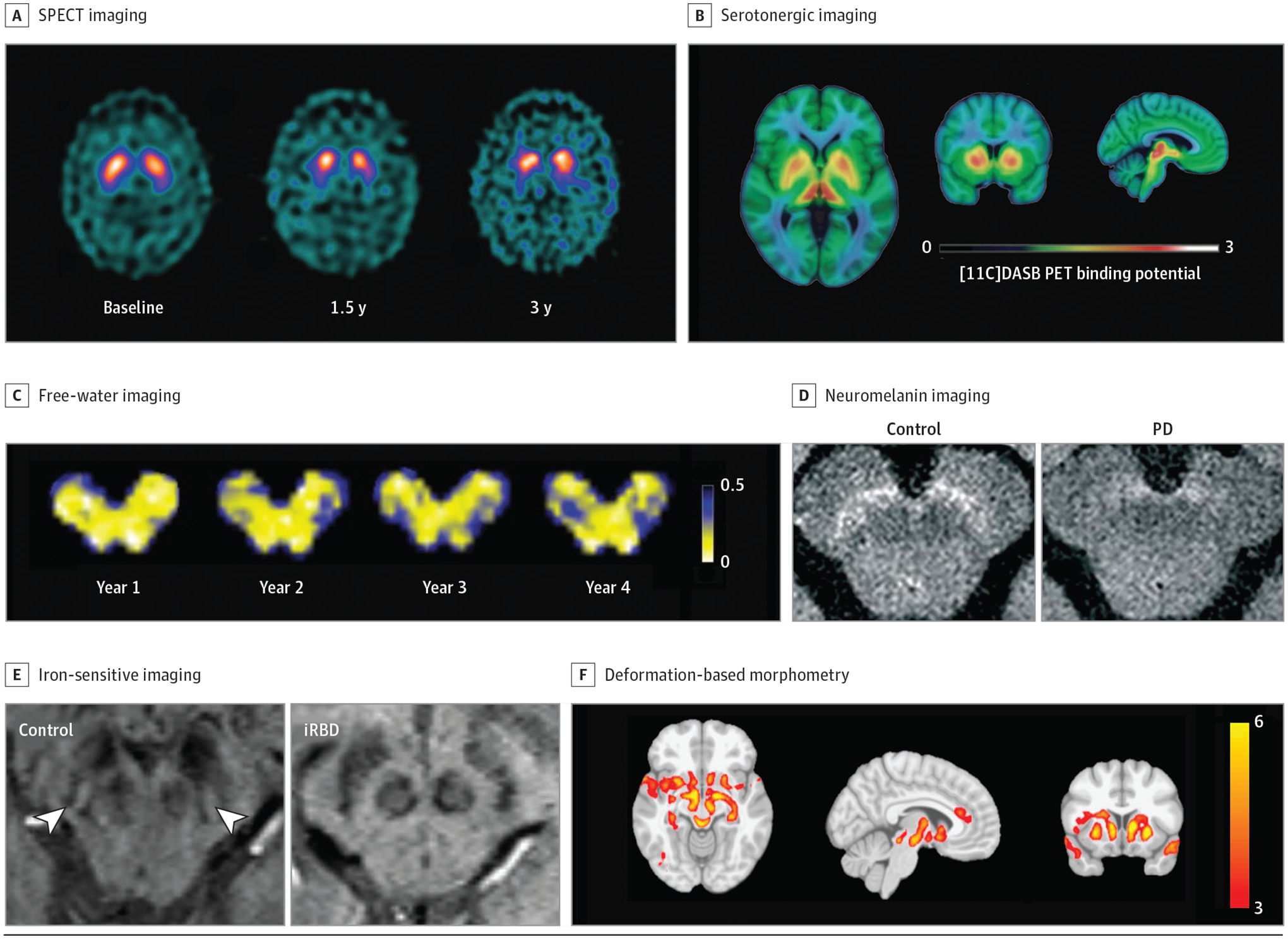
Importance: Imaging biomarkers in Parkinson disease (PD) are increasingly important for monitoring progression in clinical trials and also have the potential to improve clinical care and management. This Review addresses a critical need to make clear the temporal relevance for diagnostic and progression imaging biomarkers to be used by clinicians and researchers over the clinical course of PD. Magnetic resonance imaging (diffusion imaging, neuromelanin-sensitive imaging, iron-sensitive imaging, T1-weighted imaging), positron emission tomography/single-photon emission computed tomography dopaminergic, serotonergic, and cholinergic imaging as well as metabolic and cerebral blood flow network neuroimaging biomarkers in the preclinical, prodromal, early, and moderate to late stages are characterized.

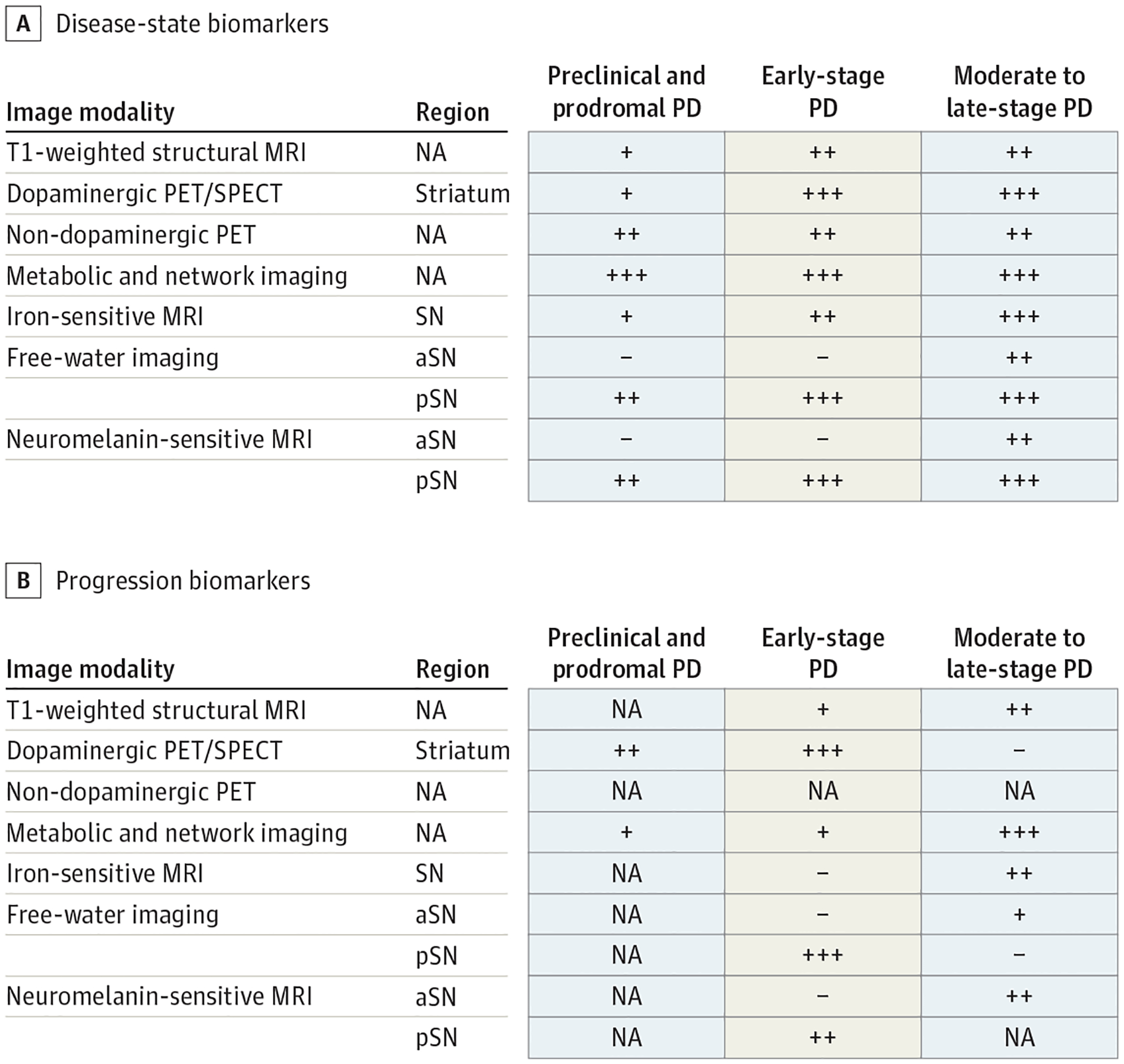
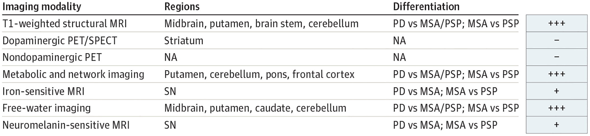
Observations: If a clinical trial is being carried out in the preclinical and prodromal stages, potentially useful disease-state biomarkers include dopaminergic imaging of the striatum; metabolic imaging; free-water, neuromelanin-sensitive, and iron-sensitive imaging in the substantia nigra; and T1-weighted structural magnetic resonance imaging. Disease-state biomarkers that can distinguish atypical parkinsonisms are metabolic imaging, free-water imaging, and T1-weighted imaging; dopaminergic imaging and other molecular imaging track progression in prodromal patients, whereas other established progression biomarkers need to be evaluated in prodromal cohorts. Progression in early-stage PD can be monitored using dopaminergic imaging in the striatum, metabolic imaging, and free-water and neuromelanin-sensitive imaging in the posterior substantia nigra. Progression in patients with moderate to late-stage PD can be monitored using free-water imaging in the anterior substantia nigra, R2* of substantia nigra, and metabolic imaging. Cortical thickness and gyrification might also be useful markers or predictors of progression. Dopaminergic imaging and free-water imaging detect progression over 1 year, whereas other modalities detect progression over 18 months or longer. The reliability of progression biomarkers varies with disease stage, whereas disease-state biomarkers are relatively consistent in individuals with preclinical, prodromal, early, and moderate to late-stage PD.

Conclusions and relevance: Imaging biomarkers for various stages of PD are readily available to be used as outcome measures in clinical trials and are potentially useful in multimodal combination with routine clinical assessment. This Review provides a critically important template for considering disease stage when implementing diagnostic and progression biomarkers in both clinical trials and clinical care settings.


Figures

Figure 1.
Figure 1.
Neuroimaging Biomarkers Neuroimaging techniques are reviewed. A, Single-photon emission computed tomography (SPECT) imaging in controls and those with early-stage, moderate, and late-stage Parkinson disease (PD; reprinted with permission from Schapira and Olanow). B, Positron emission tomography (PET) fluorodopa imaging in controls and those with early-stage and late-stage PD (reprinted with permission from Schapira and Olanow). C, Free-water imaging longitudinally over 4 years in an individual with de novo PD (provided with courtesy of Roxanna Burciu, PhD [Department of Kinesiology and Applied Physiology, University of Delaware, Newark], and used with permission). D, Neuromelanin-sensitive imaging in a healthy control and patient with PD (disease duration of 4 years; reprinted with permission from Biondetti et al). E, Absence of the nigrosome formation hypointensity using iron-sensitive imaging in a patient with idiopathic rapid eye movement sleep behavior disorder (reprinted with permission from De Marzi et al). F, Distribution of atrophy in a patient with PD measured by deformation-based morphometry (reprinted with permission from Zeighami et al).
Figure 2.
Figure 2.
Disease State and Progression Biomarkers A, Evidence for disease-state biomarkers in patients with preclinical and prodromal, early-stage, and moderate to late-stage Parkinson disease. B, Evidence for progression biomarkers in patients with preclinical and prodromal, early-stage, and moderate to late-stage Parkinson disease. + Indicates the effect was shown by only 1 article or that there are inconsistent results across studies; ++, effect shown in 2 to 3 articles using a single-site cohort; +++, effect shown consistently in 3 or more articles or in a multisite cohort; −, no effect of biomarker was shown. Gray-colored squares are used where there are no studies available. aSN indicates anterior substantia nigra; MRI, magnetic resonance imaging; NA, not applicable; PET, positron emission tomography; pSN, posterior substantia nigra; SN, substantia nigra; SPECT, single-photon emission computed tomography.
Figure 3.
Figure 3.
Biomarkers in Differentiating Parkinson Disease (PD) From Atypical Parkinsonisms + Indicates the effect was shown by only 1 article or that there are inconsistent results across studies; +++, effect shown consistently in 3 or more articles or in a multisite cohort; −, no effect of biomarker was shown. MRI indicates magnetic resonance imaging; MSA, multiple system atrophy; NA, not applicable; PET, positron emission tomography; PSP, progressive supranuclear palsy; SN, substantia nigra; SPECT, single-photon emission computed tomography.

References

    1. Schapira AH, Olanow CW. Neuroprotection in Parkinson disease: mysteries, myths, and misconceptions. JAMA. 2004;291(3):358–364. doi:10.1001/jama.291.3.358 - DOI - PubMed
    1. Biondetti E, Gaurav R, Yahia-Cherif L, et al. Spatiotemporal changes in substantia nigra neuromelanin content in Parkinson’s disease. Brain. 2020;143(9):2757–2770. doi:10.1093/brain/awaa216 - DOI - PubMed
    1. De Marzi R, Seppi K, Högl B, et al. Loss of dorsolateral nigral hyperintensity on 3.0 tesla susceptibility-weighted imaging in idiopathic rapid eye movement sleep behavior disorder. Ann Neurol. 2016;79(6):1026–1030. doi:10.1002/ana.24646 - DOI - PubMed
    1. Zeighami Y, Ulla M, Iturria-Medina Y, et al. Network structure of brain atrophy in de novo Parkinson’s disease. Elife. 2015;4:4. doi:10.7554/eLife.08440 - DOI - PMC - PubMed
    1. Iranzo A, Stefani A, Niñerola-Baizan A, et al.; Sleep Innsbruck Barcelona (SINBAR) Group. Left-hemispheric predominance of nigrostriatal deficit in isolated REM sleep behavior disorder. Neurology. 2020;94(15):e1605–e1613. doi:10.1212/WNL.0000000000009246 - DOI - PubMed

Publication types