Review
. 2022 Jan 20;34(1):228-246.
doi: 10.1093/plcell/koab218.

The bryophytes Physcomitrium patens and Marchantia polymorpha as model systems for studying evolutionary cell and developmental biology in plants

Affiliations
Review

The bryophytes Physcomitrium patens and Marchantia polymorpha as model systems for studying evolutionary cell and developmental biology in plants

Satoshi Naramoto et al. Plant Cell. .

Abstract

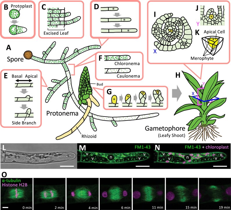
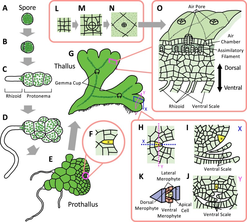
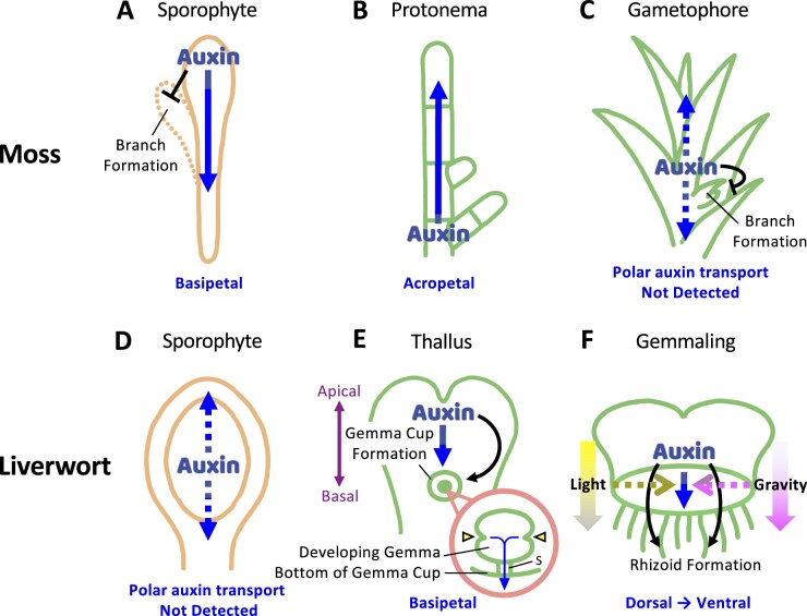
Bryophytes are nonvascular spore-forming plants. Unlike in flowering plants, the gametophyte (haploid) generation of bryophytes dominates the sporophyte (diploid) generation. A comparison of bryophytes with flowering plants allows us to answer some fundamental questions raised in evolutionary cell and developmental biology. The moss Physcomitrium patens was the first bryophyte with a sequenced genome. Many cell and developmental studies have been conducted in this species using gene targeting by homologous recombination. The liverwort Marchantia polymorpha has recently emerged as an excellent model system with low genomic redundancy in most of its regulatory pathways. With the development of molecular genetic tools such as efficient genome editing, both P. patens and M. polymorpha have provided many valuable insights. Here, we review these advances with a special focus on polarity formation at the cell and tissue levels. We examine current knowledge regarding the cellular mechanisms of polarized cell elongation and cell division, including symmetric and asymmetric cell division. We also examine the role of polar auxin transport in mosses and liverworts. Finally, we discuss the future of evolutionary cell and developmental biological studies in plants.


Figures

Figure 1
Figure 1
Illustration of gametophyte development in P. patens. Protonemata germinate from spores (A) or emerge from protoplasts (B) and exhibit filamentous tip growth, which is dependent on polarized cell elongation and subsequent asymmetric cell divisions of apical stem cells located at the tip of each filament. Protonemata can also regenerate from excised cells and tissues upon physical damage (C). Concomitant with tip growth (D), protonemata undergo side branching by performing additional asymmetric cell divisions at the apical end of each cell (except for the apical cell) (E). At a still poorly defined time frame during filamentous growth, chloronemata, a type of juvenile protonemata, transition to caulonemata, which serve as precursor cells that have the potential to generate buds (F). While branching produces new apical stem cells that exhibit 1D growth, bud formation accompanies the formation of a single tetrahedral apical stem cell that undergoes 3D growth, a critical step for gametophore formation (G–K). This single apical stem cell in the shoot displays a spiral pattern of asymmetric cell division and produces merophytes (K), whose subsequent cell division activity results in the formation of leaves (I and J). Most of the tissues generated under these developmental processes have simple tissue structures and are a single cell layer thick (Menand et al., 2007; Kofuji and Hasebe, 2014; Falz and Müller-Schussele, 2019). These characteristics present significant advantages for studies in cell and developmental biology and prompted researchers to study the mechanisms of tip growth, asymmetric cell divisions, cell differentiation, and other cell biological phenomena. For example confocal images, a protonemata tip cell stained with FM1–43 (L–N) and time-lapse images of a dividing protonemata tip cell co-expressing α-tubulin:GFP and histoneH2B:RFP (O) are shown (Hiwatashi et al., personal communication). Bright-field images (L) are shown in gray, fluorescence images of FM1-43 (M and N) and α-tubulin:GFP (O) are shown in green, and autofluorescence of chloroplasts (N), as well as fluorescence images of histone H2B:RFP (O), are shown in magenta. “v” and “n” indicates vacuoles and nucleus, respectively. Yellow-colored cells with an asterisk indicate apical stem cells. The square labeled X or Y in (H) indicates a horizontal or vertical section of the shoot apical meristem. The schematic illustration of the horizontal section (I) or vertical section (J) was drawn based on Harrison et al., 2009 and it is labeled as X or Y, respectively. Scale bars 20 µm (L–N) and 5 µm (O).
Figure 2
Figure 2
Illustration of gametophyte development in M. polymorpha. Marchantia polymorpha produces protonemata following spore germination (A–C). Spores exhibit asymmetric cell division, producing one large and one small cell (A and B). While the small cell directly differentiates into a rhizoid, the large cell undergoes several rounds of transverse cell division, leading to the generation of small protonemata (C). In contrast to the filamentous protonemata seen in P. patens, protonemata in M. polymorpha subsequently divide irregularly and generate cell clusters with spherical structures (D). They further generate an apical stem cell that undergoes oblique cell division with two cutting faces, leading to the formation of a 2D heart-shaped body called the prothallus (E and F). Alongside the growth of the prothallus, the apical stem cell eventually begins to exhibit cell division with four cutting faces (G–K) and produces 3D vegetative thalli containing dorsal and ventral tissues (L–O). Rhizoids differentiate in ventral tissues and undergo tip growth (O), while air chambers and gemma cups differentiate in the dorsal tissues (G, L–N). Thalli also undergo periodic dichotomous branching via doubling apical meristems (G). Marchantia polymorpha has recently established as a model organism. Its simple genome structure with low gene redundancy as well as the availability of Agrobacterium-mediated T-DNA transformation provide it with significant advantages for performing molecular biology, including the isolation of large populations of mutants and the comprehensive establishment of fluorescent protein-tagged organelle marker lines (Kanazawa et al., 2016; Minamino et al., 2018). Yellow-colored cells with an asterisk indicate an apical stem cell. The square labeled X or Y in (G) indicates a vertical longitudinal or vertical transverse section of the apical meristem. K, Shows the apical cell and the merophytes that are cut off from the apical cell. X and Y in (H–J) indicate a vertical longitudinal (I) and vertical transverse (J) section of the apical stem cell. H, Shows a horizontal section of the apical stem cell. L–N, Show the developmental process of air pores, which undergo several rounds of regular cell division. The schematic illustrations of the prothallus are drawn based on Kny 1890.
Figure 3
Figure 3
Current model for tip growth in P. patens. A, Cytoskeletal structures and their regulatory proteins in a protonemata apical stem cell. Cytoskeletons play crucial roles in tip growth of protonemata. While actin filaments mediate polarized elongation, microtubules regulate the directionality of tip growth. Actin filaments are enriched at apical regions and form cluster-like structures. In contrast, microtubules align along the longitudinal axis of protonemata with their plus end directed to the apical region. The plus ends of microtubules are converged by kinesin motor protein KINID1 and KCH just beneath apical actin clusters. Actin clusters and microtubule convergence are thought to be interconnected, where the microtubule spatially restricts actin polymerization and vesicle clustering. The interconnection between actin clusters and microtubule convergence is mediated by cytoskeletal motor proteins, including myosin VIII and KCH. B, Proposed model for the positive feedback loop between actin filaments and microtubules. The convergence of forward-oriented microtubules confines the distribution of class II formins that generate actin filaments. Actin filaments thereafter induce microtubule convergence through the function of myosin VIII. This positive feedback loop ensures persistent polarized growth.
Figure 4
Figure 4
Illustration of the cytoskeletal organization in a dividing plant cell. A, Stages of plant cytokinesis in flowering plants. The PPB forms during the G2 phase at the future division site and persists throughout prophase. Although the PPB disassembles as the mitotic spindle forms during the transition to metaphase, the PPB leaves behind information that determines the future division site. This information, the PPB memory, is composed of several proteins, such as the motor proteins POK1, KCBP, and myosin VIII. During cytokinesis, two antiparallel cytoskeletal structures form a phragmoplast with their plus ends facing the nascent cell plate. The cell plate and phragmoplast expand gradually until they reach the PM. When the phragmoplast reaches the PM, the cell plate and parental membrane fuse, completing cytokinesis. γ-tubulin complexes and Augmin are thought to be involved in organizing the spindle and phragmoplast microtubule arrays, where Augmin activates the γ-tubulin ring complex to generate spindle and phragmoplast microtubules (Ho et al., 2011b; Hofmann, 2012). B, Proposed mechanism of phragmoplast guidance. MAP65 resides between interdigitating (antiparallel overlapping) microtubules in the phragmoplast midplane, where it confines deposition of the cell plate membrane to a well-defined plane. Class II formin, localized at the phragmoplast midzone, generates actin filaments, which interact with myosin VIII localized at the CDS and peripheral microtubules. While peripheral microtubules with plus ends associated with myosin VIII translocate on actin filaments and are incorporated into the expanding phragmoplast, cortical myosin VIII reels the growing phragmoplast toward the CDS based on its motor activity. In addition to myosin VIII, the myosin/kinesin-chimera protein KCBP is also involved in phragmoplast guidance. KCBP localizes at the CDS and interacts with microtubules at the phragmoplast periphery, whereby KCBP reels in the growing phragmoplast to ensure the correct landing of the phragmoplast at the CDS. In P. patens, microtubule overlap zones are thought to be established by the microtubule cross-linking protein Map65 and their regions confined by kinesin-4, which in turn recruits the exocyst complex to define the region where vesicles are delivered to build the new cell plate (Kosetsu et al., 2013; de Keijzer et al., 2017; Tang et al., 2019). Although PPBs do not exist in some cell types in P. patens, the cellular functions and structures of phragmoplasts are conserved between P. patens and flowering plants. This suggests that homologs of the kinesin-4 and exocyst complex in flowering plants are also involved in defining the regions of cell plate formation.
Figure 5
Figure 5
Auxin transport in gametophytic and sporophytic tissue of mosses and liverworts. A–C, Transport patterns of auxin and its role in moss development. Polar auxin transport has been experimentally measured in sporophytic tissue in P. patens (A) but not in gametophytic tissue in protonemata (B) or the leafy gametophore (C). Polar auxin transport inhibits branch formation in sporophytic tissues, which is reminiscent of the apical dominance mediated by polar auxin transport in flowing plants. In protonemata, acropetal auxin transport is expected due to the apical localization of PIN proteins (B). It is also thought that auxin is diffusively transported through plasmodesmata, which may inhibit shoot branching in P. patens (C). Note that bidirectional polar auxin transport has also been reported in Polytrichum (another moss genus) sporophytes. The different sensitivities of polar auxin transport inhibitors between acropetal and basipetal transport suggest that these processes are regulated by different cellular machineries (Poli et al., 2003). D–F, Transport patterns of auxin and its role in liverwort development. Polar auxin transport is not detected in sporophytic tissue (D) but is detected in gametophytic tissue in liverworts (E and F). In M. polymorpha, auxin is basipetally transported along the midvein, which is thought to regulate gemma cup formation (E). Within gemma cups, the growth and development of gemma, including the number and position of apical notches (E, inset in pink circle), are regulated by polar auxin transport. The expression pattern of ProGH3:GUS, an auxin response marker, suggests that auxin accumulates at the bottom of gemma cups (Ishizaki et al., 2012). Therefore, auxin may be transported from apical notches to the stalk attached to the bottom of the gemma cup (E, inset in the pink circle). In addition to basipetal auxin transport, auxin may be transported from dorsal tissues to ventral tissues during the germination process of gemmalings (F). This auxin transport toward ventral tissues determines dorsoventral tissue patterning, which in turn promotes rhizoid formation at ventral tissues (F). Interfering with either polar auxin transport or light and gravity signals induces rhizoid formation from both dorsal and ventral tissues (Halbsguth and Kohlenbach, 1953; Allsopp et al., 1968). This suggests that light and gravity signals are translated into polar auxin transport to specify dorsoventral patterning of gemmalings. Blue arrow indicates the direction of auxin flow. Arrowhead in the inset of (E) indicates the apical notch. Stalk is abbreviated as “S.”

Comment in

  • Back to the roots: A focus on plant cell biology.
    Weijers D, Bezanilla M, Jiang L, Roeder AHK, Williams M. Weijers D, et al. Plant Cell. 2022 Jan 20;34(1):1-3. doi: 10.1093/plcell/koab278. Plant Cell. 2022. PMID: 34755878 Free PMC article. No abstract available.

References

    1. Adamowski M, Friml J (2015) PIN-dependent auxin transport: action, regulation, and evolution. Plant Cell 27: 20–32 - PMC - PubMed
    1. Allsopp A, Pearman C, Rao AN (1968) The effects of some growth substances and inhibitors on the development of marchantia gemmae. Phytomorphology 18: 84–94
    1. Aoyama T, Hiwatashi Y, Shigyo M, Kofuji R, Kubo M, Ito M, Hasebe M (2012) AP2-type transcription factors determine stem cell identity in the moss Physcomitrella patens. Development 139: 3120–3129 - PubMed
    1. Augustine RC, Vidali L, Kleinman KP, Bezanilla M (2008) Actin depolymerizing factor is essential for viability in plants, and its phosphoregulation is important for tip growth. Plant J 54: 863–875 - PubMed
    1. Bascom C Jr, Burkart GM, Mallett DR, O’Sullivan JE, Tomaszewski AJ, Walsh K, Bezanilla M (2019) Systematic survey of the function of ROP regulators and effectors during tip growth in the moss Physcomitrella patens. J Exp Bot 70: 447–457 - PMC - PubMed

Publication types

LinkOut - more resources