. 2021 Dec;147(12):3503-3516.
doi: 10.1007/s00432-021-03765-6. Epub 2021 Aug 30.

Peritumoral edema status of glioblastoma identifies patients reaching long-term disease control with specific progression patterns after tumor resection and high-dose proton boost

Affiliations

Peritumoral edema status of glioblastoma identifies patients reaching long-term disease control with specific progression patterns after tumor resection and high-dose proton boost

Hsiang-Kuang Tony Liang et al. J Cancer Res Clin Oncol. 2021 Dec.

Abstract

Background: Glioblastoma peritumoral edema (PE) extent is associated with survival and progression pattern after tumor resection and radiotherapy (RT). To increase tumor control, proton beam was adopted to give high-dose boost (> 90 Gy). However, the correlation between PE extent and prognosis of glioblastoma after postoperative high-dose proton boost (HDPB) therapy stays unknown. We intend to utilize the PE status to classify the survival and progression patterns.

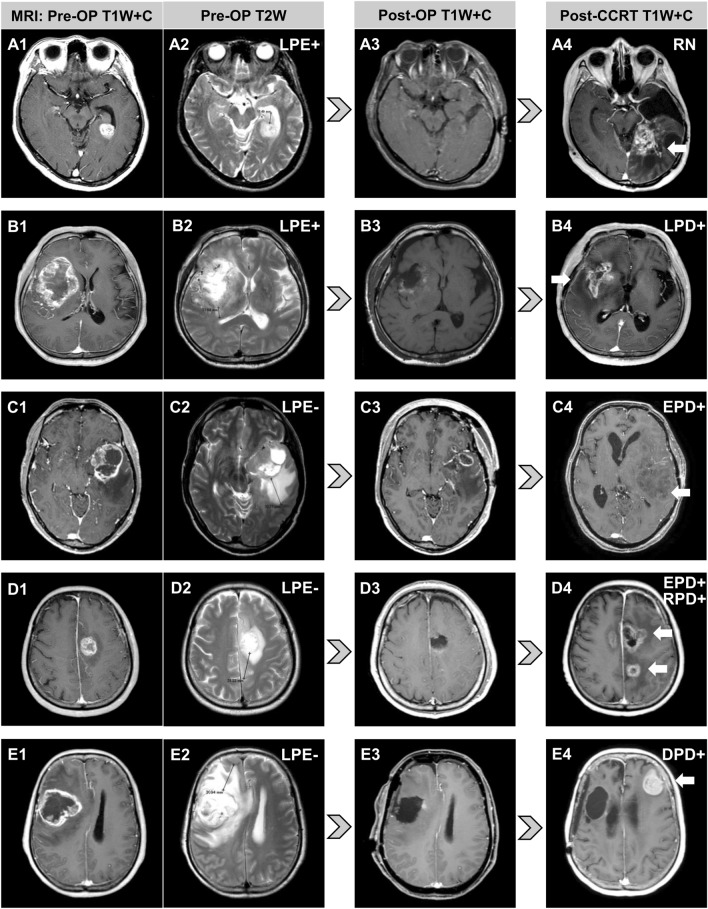
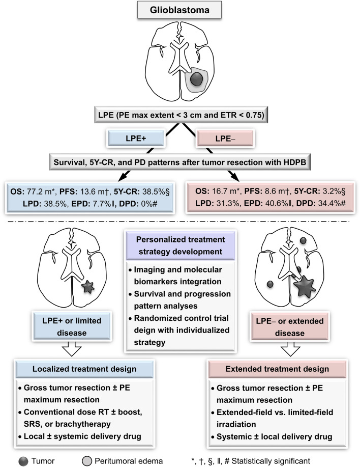
Methods: Patients receiving HDPB (96.6 GyE) were retrospectively evaluated. Limited peritumoral edema (LPE) was defined as PE extent < 3 cm with a ratio of PE extent to tumor maximum diameter of < 0.75. Extended progressive disease (EPD) was defined as progression of tumors extending > 1 cm from the tumor bed edge.

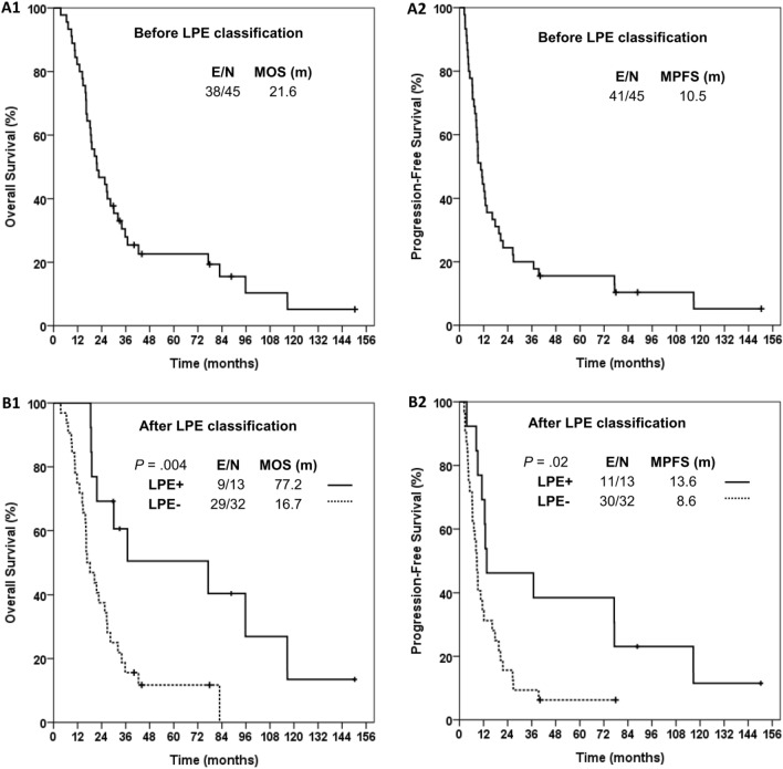
Results: After long-term follow-up (median 88.7, range 63.6-113.8 months) for surviving patients with (n = 13) and without (n = 32) LPE, the median overall survival (OS) and progression-free survival (PFS) were 77.2 vs. 16.7 months (p = 0.004) and 13.6 vs. 8.6 months (p = 0.02), respectively. In multivariate analyses combined with factors of performance, age, tumor maximum diameter, and tumor resection extent, LPE remained a significant factor for favorable OS and PFS. The rates of 5-year complete response, EPD, and distant metastasis with and without LPE were 38.5% vs. 3.2% (p = 0.005), 7.7% vs. 40.6% (p = 0.04), and 0% vs. 34.4% (p = 0.02), respectively.

Conclusions: The LPE status effectively identified patients with relative long-term control and specific progression patterns after postoperative HDPB for glioblastoma.

Keywords: Dose-escalated radiotherapy; Glioblastoma; Imaging biomarker; Peritumoral edema; Personalized treatment; Proton beam therapy.


Conflict of interest statement

No competing interests.

Figures

Fig. 1
Fig. 1
A Radiotherapy protocol in the current study. The radiotherapy courses, dose prescription and target definitions. B Method of measuring peritumoral edema extent in our study. First, we selected the images that presented the tumor’s midplane among axial, sagittal, and coronal sections, respectively. Then, we created tangential lines (red dash line) to the tumor edge and then measured the PE maximum extent from the tumor edge to the PE area edge along their normal lines (red line with arrowhead). CTV clinical target volume, fr fractions, max maximum, PE peritumoral edema
Fig. 2
Fig. 2
Survival analyses with/without classification of peritumoral edema status. Kaplan–Meier estimates of overall survival and progression-free survival for patients before (A1 and A2) and after (B1 and B2) LPE classification, respectively. E event, LPE limited peritumoral edema, MOS median overall survival, MPFS median progression-free survival, N number
Fig. 3
Fig. 3
Diverse progression patterns observed after tumor resection and high-dose proton boost for five glioblastoma patients according to their peritumoral edema statuses. The arrows mark tumor progression. A LPE + patient with radiation necrosis only. B LPE + patient with tumor progression confined to the tumor bed. C LPE − patient with EPD. D LPE − patient with EPD, extending into the contralateral hemisphere and regional PD. E LPE − patient with distant tumor progression at the contralateral frontal lobe. DPD distant progressive disease, EPD extended progressive disease, LPE limited peritumoral edema, OP operative, RN radiation necrosis, RPD regional progressive disease, T1W + C contrast-enhanced T1-weighted magnetic resonance imaging
Fig. 4
Fig. 4
Utilizing peritumoral edema status to facilitate clinical trial design for developing personalized treatment strategies of glioblastoma. 5Y-CR 5-year complete response, DPD distant progressive disease, EPD extended progressive disease, ETR edema-to-tumor ratio, HDPB high-dose proton boost, LPE limited peritumoral edema, OS overall survival, PE peritumoral edema, PFS progression-free survival, SRS stereotactic radiosurgery

References

    1. Adeberg S, Harrabi SB, Bougatf N et al (2016) Intensity-modulated proton therapy, volumetric-modulated arc therapy, and 3D conformal radiotherapy in anaplastic astrocytoma and glioblastoma : a dosimetric comparison. Strahlenther Onkol 192(11):770–779 - DOI - PubMed
    1. Al-Holou WN, Hodges TR, Everson RG et al (2019) Perilesional resection of glioblastoma is independently associated with improved outcomes. Neurosurgery. 10.1093/neuros/nyz008 - DOI - PMC - PubMed
    1. Ashby LS, Smith KA, Stea B (2016) Gliadel wafer implantation combined with standard radiotherapy and concurrent followed by adjuvant temozolomide for treatment of newly diagnosed high-grade glioma: a systematic literature review. World J Surg Oncol 14(1):225 - DOI - PMC - PubMed
    1. Bangalore Yogananda CG, Shah BR, Vejdani-Jahromi M et al (2020) A novel fully automated MRI-based deep-learning method for classification of IDH mutation status in brain gliomas. Neuro Oncol 22(3):402–411 - PMC - PubMed
    1. Barbarite E, Sick JT, Berchmans E et al (2017) The role of brachytherapy in the treatment of glioblastoma multiforme. Neurosurg Rev 40(2):195–211 - DOI - PubMed