. 2020 Jul 4;6(7):64.
doi: 10.3390/jimaging6070064.

Optimization of Breast Tomosynthesis Visualization through 3D Volume Rendering

Affiliations

Optimization of Breast Tomosynthesis Visualization through 3D Volume Rendering

Ana M Mota et al. J Imaging. .

Abstract

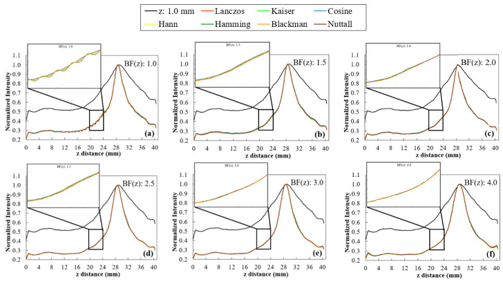
3D volume rendering may represent a complementary option in the visualization of Digital Breast Tomosynthesis (DBT) examinations by providing an understanding of the underlying data at once. Rendering parameters directly influence the quality of rendered images. The purpose of this work is to study the influence of two of these parameters (voxel dimension in z direction and sampling distance) on DBT rendered data. Both parameters were studied with a real phantom and one clinical DBT data set. The voxel size was changed from 0.085 × 0.085 × 1.0 mm3 to 0.085 × 0.085 × 0.085 mm3 using ten interpolation functions available in the Visualization Toolkit library (VTK) and several sampling distance values were evaluated. The results were investigated at 90º using volume rendering visualization with composite technique. For phantom quantitative analysis, degree of smoothness, contrast-to-noise ratio, and full width at half maximum of a Gaussian curve fitted to the profile of one disk were used. Additionally, the time required for each visualization was also recorded. Hamming interpolation function presented the best compromise in image quality. The sampling distance values that showed a better balance between time and image quality were 0.025 mm and 0.05 mm. With the appropriate rendering parameters, a significant improvement in rendered images was achieved.

Keywords: breast tomosynthesis; visualization; volume rendering.


Conflict of interest statement

The authors declare no conflict of interest.

Figures

Figure 1
Figure 1
(a) Acrylic phantom simulating breast tissue and high density lesions (aluminum disks of different diameters and 1 mm thickness). (b) Scheme of the disks in the first column (top to bottom): 5.0 mm, 3.0 mm, 1.0 mm, 0.5 mm, 2.0 mm, and 4.0 mm, respectively.
Figure 2
Figure 2
Illustrative scheme of visualization at 0° and 90°.
Figure 3
Figure 3
Computation time required for rendering the original data (z: 1.0 mm) and data after rescaling with linear, cubic, nearest interpolators and Lanczos, Kaiser, Cosine, Hann, Hamming, Blackman, and Nuttal window functions (with sampling distance 1.0 mm).
Figure 4
Figure 4
Profile of the 5.0 mm disk obtained at 90° for the original data (z: 1.0 mm) and after rescaling with the linear, cubic, and nearest interpolators. Zoom-in of a range with large intensity variation.
Figure 5
Figure 5
Profiles of the 5.0 mm disk obtained at 90º for the original data (z: 1.0 mm) and after rescaling with Lanczos, Kaiser, Cosine, Hann, Hamming, Blackman, and Nuttall window functions with WHW values of 1, 3, 5, 8, 13, and 16 ((a) to (f), respectively). Zoom-in of a range with large intensity variation.
Figure 6
Figure 6
Results obtained with Lanczos, Kaiser, Cosine, Hann, Hamming, Blackman, and Nuttall window functions for the profile at 90°, by changing WHW values (from 1 to 16). (a) Smoothness values as a function of total time and (b) FWHM of the 5.0 mm disk at 90°.
Figure 7
Figure 7
Profiles of the 5-mm disk obtained at 90° for the original data (z: 1.0 mm) and after rescaling with Lanczos, Kaiser, Cosine, Hann, Hamming, Blackman, and Nuttall window functions with BF(z) values of 1.0, 1.5, 2.0, 2.5, 3.0, and 4.0 (af). Zoom-in of a range with large intensity variation.
Figure 8
Figure 8
Results obtained with Lanczos, Kaiser, Cosine, Hann, Hamming, Blackman, and Nuttall window functions for the profile at 90°, by changing BF(z) values (from 1.0 to 4.0). (a) Smoothness values as a function of total time and (b) FWHM of the 5.0 mm disk at 90°.
Figure 9
Figure 9
Computation time required for rendering the original data (black) and data after rescaling (gray) taking into account the different sampling distance values. For each sampling distance, each gray value was obtained by averaging the rendering times recorded for each interpolator considered here (cubic, Hamming with WHW = 5, Hamming with BF (z) = 2 and Hamming with WHW = 5 and BF (z) = 2).
Figure 10
Figure 10
Profiles of the 5.0 mm disk obtained at 90°, for some values of sampling distance tested (0.010, 0.025, 0.050, 0.100, 0.170, 0.195, and 1.0 mm), with the original data (a) and after rescaling with cubic (b), Hamming with WHW = 5 (c), Hamming with BF (z) = 2 (d), and Hamming with WHW = 5 and BF (z) = 2 (e).
Figure 11
Figure 11
Smoothness (a), CNR (b) and FWHM (c) plotted as a function of sampling distance values for original data and data after interpolation with cubic, Hamming with WHW = 5, Hamming with BF (z) = 2 and Hamming with WHW = 5 and BF (z) = 2. Results obtained for rendered images at 90°.
Figure 12
Figure 12
Volume rendering images at 0º and 90º for the 5-mm disk obtained for the original data with default visualization (top row) and interpolated data with Hamming window with BF (z) = 2 and sampling distance 0.025 mm (bottom row).
Figure 13
Figure 13
2D displays of composite volume rendering visualization obtained at 0º and 90º ((a,b), respectively) for original data with default sampling distance (1.0 mm) (a1,b1) and interpolated data with sampling distance 0.025 mm (a2,b2).

References

    1. Ferlay J., Colombet M., Soerjomataram I., Dyba T., Randi G., Bettio M., Gavin A., Visser O., Bray F. Cancer incidence and mortality patterns in Europe: Estimates for 40 countries and 25 major cancers in 2018. Eur. J. Cancer. 2018;103:356–387. doi: 10.1016/j.ejca.2018.07.005. - DOI - PubMed
    1. Siegel R.L., Miller K.D., Jemal A. Cancer statistics, 2020. CA Cancer J. Clin. 2020;70:7–30. doi: 10.3322/caac.21590. - DOI - PubMed
    1. Berry D.A., Cronin K.A., Plevritis S.K., Fryback D.G., Clarke L., Zelen M., Mandelblatt J.S., Yakovlev A.Y., Habbema J.D., Feuer E.J. Effect of screening and adjuvant therapy on mortality from breast cancer. N. Engl. J. Med. 2005;353:1784–1792. doi: 10.1056/NEJMoa050518. - DOI - PubMed
    1. Independent UK Panel on Breast Cancer Screening The benefits and harms of breast cancer screening: An independent review. Lancet. 2012;380:1778–1786. doi: 10.1016/S0140-6736(12)61611-0. - DOI - PubMed
    1. Poplack S.P., Tosteson T.D., Kogel C.A., Nagy H.M. Digital breast tomosynthesis: Initial experience in 98 women with abnormal digital screening mammography. AJR. 2007;189:616–623. doi: 10.2214/AJR.07.2231. - DOI - PubMed

LinkOut - more resources