Clinical Trial
. 2021 Oct:172:105856.
doi: 10.1016/j.phrs.2021.105856. Epub 2021 Aug 28.

Simvastatin modulates estrogen signaling in uterine leiomyoma via regulating receptor palmitoylation, trafficking and degradation

Affiliations
Clinical Trial

Simvastatin modulates estrogen signaling in uterine leiomyoma via regulating receptor palmitoylation, trafficking and degradation

Sadia Afrin et al. Pharmacol Res. 2021 Oct.

Abstract

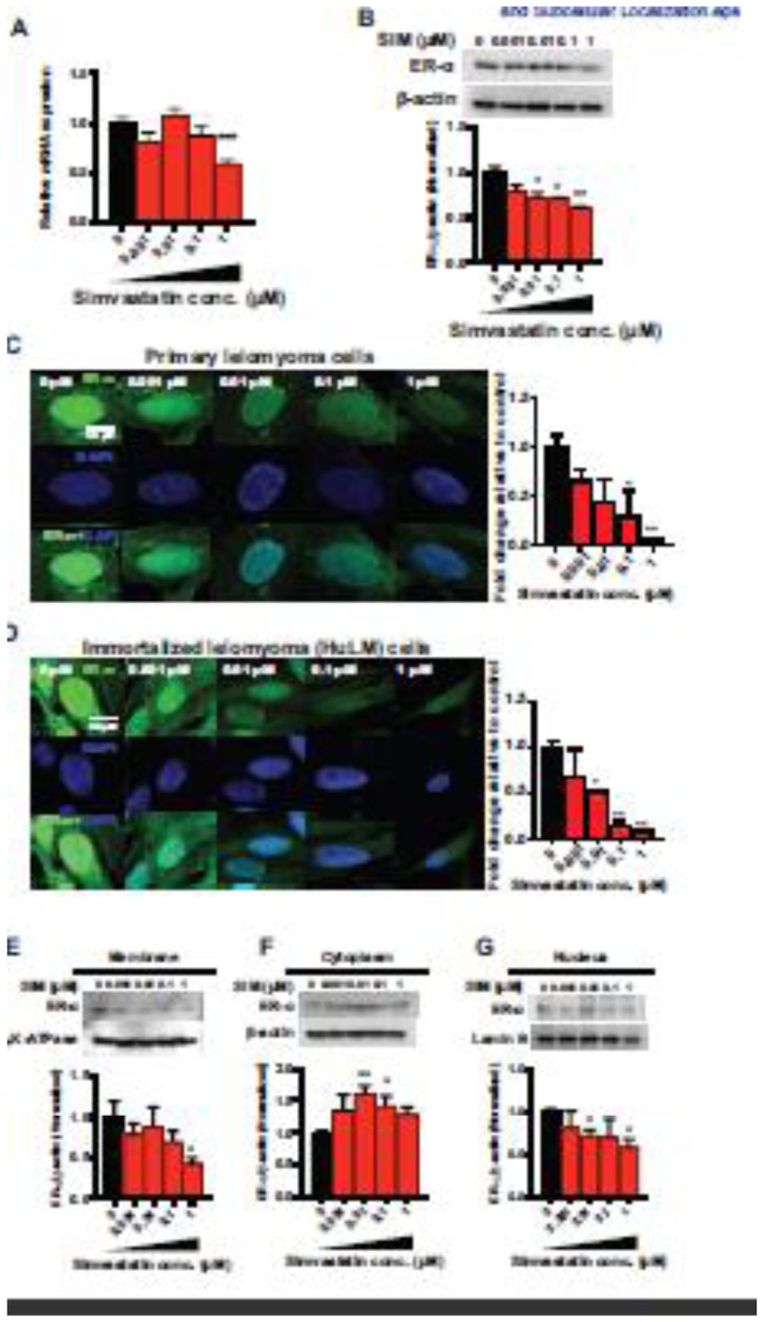
Uterine leiomyomas or fibroids are the most common tumors of the female reproductive tract. Estrogen (E2), a steroid-derived hormone, and its receptors (ERs), particularly ER-α, are important drivers for the development and growth of leiomyomas. We previously demonstrated that simvastatin, a drug used for hyperlipidemia, also possesses anti-leiomyoma properties. The aim of this work is to investigate the impact of simvastatin on ER-α signaling in leiomyoma cells, including its expression, downstream signaling, transcriptional activity, post-translational modification, trafficking and degradation. Primary and immortalized human uterine leiomyoma (HuLM) cells were used for in vitro experiments. Immunodeficient mice xenografted with human leiomyoma tissue explants were used for in vivo studies. Leiomyoma samples were obtained from patients enrolled in an ongoing double-blinded, phase II, randomized controlled trial. Here, we found that simvastatin significantly reduced E2-induced proliferation and PCNA expression. In addition, simvastatin reduced total ER-α expression in leiomyoma cells and altered its subcellular localization by inhibiting its trafficking to the plasma membrane and nucleus. Simvastatin also inhibited E2 downstream signaling, including ERK and AKT pathways, E2/ER transcriptional activity and E2-responsive genes. To explain simvastatin effects on ER-α level and trafficking, we examined its effects on ER-α post-translational processing. We noticed that simvastatin reduced ER-α palmitoylation; a required modification for its stability, trafficking to plasma membrane, and signaling. We also observed an increase in ubiquitin-mediated ER-α degradation. Importantly, we found that the effects of simvastatin on ER-α expression were recapitulated in the xenograft leiomyoma mouse model and human tissues. Thus, our data suggest that simvastatin modulates several E2/ER signaling targets with potential implications in leiomyoma therapy and beyond.

Keywords: Estrogen signaling; Palmitoylation; Receptor degradation; Receptor trafficking; Simvastatin; Uterine leiomyoma.


Conflict of interest statement

Competing interests: The authors declare no conflict of interest.

Figures

Fig. 1
Fig. 1. Reduction of estrogen (E2)-induced proliferation in simvastatin-treated leiomyoma cells.
A, Human leiomyoma cells (HuLM) were treated with the indicated amounts of simvastatin for 48 h. B, HuLM cells were treated with E2 (10 nM) alone or in combination with different simvastatin doses for 48 h. Dimethyl sulfoxide (DMSO) was used as a vehicle control. Cell viability was assessed by an MTT assay. C, HuLM cells were treated with E2 (10 nM) at the indicated time points and PCNA protein expression levels were detected by Western blotting, and β-actin was used as a loading control. D, HuLM cells were treated with the indicated amounts of simvastatin for 48 h and PCNA protein expression levels were detected by Western blotting, and β-actin was used as a loading control. E, HuLM cells were treated with E2 (10 nM) and simvastatin (1 μM), alone and in combination, for 48 h and PCNA protein expression levels were detected by Western blotting, and β-actin was used as a loading control. The data are presented as the mean ± SEM from three independent experiments. *, p < 0.05; **, p < 0.01; ***, p < 0.001 vs. the control; #, p < 0.05 vs. E2.
Fig. 2
Fig. 2. ER-α expression and cellular localization after simvastatin treatment.
HuLM cells were treated with the indicated amounts of simvastatin for 48 h. DMSO was used as a vehicle control. A, mRNA expression levels of ESR1 were quantitated by RT-qPCR. RPLP0 was amplified under the same RT-qPCR conditions used for normalizing data. B, The ER-α protein expression levels were detected by Western blotting, and β-actin was used as a loading control. Cellular localization was assessed in the membrane, cytoplasm, and nucleus. C, D, Immunocytochemistry staining was performed to confirm the cellular localization of ER-α (green fluorescence) and DAPI (blue fluorescence). All images were captured with same time exposure using a confocal microscope (63× magnification). Scale bar, 50 μm. ER-α expression in the membrane (E), cytoplasm (F), and nuclear (G) cellular fractions was determined using Western blot analysis. β-Actin, Na,K-ATPase, and Lamin B were used as a loading control for whole cells, membrane and nucleus fractions. The data are presented as the mean ± SEM of three independent experiments. *, p < 0.05; **, p < 0.01 vs. the control.
Fig. 3
Fig. 3. Suppression of E2-induced ER signaling after simvastatin treatment.
A, D, G, HuLM cells were treated with E2 (10 nM) at the indicated time points. A, phosphorylated to total ERK1/2 ratio (pERK1/2/ERK1/2), (D) phosphorylated to total AKT ratio (pAKT/AKT), and (G) COL1A1 protein expression levels were detected by Western blotting, and β-actin was used as a loading control. B and E, HuLM cells were treated with the indicated amounts of simvastatin for 48 h. The phosphorylated to total ERK1/2 ratio (pERK1/2/ERK1/2) (B) and phosphorylated to total AKT ratio (pAKT/AKT) (E) of the protein expression levels were determined by Western blotting, and β-actin was used as a loading control. C, F, and H, HuLM cells were treated with E2 (10 nM) and simvastatin (1 μM) alone and in combination for 48 h. C, pERK1/2/ERK1/2, F, pAKT/AKT, and H, COL1A1 protein expression levels were detected by Western blotting, and β-actin was used as a loading control. I, The transcriptional activity of E2/ER was reduced by simvastatin treatment. HuLM cells were transfected with plasmids expressing the ERETK-LUC reporter and human ER-α. Cells were treated with the vehicle, simvastatin, E2, and E2 + simvastatin, as indicated, for 48 h. Luciferase activity was determined by a luminometer and normalized by protein concentration. The results are expressed as the fold change relative to the vehicle control. The data are presented as the mean ± SEM of three independent experiments. *, p < 0.05; **, p < 0.01 vs. the control; #, p < 0.05 vs. E2; §, p < 0.05 vs. SIM; ns, non-significant.
Fig. 4
Fig. 4. Effect of simvastatin on ER-α palmitoylation and ubiquitination.
A, In autopalmitoylation, the DHHC domain is acylated at cysteine residues using palmitate from palmitoyl-CoA, and in protein S-palmitoylation, palmitoyl is transferred to the thiol of a cysteine in a protein substrate. B, HuLM cells were treated with simvastatin (0.1 μM) for 48 h. DMSO was used as a vehicle control. An acyl-RAC assay was used to detect S-acylation of the ER-α proteins. Western blotting of ER-α -specific antibodies shows S-acylation of these proteins in the hydroxylamine-treated sample (+HAM lane). A sample treated with sodium chloride (−HAM lane) served as the negative control. Untreated lysates are shown in the input lane, act as a loading control. C, Cells were treated with 50 μg/mL cycloheximide (CHX) for 16 h in CHX group, 0.1 μM simvastatin for 48 h in SIM group, and 0.1 μM simvastatin for 32 h and were treated for an additional 16 h in the presence of CHX in the CHX+SIM group. ER-α protein expression levels were detected in the whole cells by Western blotting, and β-actin was used as a loading control. D, Cells were treated with MG132 (10 μM) for 2 h in MG132 group, 0.1 μM simvastatin for 48 h in SIM group, and 0.1 μM simvastatin for 46 h and cells were treated for an additional 2 h in the presence of MG132 in MG132+SIM group. ER-α protein expression levels were detected in whole cells by Western blotting, and β-actin was used as a loading control. Ubiquitination of ER-α was identified using the Signal-Seeker Ubiquitination Detection Kit. HuLM cells were grown to 70% confluency and treated with MG132 (10 μM/ 2h) prior to treatment with 0.1 μM simvastatin or DMSO (control) for 48 h. Western blot analysis was performed to detect ER-α ubiquitination. The membrane was probed with anti- ER-α (E, left panel) and re-probed with anti-ubiquitin-HRP antibodies (E, right panel). Input lane served as a marker for the unmodified protein band. *, p < 0.05; **, p < 0.01 vs. the control.
Fig. 5
Fig. 5. Immunohistochemical (IHC) staining showing decreased expression of ER-α in simvastatin-treated xenograft mouse tissue.
A, Diagram illustrating the experiment’s design. The mice were injected with estrogen and progesterone pellets after adjusting to their environment, followed five days later by leiomyoma xenografts. Treated animals (n = 10) received simvastatin (20 μg/g body weight/d) subcutaneously, and the control animals (n =10) received the vehicle for 28 days. After the animals were sacrificed, the tissues were collected and placed in 10% buffered formalin and then processed to prepare the tissue sections. B, The tissues were stained with an ER-α antibody. Expression was then graded using an arbitrary grading system that was generated by the image analysis software and was correlated with the percentage of positively stained cells and the intensity of the staining in 10 separate high-power fields (20×) per slide. The expression is presented as arbitrary numbers. Scale bars, 50 μm. Data are presented of 20 independent slides from the treatment group and 20 slides from the control group. Representative images are at 40x magnification. Data are presented as the means ± SEM. * p < 0.05 vs. the control.
Fig. 6
Fig. 6. Immunohistochemical (IHC) staining showing decreased expression of ER-α in simvastatin-treated clinical tissue.
A, Patients with uterine fibroids were recruited for treatment with simvastatin (40 mg daily) or a placebo (starch 1500 encapsulated) for a total of 12 weeks. At the end of the treatment, fibroid samples were collected after the surgery to evaluate the effects of simvastatin. The tissues were fixed with a 10% buffered formalin solution for 24 h and kept in 70% ethanol at 4 °C. Tissues were then stained with ER-α antibody. B, Representative images of IHC staining of the ER-α antibody. Scale bars, 50 μm. Optical density (OD) was measured and quantified in 20× images using ImageJ software with automated macro instructions. C, Fifteen histologically similar fields were randomly selected from each slide to analyze OD. * p < 0.05 vs. the placebo.
Fig. 7
Fig. 7. Summary of results.
This figure represents a summary of the ER-α palmitoylation, ubiquitination, and signaling pathways, all of which alter ER-α downstream regulation. After binding to its receptor, estrogen initiates downstream signaling pathways that include the genomic, where the E2/ER complex translocates to the nucleus to induce the transcription of estrogen responsive genes, and nongenomic signaling pathways, which includes ERK and AKT phosphorylation. ER-α can undergo palmitoylation, and this post-translational modification step was previously shown to be necessary for ER-α membrane localization, its downstream signaling, and protection against degradation. Simvastatin is implicated in several steps with red, stunted arrows denoting simvastatin suppression and blue arrows indicating simvastatin induction. ER-α: estrogen receptor α, E2: estrogen, Palm: palmitate, Hsp90: heat shock protein 90, ERE: estrogen response element, CoR: coregulators, and TF: transcription factors.

References

    1. Cramer SF, Patel A, The frequency of uterine leiomyomas, Am. J. Clin. Pathol 94(4) (1990) 435–438. - PubMed
    1. Baird DD, Dunson DB, Hill MC, Cousins D, Schectman JM, High cumulative incidence of uterine leiomyoma in black and white women: ultrasound evidence, Am. J. Obstet. Gynecol 188(1) (2003) 100–107. - PubMed
    1. Kasai T, Shozu M, Murakami K, Segawa T, Shinohara K, Nomura K, Inoue M, Increased Expression of Type I 17β-Hydroxysteroid Dehydrogenase Enhances in Situ Production of Estradiol in Uterine Leiomyoma, J. Clin. Endocrinol. Metab 89(11) (2004) 5661–5668. - PubMed
    1. Walker CL, Stewart EA, Uterine fibroids: the elephant in the room, Science 308(5728) (2005) 1589–1592. - PubMed
    1. Yang Q, Mas A, Diamond MP, Al-Hendy A, The Mechanism and Function of Epigenetics in Uterine Leiomyoma Development, Reprod. Sci 23(2) (2016) 163–175. - PMC - PubMed

Publication types

MeSH terms