. 2021 Oct 6;29(10):2898-2909.
doi: 10.1016/j.ymthe.2021.08.031. Epub 2021 Aug 28.

AAV integration in human hepatocytes

Affiliations

AAV integration in human hepatocytes

Dhwanil A Dalwadi et al. Mol Ther. .

Abstract

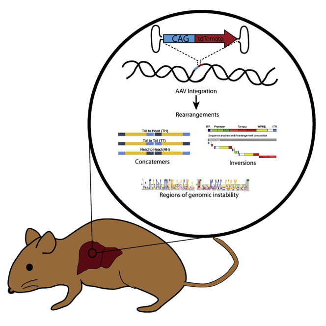
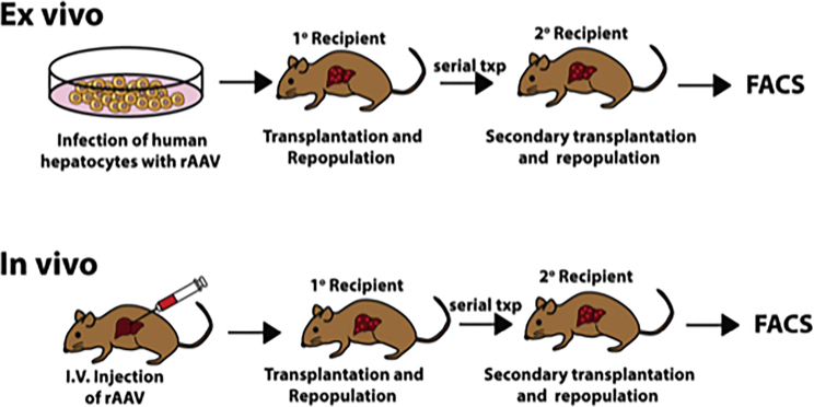
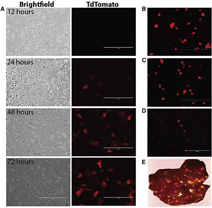
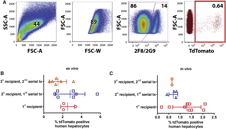
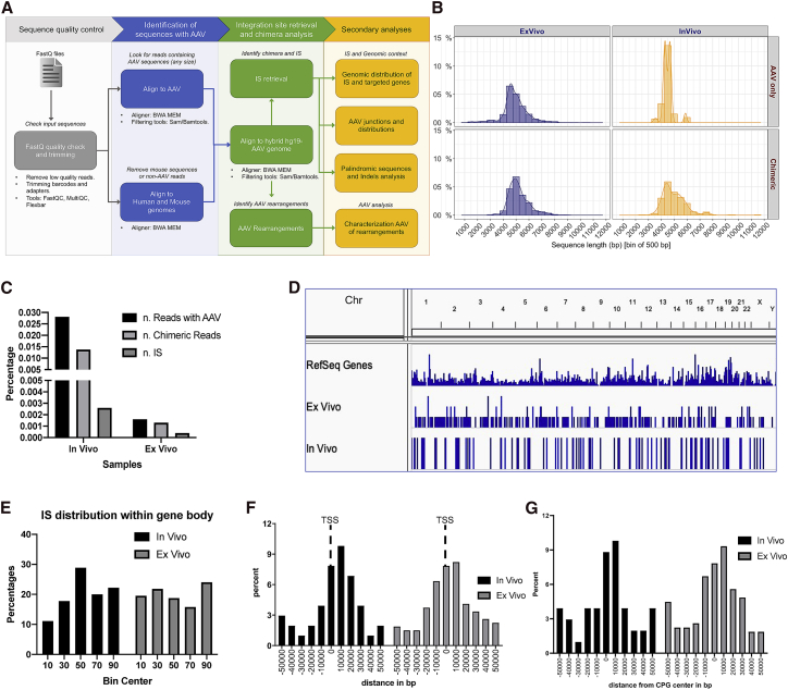
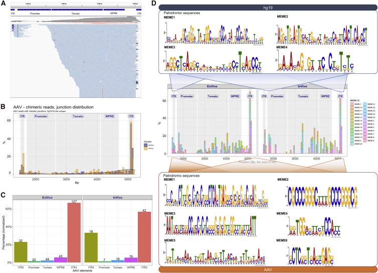
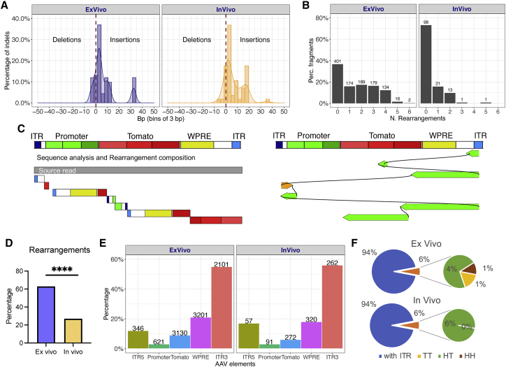
Recombinant adeno-associated viral (rAAV) vectors are considered promising tools for gene therapy directed at the liver. Whereas rAAV is thought to be an episomal vector, its single-stranded DNA genome is prone to intra- and inter-molecular recombination leading to rearrangements and integration into the host cell genome. Here, we ascertained the integration frequency of rAAV in human hepatocytes transduced either ex vivo or in vivo and subsequently expanded in a mouse model of xenogeneic liver regeneration. Chromosomal rAAV integration events and vector integrity were determined using the capture-PacBio sequencing approach, a long-read next-generation sequencing method that has not previously been used for this purpose. Chromosomal integrations were found at a surprisingly high frequency of 1%-3% both in vitro and in vivo. Importantly, most of the inserted rAAV sequences were heavily rearranged and were accompanied by deletions of the host genomic sequence at the integration site.

Keywords: FRGN; capture sequencing; genotoxicity; rAAV; random integration.


Conflict of interest statement

Declaration of interests M.G. has a financial interest in Yecuris Corp. (Tigard, Oregon), a company that has commercialized the humanized mouse liver model used in this work. MG also is a consultant for LogicBio Therapeutics. Other authors declare no competing interests.

Figures

None
Graphical abstract
Figure 1
Figure 1
Ex vivo and in vivo model for interrogating AAV integration frequency in human hepatocytes In the ex vivo approach, human hepatocytes were transduced with AAV-CAG-tdTomato, then transplanted into FRGN mice. Following humanization, hepatocytes were harvested and serially transplanted. In the in vivo approach, highly liver humanized mice were infected with AAV. 2 weeks after infection, hepatocytes were harvested and serially transplanted.
Figure 2
Figure 2
Ex vivo transduction of human hepatocytes with rAAV-CAG-tdTomato (A) Hepatocytes at 12, 24, 48, and 72 h post-AAV transduction (DJ serotype). (B) Hepatocytes at 24 h post-AAV-tdTomato infection (DJ serotype). (C) Hepatocytes at 24 h post-AAV-tdTomato infection (LK03 serotype). (D) Hepatocytes recovered from mouse liver repopulated with AAVDJ-CAG-tdTomato-infected human hepatocytes, imaged 24 h after perfusion and plating. (E) Mouse liver repopulated with human hepatocytes infected with AAV-tdTomato 4 months prior, shown illuminated with RFP (red fluorescent protein) flashlight.
Figure 3
Figure 3
AAV genome persists in 0.6% to 2.8% of human hepatocytes in a liver injury mouse model (A) Representative flow analysis. Hepatocytes perfused from humanized mice were stained with OC2-CF8 and OC2-2G9 antibodies, which have affinity for mouse cells but not human hepatocytes. Using this gating strategy, human hepatocytes were analyzed for tomato reporter gene expression. (B and C) AAV integration frequency in human hepatocytes transduced (B) ex vivo or (C) in vivo. Data presented as mean ± SD. In both approaches, there is a decrease in tdTomato-positive hepatocytes with serial transplantation, but the variance was such that the difference is not statistically significant by one-way ANOVA.
Figure 4
Figure 4
rAAV genomic integration frequency and distribution profile in human hepatocytes (A) Schematic workflow of integration site analysis and bioinformatics procedures for rearrangement characterization. (B) Distribution of raw-reads length per sample. In rows, the two groups are “Chimeric” (meaning reads containing AAV and also aligning to target genome) and “AAV only” (meaning reads with AAV sequence but no hit on target genome). (C) Summary of raw reads. Considering all raw reads, only a fraction contained AAV sequences (“n. Reads with AAV”), and within this subset, only a portion contained reads with a sequence from AAV and sequence from human genome (hg19), for this reason, called “chimeric reads.” From the latter subset, we identified unique IS (integration sites) “n. IS.” (D) Genome-wide distribution of integration sites. (E) Integration site distribution within gene bodies (normalized by gene size): each gene interval has been quantified from transcription start site (TSS) up to the end of its coding region, and this interval is considered as 100%, then normalized in bins of 10%. (F) Integration site distribution around TSSs and (G) CpG islands.
Figure 5
Figure 5
Sequence analysis and distribution of rAAV regions involved in integration events (A) Representative example of sequence alignments on the rAAV genome. Top track shows the position of genetic elements (5′ and 3′ ITRs, promoter, tomato, and WPRE) within rAAV; bottom track shows a portion of the alignments. (B) Overall frequency distribution (in percent) of integration site breakpoints along the rAAV genome. (C) Frequency (percentage) of integration site breakpoints within rAAV, normalized by the length of each genetic element (numbers on top of each bar represent the number of observations). (D) Recurrent motif analysis on the 100-bp interval surrounding each integration site breakpoint. Top panel: palindromic sequences identified in the human sequence (Hg19). Middle panel: frequency distribution (in percent) of palindromes found across the rAAV genome (genetic element within the AAV genome is indicated). Bottom panel: 6 out 24 palindromic sequences identified in the rAAV genome are shown. (See Figure S2 for remaining motifs.)
Figure 6
Figure 6
Rearrangements of the rAAV genome in ex vivo- and in vivo-transduced human hepatocytes (A) Analysis of insertions and deletions (indels) in chimeric reads. (B) Percentage (y axis) of AAV genomes with zero to six rearranged fragments (in x axis). (C) Representative examples of rAAV genomic rearrangements from the source read (on the left side) and aligned to the AAV reference genome (on the right side). The AAV sequence is annotated with different features and use of distinct colors. Within each feature, we used color scales to split longer features in shorter segments. 5′ ITR is colored in blue, whereas 3′ ITR in light blue; promoter in green; tdTomato in red; WPRE in yellow. Starting from the source read (in gray), each rearrangement is plotted as a single rectangle composed by colored segments that indicate the AAV mapping position and orientation. On the right side, a linear representation of all aligned rearrangements. Rearrangements are plotted under the reference genome in the corresponding alignment position and orientation (positive orientation in orange; negative orientation in green). Consecutive rearrangements are visualized in order of appearance and connected by curved links. (D) Frequency of rAAV sequences with one or more rearrangements observed in ex vivo and in in vivo datasets (p < 0.0001 by two-tailed Fisher’s exact test). (E) Percentage of AAV features covered by rearrangements normalized by each feature size. On top of each bar, the absolute number of rearrangements. (F) AAV concatemer analysis. Nested pie chart of the percentage of AAV concatemer for in vivo and ex vivo datasets, showing the proportion of rearrangements having at least one ITR and the observed concatemers in the three classes: head to tail (HT), tail to tail (TT), and head to head (HH).

References

    1. Shahryari A., Saghaeian Jazi M., Mohammadi S., Razavi Nikoo H., Nazari Z., Hosseini E.S., Burtscher I., Mowla S.J., Lickert H. Development and Clinical Translation of Approved Gene Therapy Products for Genetic Disorders. Front. Genet. 2019;10:868. - PMC - PubMed
    1. Anguela X.M., High K.A. Entering the Modern Era of Gene Therapy. Annu. Rev. Med. 2019;70:273–288. - PubMed
    1. Podsakoff G., Wong K.K., Jr., Chatterjee S. Efficient gene transfer into nondividing cells by adeno-associated virus-based vectors. J. Virol. 1994;68:5656–5666. - PMC - PubMed
    1. Daya S., Berns K.I. Gene therapy using adeno-associated virus vectors. Clin. Microbiol. Rev. 2008;21:583–593. - PMC - PubMed
    1. Chandler R.J., Sands M.S., Venditti C.P. Recombinant Adeno-Associated Viral Integration and Genotoxicity: Insights from Animal Models. Hum. Gene Ther. 2017;28:314–322. - PMC - PubMed

Publication types

LinkOut - more resources