May-Thurner Syndrome: An Anatomic Predisposition to Deep Vein Thrombosis
- PMID: 34462701
- PMCID: PMC8389860
- DOI: 10.7759/cureus.16682
May-Thurner Syndrome: An Anatomic Predisposition to Deep Vein Thrombosis
Abstract
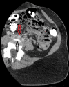
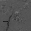
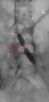
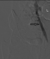
May-Thurner syndrome (MTS) is a rare clinical condition caused by extrinsic compression of the left common iliac vein by the right common iliac artery, leading to venous stasis and predisposing to thrombus formation. Here, we present the case of a 39-year-old female with no obviously known other risk factors predisposing to thrombosis who presented with severe left leg pain and swelling for a week. The international normalized ratio was elevated and the venous Doppler study showed extensive thrombosis extending from the left common iliac vein to the common femoral vein and the popliteal vein. She was diagnosed with MTS and treated with catheter-directed mechanical thrombolysis and thrombectomy, along with angioplasty of the left common iliac vein and external iliac vein, with near-complete resolution post-treatment. MTS should be suspected in patients who present with unilateral limb thrombosis regardless of the presence of predisposing factors. Timely management with endovascular procedures is necessary to help prevent other potential life-threatening complications.
Keywords: deep vein thrombosis; endovascular procedure; iliac artery; iliac vein; may-thurner syndrome; mechanical thrombolysis.
Copyright © 2021, Mir et al.
Conflict of interest statement
The authors have declared that no competing interests exist.
Figures


References
-
- The cause of the predominantly sinistral occurrence of thrombosis of the pelvic veins. May R, Thurner J. Angiology. 1957;8:419–427. - PubMed
-
- Systematic review of May-Thurner syndrome with emphasis on gender differences. Kaltenmeier CT, Erben Y, Indes J, Lee A, Dardik A, Sarac T, Ochoa Chaar CI. J Vasc Surg Venous Lymphat Disord. 2018;6:399–407. - PubMed
-
- Arterial compression of the right common iliac vein; an unusual anatomical variant. Molloy S, Jacob S, Buckenham T, Khaw KT, Taylor RS. Cardiovasc Surg. 2002;10:291–292. - PubMed
-
- Unusual cases of right-sided and left-sided May-Thurner syndrome. Vijayalakshmi IB, Setty HS, Narasimhan C. Cardiol Young. 2015;25:797–799. - PubMed
-
- "Right-sided" May-Thurner syndrome. Abboud G, Midulla M, Lions C, El Ngheoui Z, Gengler L, Martinelli T, Beregi JP. Cardiovasc Intervent Radiol. 2010;33:1056–1059. - PubMed
Publication types
LinkOut - more resources
Full Text Sources
