Review
. 2021 Sep;11(9):210074.
doi: 10.1098/rsob.210074. Epub 2021 Sep 1.

Unravelling the mystery of female meiotic drive: where we are

Affiliations
Review

Unravelling the mystery of female meiotic drive: where we are

Frances E Clark et al. Open Biol. 2021 Sep.

Abstract

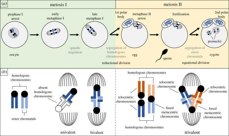
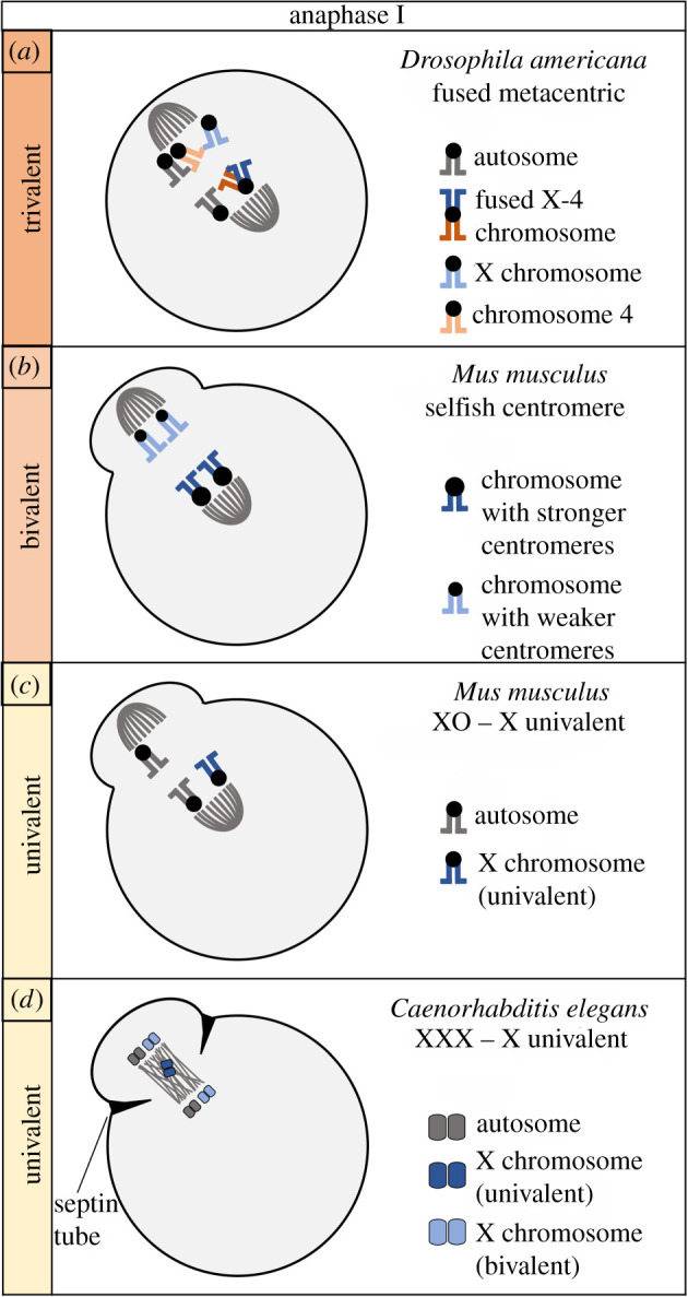
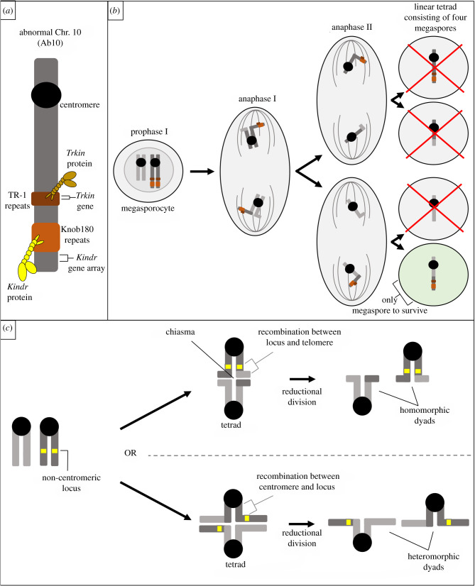
Female meiotic drive is the phenomenon where a selfish genetic element alters chromosome segregation during female meiosis to segregate to the egg and transmit to the next generation more frequently than Mendelian expectation. While several examples of female meiotic drive have been known for many decades, a molecular understanding of the underlying mechanisms has been elusive. Recent advances in this area in several model species prompts a comparative re-examination of these drive systems. In this review, we compare female meiotic drive of several animal and plant species, highlighting pertinent similarities.

Keywords: chromosome segregation; female meiosis; meiotic drive; selfish genetic elements.


Conflict of interest statement

We declare we have no competing interests.

Figures

Figure 1.
Figure 1.
Female meiosis in animals. (a) Typical progression through animal oogenesis is depicted for a cell with two chromosome pairs (n = 2). (b) Homologous chromosomes can remain unpaired (univalent) or pair (form a bivalent or trivalent) during meiosis I.
Figure 2.
Figure 2.
Comparison of female meiotic drive systems. Meiosis I is depicted for four examples of female meiotic drive in three animal species. Drive involving trivalents in Drosophila (a) and bivalents in mice (b) may be mechanistically distinct from the drive of univalent chromosomes in mice (c) and worms (d).
Figure 3.
Figure 3.
Molecular mechanisms of centromere drive in mice. (a) Stronger centromeres build larger kinetochores which recruit more destabilizers compared to weaker centromeres in mice. (b) In addition to this centromere asymmetry, cortical positioning of the spindle induces spindle asymmetry in microtubule tyrosination, facilitating directional flipping until stronger centromeres are preferentially oriented in late metaphase I. (c) In a Mus musculus hybrid, heterozygous for chromosome 4 and 17 centromere size, studied by Wu et al. [39], asymmetry is seen in microtubule and MTOC density. In this system, larger centromeres preferentially orient prior to the completion of spindle migration.
Figure 4.
Figure 4.
Molecular mechanisms of the drive of knob domains in maize. (a) Kinesin-14 motor proteins, Kindr and Trkin, bind to knob180 and TR-1 repeats, respectively, in meiosis I and II. (b) Kindr and Trkin travel faster on microtubules, pulling the knob towards the upper and lower megaspores. This increases the likelihood that the knob domain will be incorporated into the lower megaspore which will become the egg. (c) When chromosomes pair, they recombine and remain attached by chiasmata. If recombination occurs between the heterozygous locus of interest and the telomere, both sister chromatids on the same side of the bivalent have the same allele, resulting in homomorphic dyads. If recombination occurs between the centromere and a heterozygous locus of interest, strands are exchanged such that each side of the bivalent now has one of each allele for that locus. This creates heteromorphic dyads.
Figure 5.
Figure 5.
Univalent drive in worms and grasshoppers. (a) Univalent X chromosomes preferentially segregate to the polar body in Caenorhabditis elegans. These univalents lag in meiosis I and are captured by the septin tube during contractile ring activity. (b) Univalent B chromosomes in Myrmeleotettix maculatus are preferentially segregated to the egg. The side of the spindle facing the egg pole is longer than the side facing the polar body pole. Univalent B chromosomes are not aligned at the metaphase plate and can be randomly found anywhere along the spindle. The B chromosome is more likely to reside on the side of the spindle facing the egg pole due to this spatial asymmetry and is therefore more likely to be incorporated into the egg.

References

    1. Rice WR. 2013. Nothing in genetics makes sense except in light of genomic conflict. Annu. Rev. Ecol. Evol. Syst. 44, 217-237. (10.1146/annurev-ecolsys-110411-160242) - DOI
    1. Werren JH. 2011. Selfish genetic elements, genetic conflict, and evolutionary innovation. Proc. Natl Acad. Sci. USA 108, 10 863-10 870. (10.1073/pnas.1102343108) - DOI - PMC - PubMed
    1. Burt A, Trivers R. 2008. Genes in conflict: the biology of selfish genetic elements, pp. 325-380. Cambridge, MA: Belknap Press.
    1. Jones RN. 1991. B-chromosome drive. Am. Nat. 137, 430-442. (10.1086/285175) - DOI
    1. Camacho JP, Sharbel TF, Beukeboom LW. 2000. B-chromosome evolution. Phil. Trans. R. Soc. B 355, 163-178. (10.1098/rstb.2000.0556) - DOI - PMC - PubMed

Publication types

LinkOut - more resources