

T cell exhaustion is associated with the risk of papillary thyroid carcinoma and can be a predictive and sensitive biomarker for diagnosis
- PMID: 34465342
- PMCID: PMC8408957
- DOI: 10.1186/s13000-021-01139-7
T cell exhaustion is associated with the risk of papillary thyroid carcinoma and can be a predictive and sensitive biomarker for diagnosis
Abstract
Background: The incidence of papillary thyroid carcinoma (PTC) has been steadily increasing over the past decades. Hashimoto's thyroiditis (HT) is the most common autoimmune disease, and is related to the pathogenesis of PTC. Programmed death-1 (PD-1) is currently used for the treatment of PTC, but there are very few studies on the clinical value of PD-1 in the diagnosis and targeted therapy of PTC.
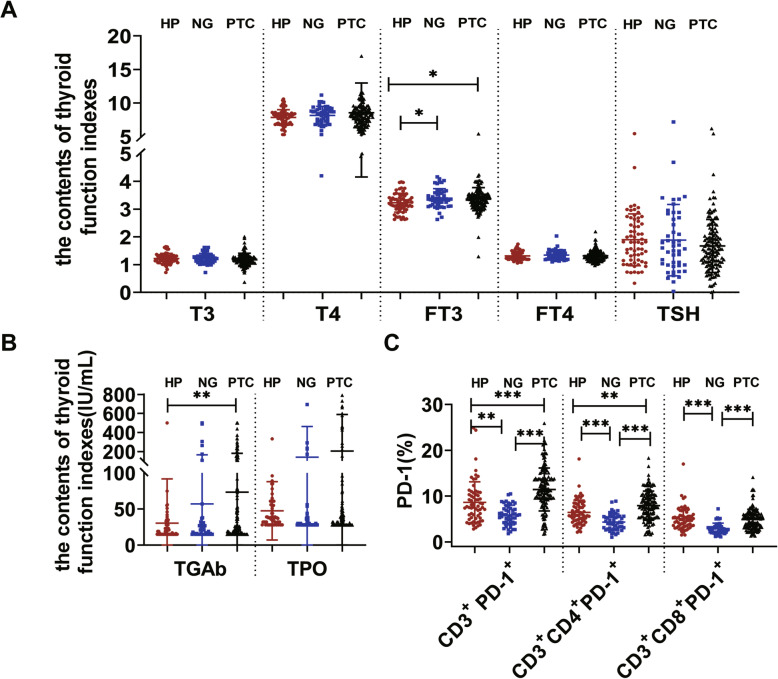
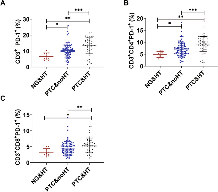
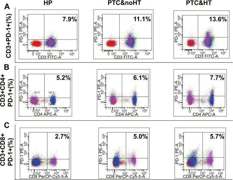
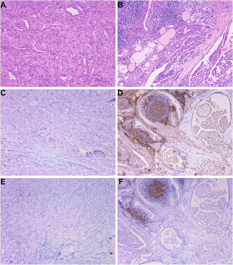
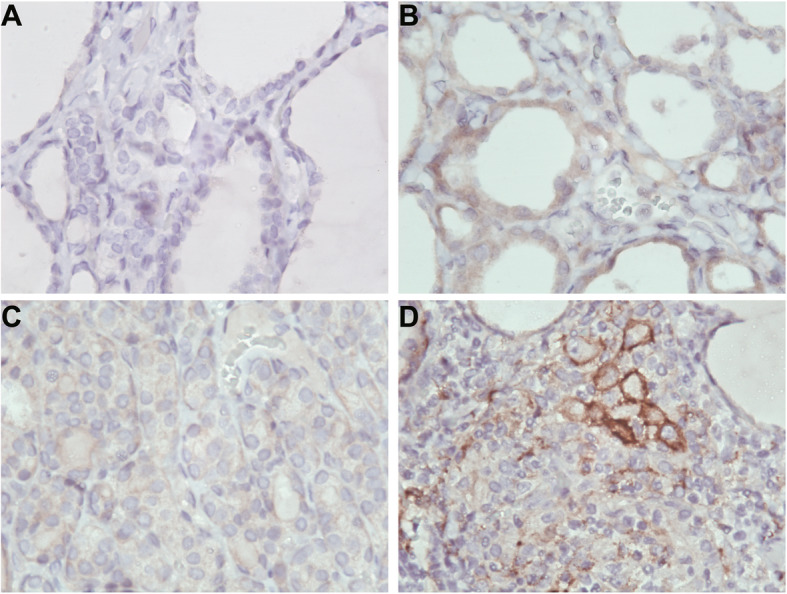
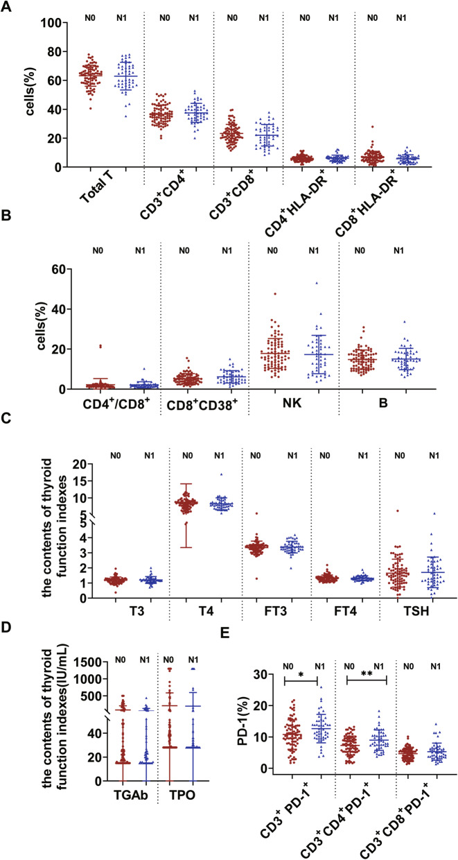
Methods: The expression of T, B, NK cells and PD-1 in the peripheral blood of 132 patients with PTC (PTC group), 48 patients with nodular goiter (NG group) and 63 healthy subjects (HP group) were detected by flow cytometry. The expression of plasma T3, T4, FT3, FT4, TSH, TGAb and TPO was detected by chemiluminescence immunoassay. Among 132 PTC, 49 PTC&HT and 83 PTC&noHT were included. Among 48 NG, 10 NG&HT and 38 NG&noHT were included. The expressions of programmed death- ligand1(PD-L1) in tumor tissues of PTC group and thyroid tissues of NG group, PD-1 and CD3 in tumor infiltration lymphocyte (TIL) were detected by immunohistochemistry.
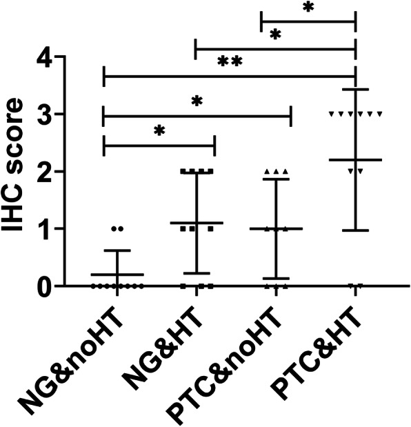
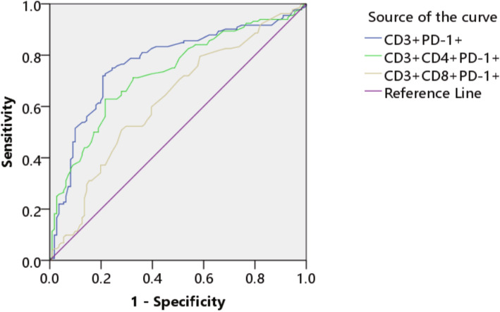
Results: The expression of FT3, TGAb, CD3+PD-1+, CD3+CD4+PD-1+ and CD3+CD8+PD-1+ in PTC and NG was significantly higher than that in the HP group. Moreover, CD3+PD-1+, CD3+CD4+PD-1+ and CD3+CD8+PD-1+ expression had significant differences between the PTC group and the NG group. In addition, the expression of TGAb, TPO, CD3+PD-1+, CD3+CD4+PD-1+ and CD3+CD8+PD-1+ in PTC&HT group was significantly higher than that in the PTC&noHT group. While, the expression of B cells, CD3+PD-1+, CD3+CD4+PD-1+ and CD3+CD8+PD-1+ in PTC&HT group was higher than that in NG&HT group. PD-1 showed a significant correlation with PTC lymph node metastasis. CD3+PD-1+ and CD3+CD4+PD-1+ was higher in N1 stage than in N0 stage. Immunohistochemical results showed that the expression of PD-1, CD3 and PD-L1 in PTC was significantly higher than that in NG.
Conclusions: T cell exhaustion might act as a biomarker for the differential diagnosis of PTC and NG. Patients with PTC&HT have obvious T cell exhaustion and increased expression of PD-1, PD-L1.Targeting the PD-1/PD-L1 pathway could be a new approach to prevent malignant transformation from HT to PTC&HT in the future.
Keywords: Biomarker; Hashimoto’s thyroiditis; Lymph node metastasis; Nodular goiter; PD-1; Papillary thyroid carcinoma.
© 2021. The Author(s).
Conflict of interest statement
No conflict of interest exits in the submission of this manuscript, and manuscript is approved by all authors for publication.
Figures
References
-
- Sung H, Ferlay J, Siegel RL, Laversanne M, Soerjomataram I, Jemal A, Bray F. Global Cancer Statistics 2020: GLOBOCAN Estimates of Incidence and Mortality Worldwide for 36 Cancers in 185 Countries. CA Cancer J Clin. 2021;71(3):209-249. - PubMed
-
- Zeng R, Zhao M, Niu H, et al. Relationship between Hashimoto's thyroiditis and papillary thyroid carcinoma in children and adolescents. Eur Rev Med Pharmacol Sci. 2018;22(22):7778–7787. - PubMed
MeSH terms
Substances
LinkOut - more resources
Full Text Sources
Medical
Research Materials
Miscellaneous
