. 2021 Aug 31;18(1):191.
doi: 10.1186/s12974-021-02241-9.

Asthmatic allergen inhalation sensitises carotid bodies to lysophosphatidic acid

Affiliations

Asthmatic allergen inhalation sensitises carotid bodies to lysophosphatidic acid

Nicholas G Jendzjowsky et al. J Neuroinflammation. .

Abstract

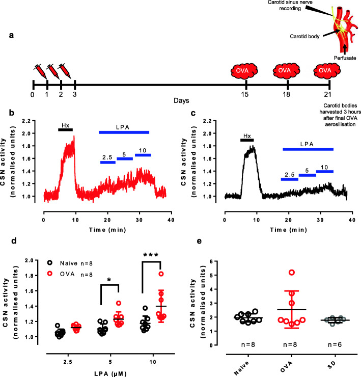
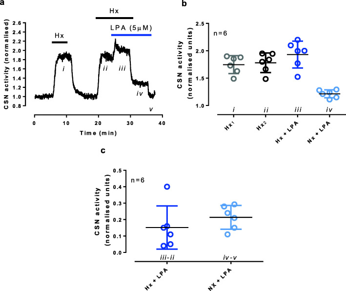
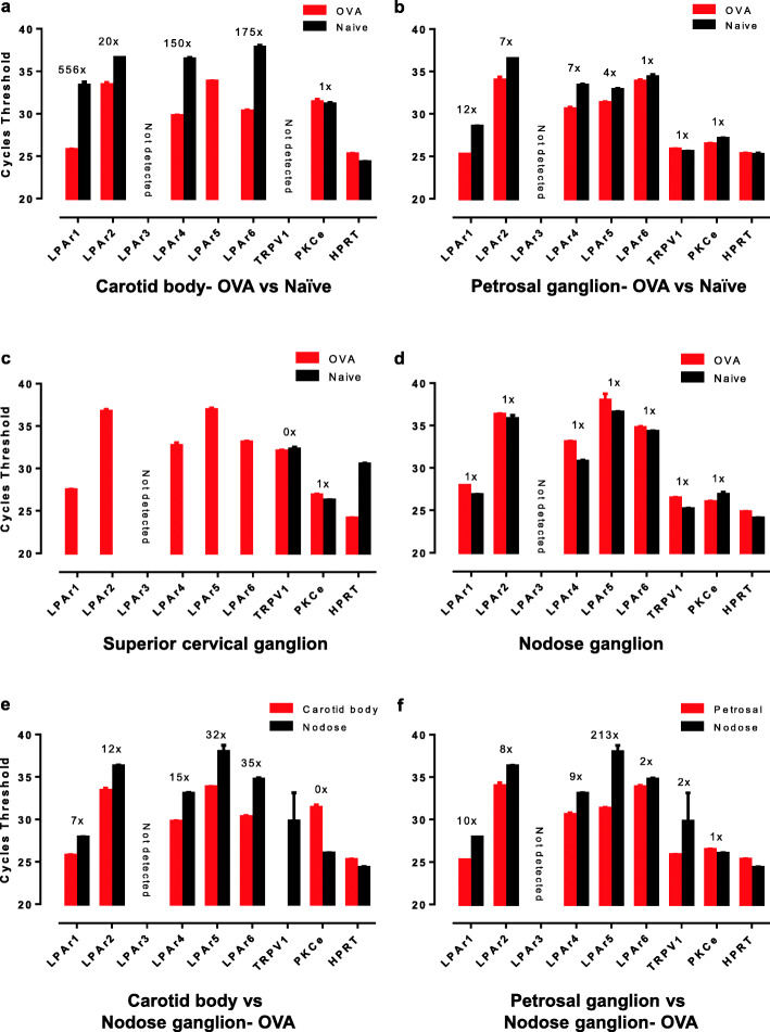
The carotid bodies are multimodal sensors that regulate various autonomic reflexes. Recent evidence demonstrates their role in immune reflex regulation. Our previous studies using the allergen (ovalbumin) sensitised and exposed Brown Norway rat model of asthma suggest that carotid bodies mediate asthmatic bronchoconstriction through a lysophosphatidic acid (LPA) receptor (LPAr)-protein kinase C epsilon (PKCε)-transient receptor potential vanilloid one channel (TRPV1) pathway. Whilst naïve carotid bodies respond to LPA, whether their response to LPA is enhanced in asthma is unknown. Here, we show that asthmatic sensitisation of Brown Norway rats involving repeated aerosolised allergen challenges over 6 days, results in an augmentation of the carotid bodies' acute sensitivity to LPA. Increased expression of LPAr in the carotid bodies and petrosal ganglia likely contributed to this sensitivity. Importantly, allergen sensitisation of the carotid bodies to LPA did not alter their hypoxic response, nor did hypoxia augment LPA sensitivity acutely. Our data demonstrate the ability of allergens to sensitise the carotid bodies, highlighting the likely role of the carotid bodies and blood-borne inflammatory mediators in asthma.

Keywords: Allergen; Asthma; Carotid body; Lysophosphatidic acid; Neuro-immune; Neuroimmunology; Petrosal ganglion; TRPV1.


Conflict of interest statement

N.G.J., A.R. and R.J.A.W declare the following competing interests. U.S. Patent Application No. 62/534,638, Status: provisional patent; “Method to Abate Acute Airway Hypersensitivity and Asthma Attacks.” Purpose: for the use of TRPV1 and LPAr blockade as a treatment for respiratory distress associated with acute asthmatic attack. Authors are also founders and/or shareholders of AazeinTx Inc., a clinical-stage University spinoff company investigating the use of a TRPV1 antagonist in asthma.

Figures

Fig. 1
Fig. 1
Asthmatic BN rats have increased carotid body sensitivity to LPA. a The ovalbumin sensitisation protocol where Brown Norway rats were sensitised (days 1-3) and exposed to aerosolised allergen (days 15, 18, 21) and experiments run 3 h after the last aerosolisation. The en bloc perfused carotid body preparation used to record chemosensory afferents in the carotid sinus nerve (CSN, a). OVA rats (b) had increased sensitivity to 5 and 10 μM LPA compared to naïve BN rats (c). d The increase in CSN activity in response to doubling doses of LPA, (2.5, 5, 10 μM, 18:1 LPA) in ovalbumin sensitised and challenged (OVA) and naïve Brown Norway (BN) rats. F1,14 (group) = 10.077, p = 0.007; F2,28 (dose) = 28.430, p < 0.001; F3,63 (groups × LPA dose) =4.469, p = 0.021; post hoc (difference between groups): 2.5 μM, p = 0.227; 5 μM, p = 0.025; 10 μM, p < 0.001. Hx = hypoxia test of viability. e The hypoxic response was similar between OVA (n = 8) and naïve (n = 8) BN rats. When comparing to hypoxic responses from carotid bodies harvested from Sprague Dawley rats (n = 6), no difference was difference between rat strain or health status. F2,19 = 1.672, p = 0.214
Fig. 2
Fig. 2
Lysophosphatidic acid stimulation is independent of hypoxic carotid body excitation. a A representative trace showing two hypoxic bouts (60 Torr, Hx) where LPA (5 μM) was given during the second hypoxic response, and normoxia (Nx) was re-established with LPA. b Summary data illustrating steady state responses during each stimulation. (c) The change in carotid sinus activity due to LPA was not different between hypoxia (iii—ii from (a)) and normoxia (iv—v from (a), n = 6), t10 = 1.015, p = 0.334
Fig. 3
Fig. 3
Lysophosphatidic acid receptors are upregulated in asthmatic rats. qPCR reveals that lysophosphatidic acid receptor (LPAr) 1, 2, 4, 5, 6 mRNA was upregulated in the carotid bodies (a), and petrosal ganglia (b) of ovalbumin sensitised and challenged (OVA) rats compared to naïve Brown Norway (BN) rats. TRPV1 was not detected in the carotid body (a) as demonstrated previously [15] and was not increased in OVA compared to naïve BN rats in the petrosal ganglion (b). PKCε was detected in all tissues analysed but was not increased in OVA compared to naïve BN rats (a-b). c LPAr 1, 2, 4, 5, 6 were only detected after 45 cycles in OVA but not naïve BN rats in the superior cervical ganglion. d No discernible increases in LPAr expression were revealed in the nodose ganglion. TRPV1 was detected in the superior cervical ganglion (c) and nodose ganglion (d) but was not increased in OVA compared to naïve BN rats. In OVA rats, the carotid body (e) and petrosal ganglion (f) demonstrated increased LPAr and TRPV1 mRNA expression in comparison to the nodose ganglion. Differences in expression were analysed by the 2∆ctt method [17] in reference to hypoxyribosyltransferase (HPRT) housekeeping gene. The 2∆ctt number indicating an increase in of red verses black bars are expressed above bars for specific receptor
Fig. 4
Fig. 4
LPAr1 expression in carotid bodies of asthmatic and naïve rats. Lysophosphatidic acid receptor subtype 1 (LPAr1) is predominantly expressed in tyrosine hydroxylase (TH) positive cells which represent glomus cells of the carotid bodies (round cells with large nuclei as imaged with DAPI nuclear stain). Carotid sinus nerves also stain positive for TH and are seen as thin neural axons. More prominent LPAr1 staining in asthmatic OVA BN rats compared to naïve BN rats is consistent with qPCR data from Fig. 3. Top images = 20× magnification, bottom images = 63× magnification

Similar articles

Cited by

References

    1. Zera T, Moraes DJA, da Silva MP, Fisher JP, Paton JFR. The logic of carotid body connectivity to the brain. Physiology. 2019;34(4):264–282. doi: 10.1152/physiol.00057.2018. - DOI - PubMed
    1. Kumar P, Prabhakar NR. Peripheral chemoreceptors: function and plasticity of the carotid body. Compr Physiol. 2012;2(1):141–219. doi: 10.1002/cphy.c100069. - DOI - PMC - PubMed
    1. Falvey A, Duprat F, Simon T, Hugues-Ascery S, Conde SV, Glaichenhaus N, Blancou P. Electrostimulation of the carotid sinus nerve in mice attenuates inflammation via glucocorticoid receptor on myeloid immune cells. J Neuroinflammation. 2020;17(1):368. doi: 10.1186/s12974-020-02016-8. - DOI - PMC - PubMed
    1. Fan J, Zhang B, Shu H-F, Zhang X-Y, Wang X, Kuang F, Liu L, Peng ZW, Wu R, Zhou Z, Wang BR. Interleukin-6 increases intracellular Ca2+ concentration and induces catecholamine secretion in rat carotid body glomus cells. J Neurosci Res. 2009;87(12):2757–2762. doi: 10.1002/jnr.22107. - DOI - PubMed
    1. Nardocci G, Martin A, Abarzúa S, Rodríguez J, Simon F, Reyes EP, Acuña-Castillo C, Navarro C, Cortes PP, Fernández R. Sepsis progression to multiple organ dysfunction in carotid chemo/baro-denervated rats treated with lipopolysaccharide. J Neuroimmunol. 2015;278:44–52. doi: 10.1016/j.jneuroim.2014.12.002. - DOI - PubMed