. 2021 Aug 17:98:100116.
doi: 10.1016/j.simyco.2021.100116. eCollection 2021 Apr.

Fusarium: more than a node or a foot-shaped basal cell

P W Crous  1   2 L Lombard  1 M Sandoval-Denis  1   3 K A Seifert  4 H-J Schroers  5 P Chaverri  6   7 J Gené  8 J Guarro  8 Y Hirooka  9 K Bensch  1 G H J Kema  2 S C Lamprecht  10 L Cai  11   12 A Y Rossman  13 M Stadler  14 R C Summerbell  15   16 J W Taylor  17 S Ploch  18 C M Visagie  19 N Yilmaz  19 J C Frisvad  20 A M Abdel-Azeem  21 J Abdollahzadeh  22 A Abdolrasouli  23   24 A Akulov  25 J F Alberts  26 J P M Araújo  27 H A Ariyawansa  28 M Bakhshi  29 M Bendiksby  30   31 A Ben Hadj Amor  1 J D P Bezerra  32 T Boekhout  1 M P S Câmara  33 M Carbia  34 G Cardinali  35 R F Castañeda-Ruiz  36 A Celis  37 V Chaturvedi  38 J Collemare  1 D Croll  39 U Damm  40 C A Decock  41 R P de Vries  1 C N Ezekiel  42 X L Fan  43 N B Fernández  44   45 E Gaya  46 C D González  47 D Gramaje  48 J Z Groenewald  1 M Grube  49 M Guevara-Suarez  50 V K Gupta  51   52 V Guarnaccia  53 A Haddaji  54 F Hagen  1 D Haelewaters  55   56 K Hansen  57 A Hashimoto  58 M Hernández-Restrepo  1 J Houbraken  1 V Hubka  59 K D Hyde  60 T Iturriaga  61 R Jeewon  62 P R Johnston  63 Ž Jurjević  64 I Karalti  65 L Korsten  66 E E Kuramae  3   67 I Kušan  68 R Labuda  69 D P Lawrence  70 H B Lee  71 C Lechat  72 H Y Li  73 Y A Litovka  74   75 S S N Maharachchikumbura  76 Y Marin-Felix  14 B Matio Kemkuignou  14 N Matočec  68 A R McTaggart  77 P Mlčoch  78 L Mugnai  79 C Nakashima  80 R H Nilsson  81 S R Noumeur  82 I N Pavlov  74   75 M P Peralta  83 A J L Phillips  84 J I Pitt  85 G Polizzi  86 W Quaedvlieg  87 K C Rajeshkumar  88 S Restrepo  89 A Rhaiem  90 J Robert  54 V Robert  1 A M Rodrigues  91 C Salgado-Salazar  92 R A Samson  1 A C S Santos  93 R G Shivas  94 C M Souza-Motta  93 G Y Sun  95 W J Swart  96 S Szoke  54 Y P Tan  94   97 J E Taylor  98 P W J Taylor  99 P V Tiago  93 K Z Váczy  100 N van de Wiele  54 N A van der Merwe  19 G J M Verkley  1 W A S Vieira  33 A Vizzini  101 B S Weir  63 N N Wijayawardene  102 J W Xia  103 M J Yáñez-Morales  104 A Yurkov  105 J C Zamora  106 R Zare  29 C L Zhang  107 M Thines  18   108   109
Affiliations

Fusarium: more than a node or a foot-shaped basal cell

P W Crous et al. Stud Mycol. .

Abstract

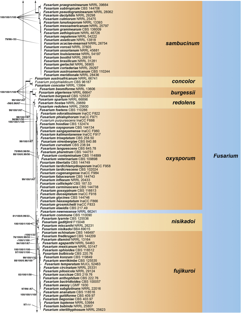
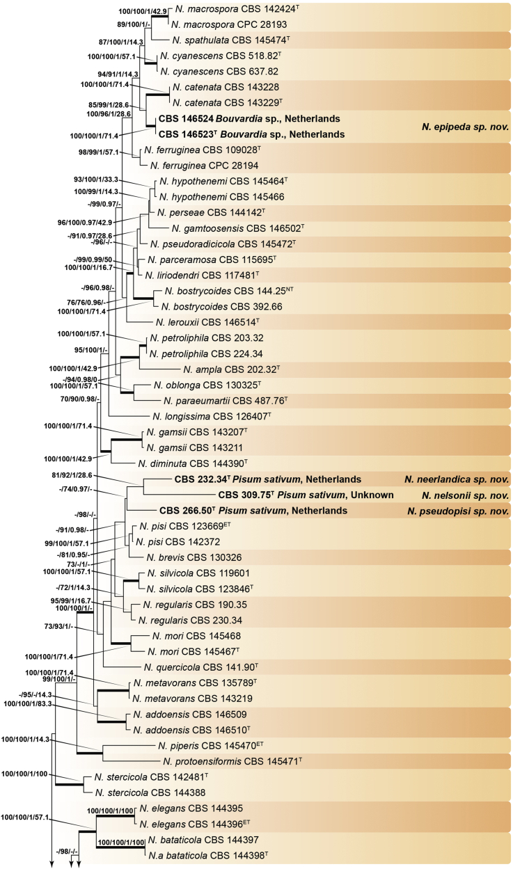
Recent publications have argued that there are potentially serious consequences for researchers in recognising distinct genera in the terminal fusarioid clade of the family Nectriaceae. Thus, an alternate hypothesis, namely a very broad concept of the genus Fusarium was proposed. In doing so, however, a significant body of data that supports distinct genera in Nectriaceae based on morphology, biology, and phylogeny is disregarded. A DNA phylogeny based on 19 orthologous protein-coding genes was presented to support a very broad concept of Fusarium at the F1 node in Nectriaceae. Here, we demonstrate that re-analyses of this dataset show that all 19 genes support the F3 node that represents Fusarium sensu stricto as defined by F. sambucinum (sexual morph synonym Gibberella pulicaris). The backbone of the phylogeny is resolved by the concatenated alignment, but only six of the 19 genes fully support the F1 node, representing the broad circumscription of Fusarium. Furthermore, a re-analysis of the concatenated dataset revealed alternate topologies in different phylogenetic algorithms, highlighting the deep divergence and unresolved placement of various Nectriaceae lineages proposed as members of Fusarium. Species of Fusarium s. str. are characterised by Gibberella sexual morphs, asexual morphs with thin- or thick-walled macroconidia that have variously shaped apical and basal cells, and trichothecene mycotoxin production, which separates them from other fusarioid genera. Here we show that the Wollenweber concept of Fusarium presently accounts for 20 segregate genera with clear-cut synapomorphic traits, and that fusarioid macroconidia represent a character that has been gained or lost multiple times throughout Nectriaceae. Thus, the very broad circumscription of Fusarium is blurry and without apparent synapomorphies, and does not include all genera with fusarium-like macroconidia, which are spread throughout Nectriaceae (e.g., Cosmosporella, Macroconia, Microcera). In this study four new genera are introduced, along with 18 new species and 16 new combinations. These names convey information about relationships, morphology, and ecological preference that would otherwise be lost in a broader definition of Fusarium. To assist users to correctly identify fusarioid genera and species, we introduce a new online identification database, Fusarioid-ID, accessible at www.fusarium.org. The database comprises partial sequences from multiple genes commonly used to identify fusarioid taxa (act1, CaM, his3, rpb1, rpb2, tef1, tub2, ITS, and LSU). In this paper, we also present a nomenclator of names that have been introduced in Fusarium up to January 2021 as well as their current status, types, and diagnostic DNA barcode data. In this study, researchers from 46 countries, representing taxonomists, plant pathologists, medical mycologists, quarantine officials, regulatory agencies, and students, strongly support the application and use of a more precisely delimited Fusarium (= Gibberella) concept to accommodate taxa from the robust monophyletic node F3 on the basis of a well-defined and unique combination of morphological and biochemical features. This F3 node includes, among others, species of the F. fujikuroi, F. incarnatum-equiseti, F. oxysporum, and F. sambucinum species complexes, but not species of Bisifusarium [F. dimerum species complex (SC)], Cyanonectria (F. buxicola SC), Geejayessia (F. staphyleae SC), Neocosmospora (F. solani SC) or Rectifusarium (F. ventricosum SC). The present study represents the first step to generating a new online monograph of Fusarium and allied fusarioid genera (www.fusarium.org).

Keywords: Apiognomonia platani (Lév.) L. Lombard; Atractium ciliatum Link; Atractium pallidum Bonord.; Calloria tremelloides (Grev.) L. Lombard; Cephalosporium sacchari E.J. Butler; Cosmosporella cavisperma (Corda) Sand.-Den., L. Lombard & Crous; Cylindrodendrum orthosporum (Sacc. & P. Syd.) L. Lombard; Dialonectria volutella (Ellis & Everh.) L. Lombard & Sand.-Den.; Fusarium aeruginosum Delacr.; Fusarium agaricorum Sarrazin; Fusarium albidoviolaceum Dasz.; Fusarium aleyrodis Petch; Fusarium amentorum Lacroix; Fusarium annuum Leonian; Fusarium arcuatum Berk. & M.A. Curtis; Fusarium aridum O.A. Pratt; Fusarium armeniacum (G.A. Forbes et al.) L.W. Burgess & Summerell; Fusarium arthrosporioides Sherb.; Fusarium asparagi Delacr.; Fusarium batatas Wollenw.; Fusarium biforme Sherb.; Fusarium buharicum Jacz. ex Babajan & Teterevn.-Babajan; Fusarium cactacearum Pasin. & Buzz.-Trav.; Fusarium cacti-maxonii Pasin. & Buzz.-Trav.; Fusarium caudatum Wollenw.; Fusarium cavispermum Corda; Fusarium cepae Hanzawa; Fusarium cesatii Rabenh.; Fusarium citriforme Jamal.; Fusarium citrinum Wollenw.; Fusarium citrulli Taubenh.; Fusarium clavatum Sherb.; Fusarium coccinellum Kalchbr.; Fusarium cromyophthoron Sideris; Fusarium cucurbitae Taubenh.; Fusarium cuneiforme Sherb.; Fusarium delacroixii Sacc.; Fusarium dimerum var. nectrioides Wollenw.; Fusarium echinatum Sand.-Den. & G.J. Marais; Fusarium epicoccum McAlpine; Fusarium eucheliae Sartory, R. Sartory & J. Mey.; Fusarium fissum Peyl; Fusarium flocciferum Corda; Fusarium gemmiperda Aderh.; Fusarium genevense Dasz.; Fusarium graminearum Schwabe; Fusarium graminum Corda; Fusarium heterosporioides Fautrey; Fusarium heterosporum Nees & T. Nees; Fusarium idahoanum O.A. Pratt; Fusarium juruanum Henn.; Fusarium lanceolatum O.A. Pratt; Fusarium lateritium Nees; Fusarium loncheceras Sideris; Fusarium longipes Wollenw. & Reinking; Fusarium lyarnte J.L. Walsh, Sangal., L.W. Burgess, E.C.Y. Liew & Summerell; Fusarium malvacearum Taubenh.; Fusarium martii f. phaseoli Burkh.; Fusarium muentzii Delacr.; Fusarium nigrum O.A. Pratt; Fusarium oxysporum var. asclerotium Sherb.; Fusarium palczewskii Jacz.; Fusarium palustre W.H. Elmer & Marra; Fusarium polymorphum Matr.; Fusarium poolense Taubenh.; Fusarium prieskaense G.J. Marais & Sand.-Den.; Fusarium prunorum McAlpine; Fusarium pusillum Wollenw.; Fusarium putrefaciens Osterw.; Fusarium redolens Wollenw.; Fusarium reticulatum Mont.; Fusarium rhizochromatistes Sideris; Fusarium rhizophilum Corda; Fusarium rhodellum McAlpine; Fusarium roesleri Thüm.; Fusarium rostratum Appel & Wollenw.; Fusarium rubiginosum Appel & Wollenw.; Fusarium rubrum Parav.; Fusarium samoense Gehrm.; Fusarium scirpi Lambotte & Fautrey; Fusarium secalis Jacz.; Fusarium spinaciae Hungerf.; Fusarium sporotrichioides Sherb.; Fusarium stercoris Fuckel; Fusarium stilboides Wollenw.; Fusarium stillatum De Not. ex Sacc.; Fusarium sublunatum Reinking; Fusarium succisae Schröt. ex Sacc.; Fusarium tabacivorum Delacr.; Fusarium trichothecioides Wollenw.; Fusarium tritici Liebman; Fusarium tuberivorum Wilcox & G.K. Link; Fusarium tumidum var. humi Reinking; Fusarium ustilaginis Kellerm. & Swingle; Fusarium viticola Thüm.; Fusarium werrikimbe J.L. Walsh, L.W. Burgess, E.C.Y. Liew & B.A. Summerell; Fusarium willkommii Lindau; Fusarium xylarioides Steyaert; Fusarium zygopetali Delacr.; Fusicolla meniscoidea L. Lombard & Sand.-Den.; Fusicolla quarantenae J.D.P. Bezerra, Sand.-Den., Crous & Souza-Motta; Fusicolla sporellula Sand.-Den. & L. Lombard; Fusisporium andropogonis Cooke ex Thüm.; Fusisporium anthophilum A. Braun; Fusisporium arundinis Corda; Fusisporium avenaceum Fr.; Fusisporium clypeaster Corda; Fusisporium culmorum Wm.G. Sm.; Fusisporium didymum Harting; Fusisporium elasticae Thüm.; Fusisporium episphaericum Cooke & Ellis; Fusisporium flavidum Bonord.; Fusisporium hordei Wm.G. Sm.; Fusisporium incarnatum Roberge ex Desm.; Fusisporium lolii Wm.G. Sm.; Fusisporium pandani Corda; Gibberella phyllostachydicola W. Yamam.; Hymenella aurea (Corda) L. Lombard; Hymenella spermogoniopsis (Jul. Müll.) L. Lombard & Sand.-Den.; Luteonectria Sand.-Den., L. Lombard, Schroers & Rossman; Luteonectria albida (Rossman) Sand.-Den. & L. Lombard; Luteonectria nematophila (Nirenberg & Hagedorn) Sand.-Den. & L. Lombard; Macroconia bulbipes Crous & Sand.-Den.; Macroconia phlogioides Sand.-Den. & Crous; Menispora penicillata Harz; Multi-gene phylogeny; Mycotoxins; Nectriaceae; Neocosmospora; Neocosmospora epipeda Quaedvl. & Sand.-Den.; Neocosmospora floridana (T. Aoki et al.) L. Lombard & Sand.-Den.; Neocosmospora merkxiana Quaedvl. & Sand.-Den.; Neocosmospora neerlandica Crous & Sand.-Den.; Neocosmospora nelsonii Crous & Sand.-Den.; Neocosmospora obliquiseptata (T. Aoki et al.) L. Lombard & Sand.-Den.; Neocosmospora pseudopisi Sand.-Den. & L. Lombard; Neocosmospora rekana (Lynn & Marinc.) L. Lombard & Sand.-Den.; Neocosmospora tuaranensis (T. Aoki et al.) L. Lombard & Sand.-Den.; Nothofusarium Crous, Sand.-Den. & L. Lombard; Nothofusarium devonianum L. Lombard, Crous & Sand.-Den.; Novel taxa; Pathogen; Scolecofusarium L. Lombard, Sand.-Den. & Crous; Scolecofusarium ciliatum (Link) L. Lombard, Sand.-Den. & Crous; Selenosporium equiseti Corda; Selenosporium hippocastani Corda; Selenosporium sarcochroum Desm; Selenosporium urticearum Corda.; Setofusarium (Nirenberg & Samuels) Crous & Sand.-Den.; Setofusarium setosum (Samuels & Nirenberg) Sand.-Den. & Crous.; Sphaeria sanguinea var. cicatricum Berk.; Sporotrichum poae Peck.; Stylonectria corniculata Gräfenhan, Crous & Sand.-Den.; Stylonectria hetmanica Akulov, Crous & Sand.-Den.; Taxonomy.


Figures

Fig. 1
Fig. 1
Timeline summarising important events in the taxonomy and nomenclature of Fusarium and related taxa.
Fig. 2
Fig. 2
Secondary metabolites from Fusarium spp. / Neocosmospora spp.
Fig. 3
Fig. 3
Some of the most important mycotoxins from Fusarium spp.
Fig. 4
Fig. 4
Secondary metabolites from fusarioid Hypocreales.
Fig. 5
Fig. 5
Basic morphological features of fusarioid fungi. A. Macroconidial shapes. A1. Slender with no significant curvature. A2. Curved with parallel walls. A3. Unequally curved. A4. Widest at the middle portion. A5. Widest at the apical third, wedge-shaped. A6. Widest at the basal portion. A7. Irregularly clavate and swollen. A8. Elongate, whip-like. A9. Distinctly curved. B. Macroconidial apex. B1. Curved. B2. Long and tapered. B3. Pointed. B4. Blunt. B5. Hooked. B6. Elongated. C. Macroconidial base. C1. Obtuse, non foot-shaped. C2. Papillate, non foot-shaped. C3. Poorly developed, foot-shaped. C4. Well-developed, foot-shaped. C5. Elongate, foot-shaped. D. Aerial phialides and microconidial organization. D1. Monophialide. D2–D5. Polyphialides. D2. Simple polyphialide. D3–D4. Polyphialides with multiple conidiogenous loci. D5. Sympodially proliferating polyphialides. D6, D7. Microconidia forming false heads. D8, D9. Microconidia in chains (D8. Dry chain. D9. Palisade). E. Sporodochial conidiophore and conidiogenous cells. F. Aerial conidiophore bearing mesoconidia. G. Mesoconidia. H. Microconidial shapes. H1. Fusiform. H2. Oval. H3. Obovoid. H4. Reniform. H5. Allantoid. H6. Clavate. H7. Napiform. H8. Pyriform. H9. Limoniform.
Fig. 6
Fig. 6
Flow diagram summarising recommended methods for the preservation, identification, and characterisation of fusarioid fungi.
Fig. 7
Fig. 7
Maximum-Likelihood (IQ-TREE-ML) consensus tree inferred from the combined ITS, LSU, rpb1, rpb2 and tef1 multiple sequence alignment of members of Nectriaceae. Numbers at the branches indicate support values (RAxML-BS / UFboot2-BS / BI-PP / gCF) above 70 % / 0.95 with thickened branches indicating full support (RAxML-BS / UFboot2-BS / gCF = 100 %; BI-PP = 1). The scale bar indicates expected changes per site. The tree is rooted to Nectria cinnabarina (CBS 125165). Arrows “F1”, “F2” and “F3” indicate the three alternative Fusarium hypotheses sensuGeiser et al. (2013). Ex-epitype, ex-isotype, ex-neotype and ex-type strains are indicated with ET, IT, NT, and T, respectively.
Fig. 8
Fig. 8
Morphological features and phylogenetic affinities of fusarioid genera of Nectriaceae and close relatives. The tree was delineated based on the phylogeny presented in Fig. 7 and does not indicate phylogenetic distances. Fully supported branches are indicated in bold. The genus Fusarium is indicated in blue. Arrows “F1”, “F2” and “F3” indicate the three alternative Fusarium hypotheses sensuGeiser et al. (2013). Fusarium. A, B. Ascomata. C–E. Ascospores. F, G. Conidiogenous cells. H–J. Macroconidia. (B. Adapted from Schroers et al. 2011). Cyanonectria. A, B. Ascomata. C–E. Ascospores. F. Conidiogenous cells. G. Macroconidia. Neocosmospora. A, B. Ascomata. C–E. Ascospores. F, G. Conidiogenous cells. H, I. Macroconidia. [A. Adapted from Sandoval-Denis & Crous (2018). G. Adapted from Sandoval-Denis et al. (2019)]. Albonectria. A, B. Ascomata. C–E. Ascospores. F, G. Conidiophores and conidiogenous cells. H. Macroconidia. Setofusarium. A, B. Ascomata. C–E. Ascospores. F–H. Setae formed on sporodochia. I. Conidiophore. J. Conidia. Geejayessia. A, B. Ascomata. C–E. Ascospores. F, G. Conidiophores and conidiogenous cells. H, I. Macroconidia. [A. Adapted from Schroers et al. (2011)]. Nothofusarium. A–D. Conidiophores and conidiogenous cells. E. Conidia. Luteonectria. A, B. Ascomata. C–D. Ascospores. F, G. Conidiophores and conidiogenous cells. H. Conidia. Rectifusarium. A–D. Conidiophores and conidiogenous cells. E, F. Conidia. Bisifusarium. A–D. Conidiophores and conidiogenous cells. E, F. Conidia. Mariannaea. A, B. Conidiophores. C, D. Conidiogenous cells. E. Conidia. Tumenectria. A, B. Ascomata. C. Ascospores. D, E. Conidiophores and conidiogenous cells. F. Conidia. [A–C. Adapted from Salgado-Salazar et al. (2016)]. Rugonectria. A, B. Ascomata. C–E. Ascospores. F, G. Conidiophores and conidiogenous cells. H. Conidia. Thelonectria. A, B. Ascomata. C, D. Ascospores. E, F. Conidiophores and conidiogenous cells. G. Conidia. Corinectria. A, B. Ascomata. C–E. Ascospores. F, G. Conidiophores and conidiogenous cells. H. Conidia. (H. Picture by C. González). Neonectria. A, B. Ascomata. C, D. Ascospores. E, F. Conidiophores and conidiogenous cells. G, H. Conidia. [A. Adapted from Chaverri et al. (2011)]. Ilyonectria. A, B. Ascomata. C, D. Ascospores. E, F. Conidiophores and conidiogenous cells. G, H. Conidia. Atractium. A, B. Conidiophores. C, D. Conidiogenous cells. E, F. Conidia. Fusicolla. A, B. Ascomata. C. Ascospores. D, E. Conidiogenous cells. F, G. Conidia. (A–C. Pictures by C. Lechat). Scolecofusarium. A. Ascomata. B, C. Ascospores. D, E. Conidiophores and conidiogenous cells. F. Conidia. Microcera. A. Ascomata. B. Ascospores. C, D. Conidiogenous cells. E, F. Conidia. (A, B. Pictures by N. Aplin, Fungi of Great Britain and Ireland). Macroconia. A, B. Ascomata. C–E. Ascospores. F, G. Conidiophores and conidiogenous cells. H, I. Conidia. (B. Picture by P. Mlčoch). Pseudofusicolla. A, B. Conidiophores and conidiogenous cells. C, D. Conidia. [A–D. Adapted from Triest et al. (2016)]. Cosmospora. A, B. Ascomata. C, D. Ascospores. E, F. Conidiophores and conidiogenous cells. G. Conidia. Dialonectria. A, B. Ascomata. C–E. Ascospores. F, G. Conidiophores and conidiogenous cells. H. Conidia. (A. Picture by P. Mlčoch). Cosmosporella. A, B. Ascomata. C–E. Ascospores. F, G. Conidiophores and conidiogenous cells. H, I. Conidia. (A–E. Pictures by P. Mlčoch). Stylonectria. A, B. Ascomata. C–E. Ascospores. F–I. Conidiophores and conidiogenous cells. J. Conidia. (A–C, E. Pictures by B. Wergen).
Fig. 8
Fig. 8
Morphological features and phylogenetic affinities of fusarioid genera of Nectriaceae and close relatives. The tree was delineated based on the phylogeny presented in Fig. 7 and does not indicate phylogenetic distances. Fully supported branches are indicated in bold. The genus Fusarium is indicated in blue. Arrows “F1”, “F2” and “F3” indicate the three alternative Fusarium hypotheses sensuGeiser et al. (2013). Fusarium. A, B. Ascomata. C–E. Ascospores. F, G. Conidiogenous cells. H–J. Macroconidia. (B. Adapted from Schroers et al. 2011). Cyanonectria. A, B. Ascomata. C–E. Ascospores. F. Conidiogenous cells. G. Macroconidia. Neocosmospora. A, B. Ascomata. C–E. Ascospores. F, G. Conidiogenous cells. H, I. Macroconidia. [A. Adapted from Sandoval-Denis & Crous (2018). G. Adapted from Sandoval-Denis et al. (2019)]. Albonectria. A, B. Ascomata. C–E. Ascospores. F, G. Conidiophores and conidiogenous cells. H. Macroconidia. Setofusarium. A, B. Ascomata. C–E. Ascospores. F–H. Setae formed on sporodochia. I. Conidiophore. J. Conidia. Geejayessia. A, B. Ascomata. C–E. Ascospores. F, G. Conidiophores and conidiogenous cells. H, I. Macroconidia. [A. Adapted from Schroers et al. (2011)]. Nothofusarium. A–D. Conidiophores and conidiogenous cells. E. Conidia. Luteonectria. A, B. Ascomata. C–D. Ascospores. F, G. Conidiophores and conidiogenous cells. H. Conidia. Rectifusarium. A–D. Conidiophores and conidiogenous cells. E, F. Conidia. Bisifusarium. A–D. Conidiophores and conidiogenous cells. E, F. Conidia. Mariannaea. A, B. Conidiophores. C, D. Conidiogenous cells. E. Conidia. Tumenectria. A, B. Ascomata. C. Ascospores. D, E. Conidiophores and conidiogenous cells. F. Conidia. [A–C. Adapted from Salgado-Salazar et al. (2016)]. Rugonectria. A, B. Ascomata. C–E. Ascospores. F, G. Conidiophores and conidiogenous cells. H. Conidia. Thelonectria. A, B. Ascomata. C, D. Ascospores. E, F. Conidiophores and conidiogenous cells. G. Conidia. Corinectria. A, B. Ascomata. C–E. Ascospores. F, G. Conidiophores and conidiogenous cells. H. Conidia. (H. Picture by C. González). Neonectria. A, B. Ascomata. C, D. Ascospores. E, F. Conidiophores and conidiogenous cells. G, H. Conidia. [A. Adapted from Chaverri et al. (2011)]. Ilyonectria. A, B. Ascomata. C, D. Ascospores. E, F. Conidiophores and conidiogenous cells. G, H. Conidia. Atractium. A, B. Conidiophores. C, D. Conidiogenous cells. E, F. Conidia. Fusicolla. A, B. Ascomata. C. Ascospores. D, E. Conidiogenous cells. F, G. Conidia. (A–C. Pictures by C. Lechat). Scolecofusarium. A. Ascomata. B, C. Ascospores. D, E. Conidiophores and conidiogenous cells. F. Conidia. Microcera. A. Ascomata. B. Ascospores. C, D. Conidiogenous cells. E, F. Conidia. (A, B. Pictures by N. Aplin, Fungi of Great Britain and Ireland). Macroconia. A, B. Ascomata. C–E. Ascospores. F, G. Conidiophores and conidiogenous cells. H, I. Conidia. (B. Picture by P. Mlčoch). Pseudofusicolla. A, B. Conidiophores and conidiogenous cells. C, D. Conidia. [A–D. Adapted from Triest et al. (2016)]. Cosmospora. A, B. Ascomata. C, D. Ascospores. E, F. Conidiophores and conidiogenous cells. G. Conidia. Dialonectria. A, B. Ascomata. C–E. Ascospores. F, G. Conidiophores and conidiogenous cells. H. Conidia. (A. Picture by P. Mlčoch). Cosmosporella. A, B. Ascomata. C–E. Ascospores. F, G. Conidiophores and conidiogenous cells. H, I. Conidia. (A–E. Pictures by P. Mlčoch). Stylonectria. A, B. Ascomata. C–E. Ascospores. F–I. Conidiophores and conidiogenous cells. J. Conidia. (A–C, E. Pictures by B. Wergen).
Fig. 9
Fig. 9
Characters for morphological identification of fusarioid genera in Nectriaceae. The rings show, from inside to outside: conidial morphology; ascospore morphology, septation and surface; colour reaction of ascomata in 3 % KOH/lactic acid (nr = no reaction); ascomata wall thickness; and general colour, appearance and wall surface of ascomata.
Fig. 10
Fig. 10
Maximum-Likelihood (IQ-TREE-ML) consensus tree inferred from the combined rpb1, rpb2 and tef1 sequence alignment of the living type strains as indicated in the nomenclator list. Numbers at the branches indicate support values (RAxML-BS / UFboot2-BS / BI-PP) above 70 % / 0.95 with thickened branches indicating full support (RAxML-BS / UFboot2-BS = 100 %; BI-PP = 1). The scale bar indicates expected changes per site. The tree is rooted to Atractium stilbaster (CBS 410.67). Names indicated in bold are in current use. Subdivision of the Fusarium clade (blue block) represent the recognised species complexes.
Fig. 10
Fig. 10
Maximum-Likelihood (IQ-TREE-ML) consensus tree inferred from the combined rpb1, rpb2 and tef1 sequence alignment of the living type strains as indicated in the nomenclator list. Numbers at the branches indicate support values (RAxML-BS / UFboot2-BS / BI-PP) above 70 % / 0.95 with thickened branches indicating full support (RAxML-BS / UFboot2-BS = 100 %; BI-PP = 1). The scale bar indicates expected changes per site. The tree is rooted to Atractium stilbaster (CBS 410.67). Names indicated in bold are in current use. Subdivision of the Fusarium clade (blue block) represent the recognised species complexes.
Fig. 11
Fig. 11
Maximum-Likelihood (IQ-TREE-ML) consensus tree inferred from the combined CaM, rpb1, rpb2, tef1, and tub2 sequence alignment of members of the Fusarium fujikuroi species complex. Numbers at the branches indicate support values (RAxML-BS / UFboot2-BS / BI-PP) above 70 % / 0.95 with thickened branches indicating full support (RAxML-BS / UFboot2-BS = 100 %; BI-PP = 1). Novel taxa are indicated in bold. The scale bar indicates expected changes per site. The tree is rooted to Fusarium curvatum CBS 744.97 and Fusarium inflexum CBS 716.74. Ex-epitype, ex-neotype and ex-type strains are indicated with ET, NT, and T, respectively.
Fig. 12
Fig. 12
Maximum-Likelihood (IQ-TREE-ML) consensus tree inferred from the combined acl1, ITS, LSU, rpb2, tef1, and tub2 sequence alignment of members of the genus Fusicolla. Numbers at the branches indicate support values (RAxML-BS / UFboot2-BS / BI-PP) above 70 % / 0.95 with thickened branches indicating full support (RAxML-BS / UFboot2-BS = 100 %; BI-PP = 1). Novel taxa are indicated in bold. The scale bar indicates expected changes per site. The tree is rooted to Macroconia leptosphaeriae CBS 100001. Ex-epitype and ex-type strains are indicated with ET, and T, respectively.
Fig. 13
Fig. 13
Maximum-Likelihood (ML) consensus tree inferred from the combined acl1, CaM, ITS, LSU, rpb1, rpb2, and tub2 sequence alignment of members of the genus Macroconia. Numbers at the branches indicate support values (RAxML-BS / UFboot2-BS / BI-PP / gCF) above 70 % / 0.95 with thickened branches indicating full support (RAxML-BS / UFboot2-BS = 100 %; BI-PP = 1). Novel taxa are indicated in bold. The scale bar indicates expected changes per site. The tree is rooted to Microcera rubra CBS 638.76. Ex-type and ex-isotype strains are indicated with T, and IT, respectively.
Fig. 14
Fig. 14
Maximum-Likelihood (IQ-TREE-ML) consensus tree inferred from the combined acl1, CaM, ITS, LSU, rpb1, rpb2, and tef1 sequence alignment of members of the genus Neocosmospora. Numbers at the branches indicate support values (RAxML-BS / UFboot2-BS / I-PP) above 70 % / 0.95 with thickened branches indicating full support (RAxML-BS / UFboot2-BS = 100 %; BI-PP = 1). Novel taxa are indicated in bold. The scale bar indicates expected changes per site. The tree is rooted to Geejayessia atrofusca NRRL 22316 and G. cicatricum CBS 125552. Ex-epitype, ex-neotype, ex-paratype and ex-type strains are indicated with ET, NT, PT, and T, respectively.
Fig. 14
Fig. 14
Maximum-Likelihood (IQ-TREE-ML) consensus tree inferred from the combined acl1, CaM, ITS, LSU, rpb1, rpb2, and tef1 sequence alignment of members of the genus Neocosmospora. Numbers at the branches indicate support values (RAxML-BS / UFboot2-BS / I-PP) above 70 % / 0.95 with thickened branches indicating full support (RAxML-BS / UFboot2-BS = 100 %; BI-PP = 1). Novel taxa are indicated in bold. The scale bar indicates expected changes per site. The tree is rooted to Geejayessia atrofusca NRRL 22316 and G. cicatricum CBS 125552. Ex-epitype, ex-neotype, ex-paratype and ex-type strains are indicated with ET, NT, PT, and T, respectively.
Fig. 15
Fig. 15
Maximum-Likelihood (IQ-TREE-ML) consensus tree inferred from the combined acl1, ITS, and rpb2 sequence alignment of members of the genus Stylonectria. Numbers at the branches indicate support values (RAxML-BS / UFboot2-BS / BI-PP) above 70 % / 0.95 with thickened branches indicating full support (RAxML-BS / UFboot2-BS = 100 %; BI-PP = 1). Novel taxa are indicated in bold. The scale bar indicates expected changes per site. The tree is rooted to Macroconia leptosphaeriae CBS 100001. Ex-epitype and ex-type strains are indicated with ET and T, respectively.
Fig. 16
Fig. 16
Albonectria spp. A–C. Ascomata on natural substrate. D. Surface view of perithecial wall in 2 % KOH. E–K. Asci and ascospores (J, K. Surface view). L–P. Conidiophores and conidiogenous cells. Q, R. Microconidia. S. Macroconidia. A, C–F, H–J. Albonectria rigidiuscula (BPI 553050). B, G, K. Albonectria rigidiuscula (BPI 1104484). L, M, P–S. Albonectria rigidiuscula (CBS 122570). N, O. Albonectria rigidiuscula (CBS 133.25). Scale bars: A–C = 100 μm; all others = 10 μm (G applies to H–K).
Fig. 17
Fig. 17
Atractium spp. A, B. Synnemata. C–G. Conidiophores and conidiogenous cells. H. Microconidia. I. Macroconidia. A–D, H. Atractium stilbaster (CBS 410.67). E–G, I. Atractium crassum (CBS 180.31). Scale bars: A = 100 μm; all others = 10 μm.
Fig. 18
Fig. 18
Bisifusarium spp. A–D, F–J. Conidiophores and conidiogenous cells. K, L. Microconidia. E, M. Macroconidia. A–E. Bisifusarium dimerum (CBS 108944). F–M. Bisifusarium delphinoides (CBS 120718). Scale bars: H, J = 5 μm; all others = 10 μm.
Fig. 19
Fig. 19
Cosmosporella spp. A–D. Ascomata on natural substrate. E. Surface view of perithecial wall. F. Asci. G–J. Ascospores. K, L. Conidiophores. M. Microconidia. N, O. Macroconidia. A–J. “Cosmospora” flavoviridis (photos P. Mlčoch). K–N. “Cosmospora” flavoviridis (CBS 124353). O. Cosmosporella cavisperma (CBS 172.31). Scale bars: A–D = 300 μm; E = 50 μm; G–J = 5 μm; all others = 10 μm.
Fig. 20
Fig. 20
Cyanonectria spp. A–C. Ascomata on natural substrate. D. Longitudinal section through perithecium in Shears. E. Surface view of perithecial wall in 2 % KOH. F, G. Asci. H–K. Ascospores (K. Surface view). L–O. Conidiogenous cells. P. Macroconidia. A–C, E–J. Cyanonectria buxi (CBS H-20380). D, K. Cyanonectria buxi (CBS H-20379). L–N. Cyanonectria buxi (CBS 130.97). O, P. Cyanonectria buxi (CBS 125551). [A, D, L. adapted from Schroers et al. (2011).] Scale bars: A–D = 100 μm; H–K = 5 μm (H applies to I and J); all others = 10 μm.
Fig. 21
Fig. 21
Dialonectria spp. A–D. Ascomata on natural substrate. E. Surface view of perithecial wall in 2 % KOH. F–H. Asci. I–M. Ascospores (L, M. Surface view). N, O. Conidiophores and conidiogenous cells. P. Macroconidia. A, B. Dialonectria episphaeria (photos P. Mlčoch). C, D, F, M. Dialonectria episphaeria (CBS H-19716). E, G, K. Dialonectria sanguinea (CBS H-2127). H–J, L. Dialonectria episphaeria (CBS H-2662). N–P. Dialonectria episphaeria (CBS 125494). Scale bars: A–D = 100 μm; I, L, M = 5 μm (I applies to J and K); all others = 10 μm.
Fig. 22
Fig. 22
Fusarium spp. A–D. Ascomata on natural substrate. E. Surface view of perithecial wall in 2 % KOH. F–H. Asci. I–M. Ascospores (M. Surface view). N–P. Conidiophores and conidiogenous cells. Q–T. Microconidia. U–A2. Macroconidia. A. Fusarium graminearum (photo P. Cannon). B, C, F. Fusarium sambucinum [adapted from Wergen (2018)]. D. Fusarium sp. (HPC 2244). E. Fusarium cf. tricinctum (CBS H-12819). G, I. Fusarium lateritium (photo P. Cannon). H, K. Fusarium equiseti (CBS H-12817). J. Fusarium sambucinum (BPI 632307). L, M. Fusarium sambucinum (CBS H-12818). N. Fusarium avenaceum (CPC 30660). O, Q. Fusarium fredkrugerii (CBS 144209). P, W. Fusarium prieskaense (CBS 146498). R. Fusarium madaense (CBS 146669). S. Fusarium globosum (CBS 428.97). T. Fusarium echinatum (CBS 146497). U. Fusarium avenaceum (CBS 408.86). V. Fusarium caeruleum (CBS 146590). X. Fusarium longicaudatum [CBS 123.73, adapted from Xia et al. (2019)]. Y. Fusarium transvaalense [CBS 144211, adapted from Sandoval-Denis et al. (2018b)]. Z. Fusarium gamsii (CBS 143610). A1. Fusarium oxysporum [CBS 144134, adapted from Lombard et al., 2019b, Lombard et al., 2019a]. A2. Fusarium convolutans [CBS 144207, adapted from Sandoval-Denis et al. (2018b)]. Scale bars: A–D = 100 μm; I–M, Q–T = 5 μm; all others = 10 μm.
Fig. 23
Fig. 23
Fusarium echinatum (CBS 146497). A–D. Aerial conidiophores. E–G. Conidiogenous cells on aerial conidiophores. H, I. Microconidia. J. Sporodochia formed on the surface of carnation leaves. K. Sporodochial conidiophores and conidiogenous cells. L. Macroconidia. Scale bars: A = 20 μm; J = 100 μm; all others = 10 μm.
Fig. 24
Fig. 24
Fusarium prieskaense (CBS 146498). A–D. Aerial conidiophores. E–G. Conidiogenous cells on aerial conidiophores. H. Microconidia. I. Sporodochia formed on the surface of carnation leaves. J–L. Sporodochial conidiophores and conidiogenous cells. M. Macroconidia. Scale bars: A, B = 20 μm; I = 100 μm; all others = 10 μm.
Fig. 25
Fig. 25
Fusicolla spp. A. Slimy macroscopic growth on natural substrate. B–E. Ascomata on natural substrate. F. Ostiolar hairs. G. Asci. H. Ascospores. I–K. Conidiophores and conidiogenous cells. L–N. Macroconidia. A. Fusicolla merismoides (photo J. Cunningham). B. Fusicolla melogrammae [CLL 16006, adapted from Crous et al. (2016)]. C–H. Fusicolla ossicola (photos N. Aplin and P. Cannon). I. Fusicolla merismoides (photo P. Cannon). J, K, M. Fusicolla aquaeductuum (CBS 734.79). L. Fusicolla violacea (CPC 38810). N. Fusicolla matuoi (CBS 581.78). Scale bars: B–E = 100 μm; F, H. 5 μm; all others = 10 μm.
Fig. 26
Fig. 26
Fusicolla quarantenae (URM 8367). A. Host. B–G. Conidiophores, conidiogenous cells and conidia. H. Macroconidia. Scale bars = 10 μm.
Fig. 27
Fig. 27
Fusicolla meniscoidea (CBS 110189). A–D. Conidiophores. E–H. Conidiogenous cells. I. Macroconidia. Scale bars: A–D, G–I = 10 μm; E, F = 5 μm.
Fig. 28
Fig. 28
Fusicolla sporellula (CBS 110191). A–C. Conidiophores. D–F. Conidiogenous cells. G. Macroconidia. Scale bars = 10 μm.
Fig. 29
Fig. 29
Geejayessia spp. A–E. Ascomata on natural substrate. F. Surface view of perithecial wall in 2 % KOH. G–I. Asci. J–M. Ascospores. N, O. Conidiophores and conidiogenous cells. P, Q. Macroconidia. A, C. Geejayessia cicatricum [CBS H-20375, adapted from Schroers et al. (2011)]. C. Geejayessia cicatricum (CBS H-20374). D, H, K, M. Geejayessia atrofusca (CBS H-20381). E–G, I, J, L. Geejayessia desmazieri (CBS H-20372). N, O, Q. Geejayessia atrofusca (CBS 502.94). P. Geejayessia cicatricum (CBS 125549). Scale bars: A = 500 μm; B, D, E = 200 μm; C = 100 μm; J–M = 5 μm; all others = 10 μm.
Fig. 30
Fig. 30
Luteonectria albida. A–C. Ascomata on natural substrate. D. Surface view of perithecial wall in lactic acid. E. Detail of ascomata hair. F. Asci. G–J. Ascospores (J. Surface view). K, L. Conidiophores and conidiogenous cells. M. Macroconidia. A, C. BPI 550103. B. BPI 1108874. D–J. BPI 1108875. K–M. CBS 102683. Scale bars: A, B = 100 μm; C = 50 μm; all others = 10 μm.
Fig. 31
Fig. 31
Macroconia spp. A–D. Ascomata on natural substrate. E. Surface view of perithecial wall in 2 % KOH. F. Asci. G–J. Ascospores (J. Surface view). K–M. Conidiophores and conidiogenous cells. N. Microconidia. O, P. Macroconidia. A. Macroconia cupularis [HMAS 97514, adapted from Luo & Zhuang (2008)]. B, C. Macroconia leptosphaeriae (photo P. Mlčoch). D. Macroconia gigas [HMAS 99592, adapted from Luo & Zhuang (2008)]. E–J. Macroconia leptosphaeriae (CBS H-15051). K, L. Macroconia phlogioides (CBS 125496). M, N. Macroconia leptosphaeriae (CBS 10001). O. Macroconia phlogioides (CBS 146500). P. Macroconia bulbipes (CBS 146679). Scale bars: A–D = 100 μm; G–J = 5 μm; all others = 10 μm.
Fig. 32
Fig. 32
Macroconia bulbipes (CBS 146679). A–D. Conidiophores and conidiogenous cells. E, F. Sporodochia formed on the agar surface. G, H. Detail of macroconidia basal cells. I. Macroconidia. Scale bars: E, F = 100 μm; G, H = 5 μm; all others = 10 μm.
Fig. 33
Fig. 33
Macroconia phlogioides (CBS 146501). A–C. Conidiophores. D, E. Conidiogenous cells. F, G. Sporodochia formed on the agar surface. H. Macroconidia. Scale bars: B, C = 20 μm; F, G = 50 μm; all others = 10 μm.
Fig. 34
Fig. 34
Microcera spp. A–C. Ascomata on natural substrate. D. Surface view of perithecial wall in 2 % KOH. E, F. Asci. G–K. Ascospores (J, K. Surface view). L–N. Conidiophores and conidiogenous cells. O–Q. Macroconidia. A. Microcera auranticola (photo N. Aplin). B, O. Microcera coccophila [adapted from Gräfenhan et al. (2011)]. C. Microcera larvarum [adapted from Gräfenhan et al. (2011)]. D, F–J. Microcera coccophila (K(M) 165807). E, K. Microcera larvarum (photo P. Cannon). L, M, Q. Microcera rubra (CBS 638.76). N, P. Microcera larvarum (CBS 169.30). Scale bars: A, B = 100 μm; G–K = 5 μm; all others = 10 μm.
Fig. 35
Fig. 35
Neocosmospora spp. A–E. Ascomata on culture. F. Surface view of perithecial wall in 2 % KOH. G–I. Asci. J–Q. Ascospores (K, M, O, Q. Surface view). R–U. Aerial conidiophores. V. Sporodochial conidiophores. W, X. Microconidia. Y–A5. Macroconidia. A, I, N, O. Neocosmospora vasinfecta (CBS 446.93). B. Neocosmospora sp. (CPC 34617). C, S, W, A1. Neocosmospora elegans (CBS 144396). D. Neocosmospora vasinfecta (CBS 863.70). E. Neocosmospora bataticola (CBS 144398). F, L, M. Neocosmospora ipomoeae (CBS 833.97). G. Neocosmospora robiniae (CBS 119601). H, J, K. Neocosmospora diminuta (CBS 144390). O, Q. Neocosmospora spinulosa (CBS H-5443). R, V, A3. Neocosmospora solani (CBS 140079). T. Neocosmospora bataticola (CBS 144398). U. Neocosmospora suttoniana (CBS 143214). X. Neocosmospora tonkinensis (CBS 115.40). Y. Neocosmospora longissima (CBS 126407). Z. Neocosmospora mori (CBS 145467). A2. Neocosmospora pseudoradicicola (CBS 145472). A4. Neocosmospora keratoplastica (CBS 490.63). A5. Neocosmospora oligoseptata (CBS 143241). [A, C, S, T, W, Y, Z, A1, A2. Adapted from adapted from Sandoval-Denis et al. (2019). R, V, A3, Adapted from Crous et al. (2019a). U, X, A4. Adapted from Sandoval-Denis & Crous (2018)]. Scale bars: A, B = 200 μm; C–E 100 μm; R–T = 20 μm; J–Q, W, X = 5 μm; all others = 10 μm.
Fig. 36
Fig. 36
Neocosmospora epipeda (CBS 146524). A–C. Aerial conidiophores and conidiogenous cells. D. Microconidia. E, F. Sporodochia formed on the surface of carnation leaves. G. Sporodochial conidiophores and conidiogenous cells. H. Macroconidia. Scale bars: A–C = 20 μm; E, F = 200 μm; D, G, H = 10 μm.
Fig. 37
Fig. 37
Neocosmospora merkxiana (CBS 146525). A–E. Aerial conidiophores and conidiogenous cells. F. Sporodochium on aerial mycelium. G, H. Chlamydospores. I, J. Sporodochial conidiophores and conidiogenous cells. K. Microconidia. L. Aerial macroconidia. M. Sporodochial macroconidia. Scale bars: A, E = 100 μm; C = 20 μm; all others = 10 μm.
Fig. 38
Fig. 38
Neocosmospora neerlandica (CBS 232.34). A–C. Conidiophores. D. Microconidia. E, F. Chlamydospores. G. Macroconidia. Scale bars: F = 5 μm; all others = 10 μm.
Fig. 39
Fig. 39
Neocosmospora nelsonii (CBS 309.75). A–D. Conidiophores and conidiogenous cells. E, F. Chlamydospores. G. Microconidia. H. Sporodochium. I, J. Sporodochial conidiophores and conidiogenous cells. K. Macroconidia. Scale bars: E, F = 5 μm; all others = 10 μm.
Fig. 40
Fig. 40
Neocosmospora pseudopisi (CBS 266.50). A–C. Conidiophores and conidiogenous cells. D. Microconidia. E. Sporodochia formed on aerial hyphae. F. Macroconidia and chlamydospores. G. Macroconidia. Scale bars: C = 20 μm; E = 100 μm; all others = 10 μm.
Fig. 41
Fig. 41
Nothofusarium devonianum (CBS 147304). A–F. Aerial conidiophores and conidiogenous cells. G–I. Sporodochia formed on the surface of carnation leaves. J–O. Sporodochial conidiophores and conidiogenous cells. P, Q. Chlamydospores. R. Macroconidia. Scale bars: B, D = 20 μm; G, H = 200 μm; O, P = 5 μm; all others = 10 μm.
Fig. 42
Fig. 42
Pseudofusicolla belgica. A, B. Conidiophores. C–G. Conidiogenous cells. H. Microconidia. I. Chlamydospores. J. Macroconidia. A, B, D–J. IHEM 2413. C. IHEM 5322. Scale bars: A = 20 μm; F, G = 5 μm; all others = 10 μm.
Fig. 43
Fig. 43
Rectifusarium spp. A–F. Conidiophores and conidiogenous cells. G. Microconidia. H. Macroconidia. A–D, H. Rectifusarium robinianum (CBS 430.91). E–G. Rectifusarium ventricosum (CBS 748.79). Scale bars = 10 μm.
Fig. 44
Fig. 44
Scolecofusarium ciliatum. A, B. Ascomata on natural substrate. C, D. Asci. E–G. Ascospores (G. Surface view). H. Pionnote on agar surface. I. Sporodochium. J–L. Conidiophores and conidiogenous cells. M. Macroconidia. A–H, J–L. CBS 146674. I. CBS 146676. M. CBS 144385. Scale bars: A, B = 100 μm; E–G = 5 μm; H = 1 mm; I = 20 μm; all others = 10 μm.
Fig. 45
Fig. 45
Setofusarium setosum. A–C. Ascomata on natural substrate. D. Surface view of perithecial wall in lactic acid. E. Ascus. F–H. Ascospores (H. Surface view). I, J. Setose sporodochia. K–M. Setae. N–P. Detail of setae (N. Base. O. Middle portion wall. P. Surface view of apical wall). Q. Conidiophore. R. Macroconidia. A–H. BPI 882043. I–R. CBS 635.92. Scale bars: A–C, I, Q = 100 μm; J–L = 20 μm; H, P = 5 μm; all others = 10 μm.
Fig. 46
Fig. 46
Stylonectria spp. A–D. Ascomata on natural substrate. E. Ascomata on culture. F. Surface view of perithecial wall in lactic acid. G, H. Asci. I–K. Ascospores. L–M. Conidiophores and conidiogenous cells. O, P. Microconidia. Q, R. Macroconidia. A, G. Stylonectria qilianshanensis [HMAS 255803, adapted from Zeng et al. (2020)]. B. Stylonectria norvegica [CLL14047, adapted from Lechat et al. (2015)]. C. Stylonectria purtonii (photo P. Mlčoch). D, I, K. Stylonectria wegeliana (photo B. Bergen). E, F, Q. Stylonectria hetmanica (CBS 147306). H, J, O. Stylonectria sp. (HPC 2668). L, M. Stylonectria corniculata (CBS 125491). N, P, R. Stylonectria applanata (CBS 125489). Scale bars: A–E = 100 μm; I–K = 5 μm; all others = 10 μm.
Fig. 47
Fig. 47
Stylonectria corniculata (CBS 125491). A–E. Conidiophores and conidiogenous cells. F. Microconidia. G. Macroconidia. Scale bars = 10 μm.
Fig. 48
Fig. 48
Stylonectria hetmanica (CBS 147305). A–C. Ascomata (A. On natural substrate. B, C. In culture). D. Surface view of perithecial wall in lactic acid. E–G. Ascospores (G. Surface view). H–K. Conidiophores and conidiogenous cells. L. Macroconidia. Scale bars: A–C = 100 μm; E–I = 5 μm; all others = 10 μm.
Fig. S
Fig. S
3Phylogenetic reconstruction from a re-analysis of the dataset presented by Geiser et al. (2021) based on Minimum Evolution, with values on the branches representing support in Minimum Evolution, Maximum Likelihood, and Bayesian Inference, respectively. An X represents a conflicting topology in the particular analysis, while a – (dash) sign means lack of support for the presented or an alternate topology. The scale bar represents the amount of substitution per site. ARSEF: Collection of entomopathogenic fungal cultures, Agricultural Research Service (ARS), US Department of Agriculture (USDA), Ithaca, NY, USA. NRRL: ARS, National Center for Agricultural Utilization Research, USDA, Peoria, IL, USA. IBT: Culture Collection of Fungi, BioCentrum, Technical University of Denmark, Lyngby Denmark.
Fig. S
Fig. S
4RAxML single gene phylogenies.

References

    1. Abbas H.K., Mirocha C.J. Isolation and purification of a hemorrhagic factor (wortmannin) from Fusarium oxysporum (N17B) Applied and Environmental Microbiology. 1988;54:1268–1274. - PMC - PubMed
    1. Abraham W.R., Hanssen H.P. Fusoxysporone-a new type of diterpene from Fusarium oxysporum. Tetrahedron. 1992;48:10559–10562.
    1. Al-Hatmi A.M.S., Ahmed S.A., van Diepeningen A.D. Fusarium metavorans sp. nov.: The frequent opportunist ‘FSSC6’. Medical Mycology. 2018;56:S144–S152. - PubMed
    1. Al-Hatmi A.M.S., Mirabolfathy M., Hagen F. DNA barcoding, MALDI-TOF, and AFLP data support Fusarium ficicrescens as a distinct species within the Fusarium fujikuroi species complex. Fungal Biology. 2016;120:265–278. - PubMed
    1. Al-Hatmi A.M.S., Normand A.C., van Diepeningen A.D. Rapid identification of clinical members of Fusarium fujikuroi complex using MALDI-TOF MS. Future Microbiology. 2015;10:1939–1952. - PubMed