Review
. 2021 Aug 12:8:724947.
doi: 10.3389/fmolb.2021.724947. eCollection 2021.

LZerD Protein-Protein Docking Webserver Enhanced With de novo Structure Prediction

Affiliations
Review

LZerD Protein-Protein Docking Webserver Enhanced With de novo Structure Prediction

Charles Christoffer et al. Front Mol Biosci. .

Abstract

Protein-protein docking is a useful tool for modeling the structures of protein complexes that have yet to be experimentally determined. Understanding the structures of protein complexes is a key component for formulating hypotheses in biophysics regarding the functional mechanisms of complexes. Protein-protein docking is an established technique for cases where the structures of the subunits have been determined. While the number of known structures deposited in the Protein Data Bank is increasing, there are still many cases where the structures of individual proteins that users want to dock are not determined yet. Here, we have integrated the AttentiveDist method for protein structure prediction into our LZerD webserver for protein-protein docking, which enables users to simply submit protein sequences and obtain full-complex atomic models, without having to supply any structure themselves. We have further extended the LZerD docking interface with a symmetrical homodimer mode. The LZerD server is available at https://lzerd.kiharalab.org/.

Keywords: LZerD; protein bioinformatics; protein structure prediction; protein-protein docking; structure modeling; symmetrical docking; web server.


Conflict of interest statement

The authors declare that the research was conducted in the absence of any commercial or financial relationships that could be construed as a potential conflict of interest.

Figures
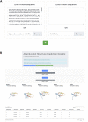
FIGURE 1
FIGURE 1
AttentiveDist panels. (A) The input page of AttentiveDist where users can input amino acid sequences of subunits to model. By clicking the “+” button, additional sequence submission field will appear. The maximum number of sequences users can submit is six. (B) the result panel of AttentiveDist. For each submitted sequence, top five scoring models are visualized. Scores of the five models are shown in a table below the visualization panel. The models are ranked by the ranksum score.
FIGURE 2
FIGURE 2
Submitting and interpreting protein docking job. (A) the job submission page. The figure represents a situation that a structure model of Protein 1 is transferred from AttentiveDist and the structure for Protein 2 will be fetched from PDB. The check box for performing C2 Symmetry docking is highlighted by rectangle in yellow. (B) docking results page. On the top of the panel, a distribution of centroids of the docked poses of the ligand structures are indicated with spheres. By clicking a sphere, the docked structure of the pose will be presented.
FIGURE 3
FIGURE 3
Input and results for unconstrained docking of IL23-IL23R. (A) the input for unconstrained LZerD. The model of IL23 was uploaded as the receptor on the left, while the model of IL23R was uploaded as the ligand on the right. The chain ID selection fields are blank since we want to use all the chains. This docking run was done without constraints, so the entire constraints section is empty. (B) the results of unconstrained docking. IL23 is shown in red, while IL23R is shown in blue. The cartoon structure shown is the top-10 model, which has an fnat of 0.28, an I-RMSD of 4.4 Å, and an L-RMSD of 7.8 Å, which is of acceptable CAPRI quality. The distribution of the top 50 ligand centroids is indicated by the orange spheres. (C) The table of model scoring information for this docking run. The ranksum score, which is used to finally rank the models, is on the right. (D) the native structure of this complex, PDB 5MZV. The non-interacting domains of IL23R and a nanobody bound to IL23 are included in the view.
FIGURE 4
FIGURE 4
Input and results for C2 symmetrical docking of bacterial AAC (2′). (A) The input for symmetrical LZerD. The subunit was uploaded by specifying the PDB ID 1M4G in the input field to fetch the structure from the PDB. A single chain is extracted from this structure by specifying “A” in the chain ID selection field. (B) The results of C2 symmetrical docking. The receptor and ligand are shown in red and blue respectively in the top-1 model conformation and are of course structurally identical. This model has an fnat of 0.88, an I-RMSD of 0.95 Å, and an L-RMSD of 1.9 Å. The distribution of the top 50 ligand centroids is indicated by the orange spheres. All output models satisfy the 5.0 Å symmetricalness criterion. (C) the native structure of this complex, both chains of PDB 1M4G. Note that some N-terminal residues of chain B (cyan) are not resolved relative to chain A (green). In fact, considering all common atoms between the two native chains, they differ by 0.8 Å RMSD. (D) Visualization of the symmetricalness criterion’s satisfaction in the top-1 model viewed along the C2 symmetry axis. The subunits are shown here in Cα trace representation. Green: the receptor; cyan: the correct ligand conformation; blue: the top-1 ligand conformation; red: the deviations of the ligand from the correct conformation. This model satisfies the symmetricalness criterion with an RMSD of 2.2 Å.
FIGURE 5
FIGURE 5
De novo subunit modeling and LZerD docking of colEdes3:Imdes3. (A) The input for de novo modeling of the subunits with AttentiveDist. The sequences were pasted into the input fields, but users can alternatively upload FASTA files. (B) The results of de novo structure prediction. Both subunits are available from this page, and colEdes3 is currently selected for display. Users can download models individually or in bulk and can forward models to LZerD by selecting the checkboxes and clicking the LZerD or Multi-LZerD button. The scoring table appears below the 3D models. (C) The top-1 AttentiveDist models superimposed to the native structure (green and cyan; PDB ID: 6ERE). The top-1 models for colEdes3 (red) and Imdes3 (blue) have RMSDs of 2.5 Å and 2.0 Å. (D) The constraint used for LZerD docking. Here, Phe68 of colEdes3 was constrained to be in contact with Tyr55 of Imdes3 by specifying a distance cutoff of 5.0 Å. (E) Results of constrained LZerD. Model 5 is shown, and is of acceptable quality with an of 0.32, an I-RMSD of 3.9 Å, and an L-RMSD of 10.7 Å. As indicated by the centroid distribution, the docking search has been focused around the binding site by the constraint. (F) Results of unconstrained LZerD. Model 13 is shown, but is not of acceptable quality, with an fnatof 0.36, an I-RMSD of 4.2 Å, and an L-RMSD of 13.1 Å. This I-RMSD barely missed the CAPRI threshold for acceptable quality. Without any constraint, the docking for this input does not produce acceptable models in the top 10. As indicated by the centroid distribution, the docked models are largely preferring a different site.

Similar articles

Cited by

References

    1. Aderinwale T., Christoffer C. W., Sarkar D., Alnabati E., Kihara D. (2020). Computational Structure Modeling for Diverse Categories of Macromolecular Interactions. Curr. Opin. Struct. Biol. 64, 1–8. 10.1016/j.sbi.2020.05.017 - DOI - PMC - PubMed
    1. Altschul S., Madden T. L., Schaffer A. A., Zhang J., Zhang Z., Miller W., et al. (1997). Gapped BLAST and PSI-BLAST: a New Generation of Protein Database Search Programs. Nucleic Acids Res. 25, 3389–3402. 10.1093/nar/25.17.3389 - DOI - PMC - PubMed
    1. Berman H. M., Westbrook J., Feng Z., Gilliland G., Bhat T. N., Weissig H., et al. (2000). The Protein Data Bank. Nucleic Acids Res. 28, 235–242. 10.1093/nar/28.1.235 - DOI - PMC - PubMed
    1. Chaudhury S., Lyskov S., Gray J. J. (2010). PyRosetta: a Script-Based Interface for Implementing Molecular Modeling Algorithms Using Rosetta. Bioinformatics 26, 689–691. 10.1093/bioinformatics/btq007 - DOI - PMC - PubMed
    1. Christoffer C., Chen S., Bharadwaj V., Aderinwale T., Kumar V., Hormati M., et al. (2021). LZerD Webserver for Pairwise and Multiple Protein-Protein Docking. Nucleic Acids Res., 49, W359, W365. 10.1093/nar/gkab336 - DOI - PMC - PubMed

LinkOut - more resources