. 2021 May 30:7:504-514.
doi: 10.1016/j.bioactmat.2021.05.016. eCollection 2022 Jan.

Study of copper-cysteamine based X-ray induced photodynamic therapy and its effects on cancer cell proliferation and migration in a clinical mimic setting

Affiliations

Study of copper-cysteamine based X-ray induced photodynamic therapy and its effects on cancer cell proliferation and migration in a clinical mimic setting

Xiangyu Chen et al. Bioact Mater. .

Abstract

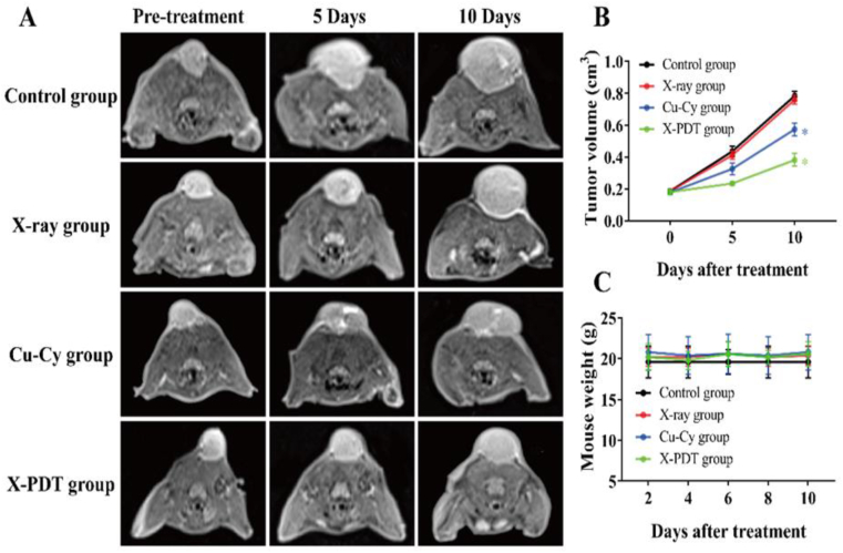
Copper-cysteamine as a new generation of sensitizers can be activated by light, X-rays, microwaves, or ultrasound to produce reactive oxygen species. X-ray induced photodynamic therapy (X-PDT) has been studied extensively; however, most of the studies reported so far were conducted in the laboratory, which is not conducive to the clinical translation conditions. In this contribution, for the first time, we investigated the treatment efficiency of copper-cysteamine (Cu-Cy) based X-PDT by mimicking the clinical conditions with a clinical linear accelerator and building deep-seated tumor models to study not only the effectiveness but also its effects on the cell migration and proliferation in the level of the cell, tissue, and animal. The results showed that, without X-ray irradiation, Cu-Cy nanoparticles (NPs) had a low toxicity in HepG2, SK-HEP-1, Li-7, and 4T1 cells at a concentration below 100 mg/L. Interestingly, for the first time, it was observed that Cu-Cy mediated X-PDT can inhibit the proliferation and migration of these cell lines in a dose-dependent manner. Antigen markers of migration and cell proliferation, proliferating cell nuclear antigen (PCNA) and E-cadherin, from tumor tissue in the X-PDT group were remarkably different from that of the control group. Furthermore, the MRI assessment showed that the Cu-Cy based X-PDT inhibited the growth of deeply located tumors in mice and rabbits (p < 0.05) without any obvious toxicities in vivo. Overall, these new findings demonstrate that Cu-Cy NPs have a safe and promising clinical application prospect in X-PDT to improve the efficiency of radiotherapy (RT) for deep-seated tumors and effectively inhibit tumor cell proliferation and migration.

Keywords: Cell migration; Copper-cysteamine; Nanoparticle; Photodynamic therapy; Proliferation; X-ray.


Conflict of interest statement

The authors declare that they have no known competing financial interests or personal relationships that could have appeared to influence the work reported in this article.

Figures

Image 1
Graphical abstract
Fig. 1
Fig. 1
(A) UV–vis absorption spectrum of the Cu-Cy NPs suspended in DI water. (B) Pictures of the Cu-Cy NPs dispersed in DI water upon 365 nm UV light (left) and room light (right). (C) The spectra of photoluminescence excitation (PLE, left) at 607 nm and emission (PL, right) at 365 nm of the Cu-Cy NPs dispersed in DI water. (D) A representative TEM image of the Cu-Cy NPs.
Fig. 2
Fig. 2
(A) Histogram of the size distribution of Cu-Cy nanoparticles used in this study. Average diameter = 43 ± 10 nm. (B) Comparison of UV–visible absorption spectra of Cu-Cy in DI water, complete cell culture media of KYSE-30 cancer cells, RPMI-1640 medium, and Ham's F-12 medium. (C) Comparison of photoluminescence excitation (PLE) and emission (PL) spectra of Cu-Cy in DI water, RPMI-1640 medium, and Ham's F-12 medium.
Fig. 3
Fig. 3
X-PDT inhibited the proliferation of tumor cells in vitro. The cell viability of HepG2, SK-HEP-1, Li-7, or 4T1 cells were calculated by CCK8 assay after treating 0, 10, 25, 50, 100, or 150 mg/L of Cu-Cy NPs with or without X-rays irradiation with a dose of 2 Gy in vitro (*p < 0.05 vs no X-ray group).
Fig. 4
Fig. 4
X-PDT inhibited the migration of tumor cells in vitro. (A) Transwell assays detected the migration of HepG2 and SK-Hep-1 after the X-PDT with low dosage (50 mg/L) and high dosage (100 mg/L) of Cu–Cy NPs, separately. (B–C) The number of migratory cells was counted and plotted on two graphs (*p < 0.05 vs control group).
Fig. 5
Fig. 5
The ROS production in Cu-Cy NPs under X-ray irradiation.
Fig. 6
Fig. 6
X-PDT enhanced the tumoricidal effect in mouse subcutaneous tumor models. (A) MR assessment of tumoricidal effect after different interventions. (B) Tumor volume changes (*p < 0.05 vs control group). (C) Body weight changes.
Fig. 7
Fig. 7
X-PDT enhanced the tumoricidal effect in rabbit VX2 tumor models. (A) MR assessment of tumoricidal effect after different interventions. (B) Tumor volume changes (*p < 0.05 vs control group). (C) Body weight changes.
Fig. 8
Fig. 8
H & E staining of heart, liver, spleen, lung, and kidney at the end of treatment. H & E staining with magnification of 200×.
Fig. 9
Fig. 9
Morphological examination and immunohistochemistry of tumors. (A) H&E staining, E-cadherin, and PCNA immunohistochemistry staining of tumor tissues. (B) The percentages of the positive expression of E-cadherin (*p < 0.05 vs control group). (C) The percentages of the positive expression of PCNA (*p < 0.05 vs control group). H&E and immunohistochemistry staining with magnification of 200 × .
Fig. 10
Fig. 10
The possible influences of X-PDT on cells: the main effects from ROS including oxidative stress, cell killing by direct injury, hypoxia formation due to the consumption of oxygen, possible induction of cell migration, and invasion as well as enhancing immunity.

References

    1. Miller K.D., Nogueira L., Mariotto A.B., Rowland J.H., Yabroff K.R., Alfano C.M., Jemal A., Kramer J.L., Siegel R.L. Cancer treatment and survivorship statistics, 2019. CA A Cancer J. Clin. 2019;69(5):363–385. - PubMed
    1. Urba S., Wolf G., Eisbruch A., Worden F., Lee J., Bradford C., Teknos T., Chepeha D., Prince M., Hogikyan N., Taylor J. Single-cycle induction chemotherapy selects patients with advanced laryngeal cancer for combined chemoradiation: a new treatment paradigm. J. Clin. Oncol. : Off. J. Am. Soc. Clin. Oncol. 2006;24(4):593–598. - PubMed
    1. Sun W., Zhou Z., Pratx G., Chen X., Chen H. Nanoscintillator-mediated X-ray induced photodynamic therapy for deep-seated tumors: from concept to biomedical applications. Theranostics. 2020;10(3):1296–1318. - PMC - PubMed
    1. Liu Z., Cao T., Xue Y., Li M., Wu M., Engle J.W., He Q., Cai W., Lan M., Zhang W. Self‐amplified photodynamic therapy through the 1O2‐mediated internalization of photosensitizers from a ppa‐bearing block copolymer. Angew. Chem. Int. Ed. 2020;59:3711–3717. - PMC - PubMed
    1. Tian J., Zhang W. Synthesis, self-assembly and applications of functional polymers based on porphyrins. Prog. Polym. Sci. 2019;95:65–117.

LinkOut - more resources