. 2021 Aug 11;24(9):102967.
doi: 10.1016/j.isci.2021.102967. eCollection 2021 Sep 24.

YAP regulates alveolar epithelial cell differentiation and AGER via NFIB/KLF5/NKX2-1

Affiliations

YAP regulates alveolar epithelial cell differentiation and AGER via NFIB/KLF5/NKX2-1

Jason J Gokey et al. iScience. .

Abstract

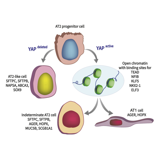
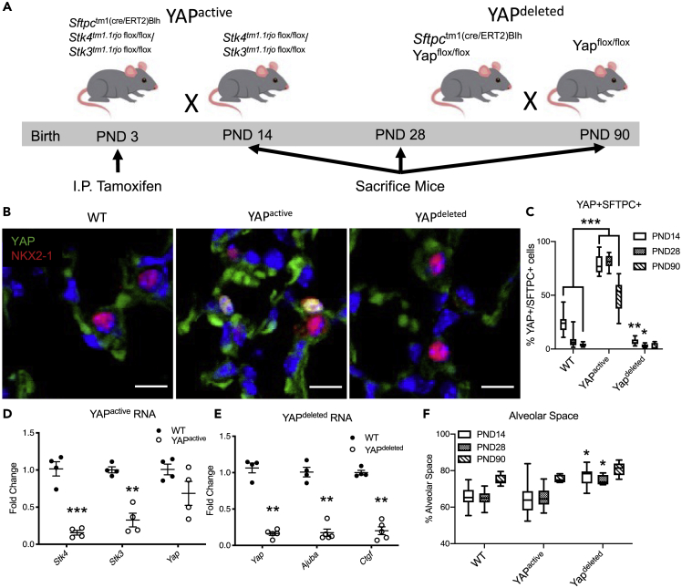
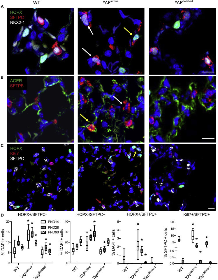
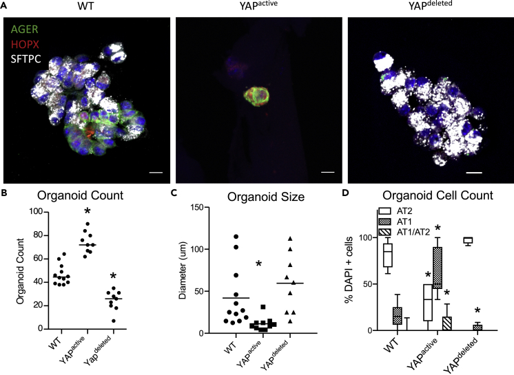
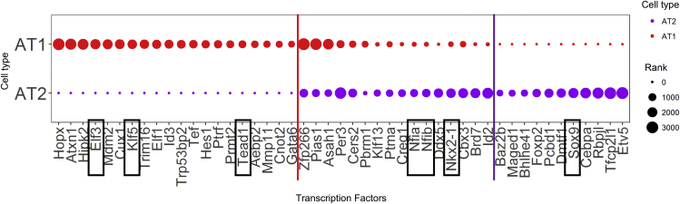
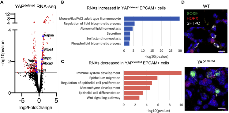
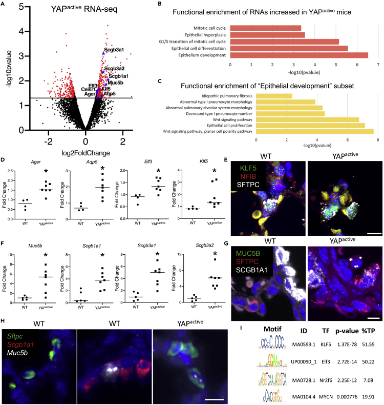
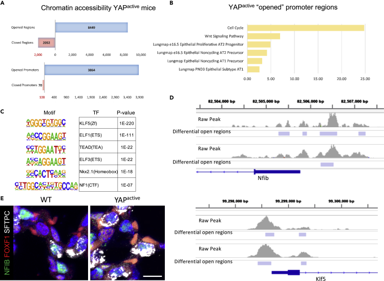
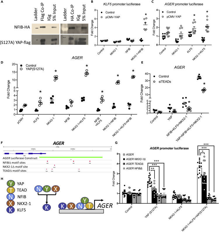
Ventilation is dependent upon pulmonary alveoli lined by two major epithelial cell types, alveolar type-1 (AT1) and 2 (AT2) cells. AT1 cells mediate gas exchange while AT2 cells synthesize and secrete pulmonary surfactants and serve as progenitor cells which repair the alveoli. We developed transgenic mice in which YAP was activated or deleted to determine its roles in alveolar epithelial cell differentiation. Postnatal YAP activation increased epithelial cell proliferation, increased AT1 cell numbers, and caused indeterminate differentiation of subsets of alveolar cells expressing atypical genes normally restricted to airway epithelial cells. YAP deletion increased expression of genes associated with mature AT2 cells. YAP activation enhanced DNA accessibility in promoters of transcription factors and motif enrichment analysis predicted target genes associated with alveolar cell differentiation. YAP participated with KLF5, NFIB, and NKX2-1 to regulate AGER. YAP plays a central role in a transcriptional network that regulates alveolar epithelial differentiation.

Keywords: Developmental genetics; Genetics; Molecular biology.


Conflict of interest statement

The authors declare no competing interests.

Figures

None
Graphical abstract
Figure 1
Figure 1
YAP activation and deletion in mouse AT2 cells (A) Strategy and timeline of inducible deletion or activation of YAP. Mice were sacrificed at postnatal day (PND) 14, 28 and 90 for analysis. (B) Immunofluorescence analysis of YAP (green) and NKX2-1 (red) and quantification of SFTPC+ AT2 cells with YAP+ nuclei at PND 14, 28, and 90 demonstrating increased nuclear YAP in YAPactive mouse lungs. (D and E) qRT-PCR analysis of YAP-related genes in isolated EPCAM+ cells following YAP activation (D) or deletion (E). (F) Quantification of alveolar space in WT, YAPactive and YAPdeleted mouse lungs at PND14, 28, and 90 demonstrating YAPdeleted mouse lungs have increased alveolar space. ∗ Indicates p < 0.05 as determined by two-way ANOVA followed by Sidak's multiple comparison test. Dot plot graph error bars represent S.E.M and whiskers of Box–Whisker plots represent min and max values. ∗∗p<0.001, ∗∗∗p<0.0001.
Figure 2
Figure 2
YAP regulates alveolar epithelial cell differentiation (A–C) Immunofluorescence analysis of proliferation (KI67), AT1 (HOPX, AGER), AT2 (SFTPC, SFTPB), and epithelial (NKX2.1) cell markers to assess alveolar epithelial cell proliferation and differentiation in YAPactive and YAPdeleted mouse lungs. (D) Quantification of HOPX+/SFTPC- AT1, HOPX-/SFTPC+ AT2, HOPX+/SFTPC+ “dual positive” (yellow arrow) and KI67+/SFTPC+ proliferating AT2 cells (red arrows) in PND14 (N = 11 WT, 12 YAPactive, 10 YAPdeleted), 28 (N = 9 WT, 9 YAPactive, 8 YAPdeleted), and 90 (N = 8 WT, 8 YAPactive, 8 YAPdeleted). SFTPC+ “doublets” are indicated with white arrows. Whiskers of box-whisker plots represent min and max values. ∗Indicates p<0.05 as determined by Two-way ANOVA followed by Sidak's multiple comparisons test.
Figure 3
Figure 3
YAP regulates growth and differentiation of alveolar organoids (A) Immunofluorescence analysis of AGER (green), HOPX (red), and SFTPC (white) of organoids generated with WT control, YAPactive, or YAPdeleted EPCAM+ cells. (B) Quantification of organoids generated per well shows YAPactive cells (N = 9) generate more organoids, while YAPdeleted cells (N = 9) produced fewer than WT cells (N = 11). (C) Analysis of average organoid size per well demonstrates YAPactive cells generated smaller organoids than those produced with YAPdeleted or WT EPCAM+ cells. (D) Quantification of cell-type specific markers demonstrate a shift in alveolar epithelial cell differentiation with more AT1 cells present in YAPactive and less AT1 cells in YAPdeleted organoids. Whiskers of box-whisker plots represent min and max values. See also Figure S1 for AEC purity during cell preparation. ∗Indicates p<0.05 as determined by Two-way ANOVA followed by Sidak's multiple comparisons test.
Figure 4
Figure 4
Predicted alveolar epithelial cell transcriptional network Prediction of alveolar epithelial cell transcriptional regulatory network (TRN) from previously published single cell RNA sequencing data. AT1 or AT2 specific differentially expressed genes were used as potential target genes and transcriptional factors commonly or selectively expressed in AT1 or AT2 were identified as potential transcription factor regulators. Transcription factors were ranked based on their nodes importance to the inferred AT1 or AT2 TRNs using a method combining the six node importance metrics as described in SINCERA, with a higher rank score corresponding to a transcription factor being more important to a given TRN. The transcription factors Elf3, Klf5 and Tead1 were predicted as AT1 selective, and Sox9 and Etv5 were AT2 selective. Nkx2-1, Nfia and Nfib were predicted as important in both AT1 and AT2 cells. See also Figure S2 for network information.
Figure 5
Figure 5
YAP deletion increases expression of AT2 cell signature genes RNA-seq analyses were performed on EPCAM+ cells isolated from PND14 YAPdeleted (N = 5) and WT (N = 4) mice. (A) Volcano plot of the differentially expressed genes (p < .05, FC > 1.5) shows increase in AT2 markers Abca3, Sftpb and Sftpa1, and decrease in known YAP targets Ajuba, Axl and Yap itself. (B) Functional enrichment analyses of the induced genes associated with lipid and surfactant production. (C) Functional enrichment analyses of the suppressed genes show decreased gene expression associated with Wnt signaling, Immune and Mesenchyme development, Epithelial cell migration and proliferation and Epithelial cell differentiation. (D) Immunofluorescence analysis of SOX9 (green) and SFTPC (white) demonstrates the presence of SOX9+/SFTPC+ epithelial cells in YAPdeleted mice. See Figure S3 for further ATAC-seq quality control.
Figure 6
Figure 6
YAP activation increases expression of AT1 and proximal epithelial cell signature genes RNA-seq analyses were performed on EPCAM+ cells isolated from D14 YAPactive (N = 6) and controls (N = 4) mice. (A) Volcano plot of the differentially expressed genes (p < .05, FC > 1.5) shows increase in AT1 markers Ager and Aqp5 as well as proximal lung airway markers Muc5b, Scgb1a1, Scgb3a1 and Scgb3a2. (B) Genes upregulated in the YAPactive epithelium are associated with mitotic cell cycle and epithelial cell differentiation and development. (C) Functional enrichment analyses of the subset of genes associated with epithelium development revealed increases in the expression of genes associated with Wnt signaling, epithelium proliferation, IPF and abnormal AT1 differentiation. (D) Realtime qRT-PCR analyses of isolated YAPactive epithelial cells demonstrate increased Ager, Aqp5, Elf3 and Klf5. ∗Indicates p<0.05 as determined by Welch's t-test. (E) Immunofluorescence analysis of NFIB (red), KLF5 (green), and SFTPC (white) shows presence of KLF5 in SFTPC+ cells including the SFTPC+ AT2 cell doublets identified in YAPactive mice. (F) qRT-PCR analysis of proximal cell signature genes demonstrate increased conducting airway epithelial cell markers in YAPactive epithelial cells. ∗Indicates p<0.05 as determined by Welch's t-test. (G) Immunofluorescence analysis of MUC5B (green), SFTPC (red), and SCGB1A1 (white), shows the presence of SCGB1A1+/SFTPC+ cells in YAPactive mice. (H) RNAscope Fluorescent in-situ hybridization of Muc5b, Scgb1a1, and Sftpc in WT and YAPactive mouse lungs demonstrating the presence of Sftpc+/Muc5b+ cells in YAPactive lungs. (I) Promoter analyses of the genes increased in the YAPactive epithelium show that transcription factors Klf5, Elf3, Nr2f6, and Mycn RNAs are increased and have enriched binding sites in YAPactive induced genes. See Table S1 for pathways down regulated in YAPactive mice and Figure S4 for immunofluorescence images.
Figure 7
Figure 7
YAP regulates chromatin accessibility of promoter regions in genes associated with alveolar differentiation ATAC-seq was performed on EPCAM+ cells isolated from PND14 YAPactive (N = 3) and WT (N = 3) mice. (A) YAP activation opened over 8400 regions of DNA and closed 2052 regions (p < .01). Over 3800 gene promoters (1.5kb of predicted transcriptional start site) were opened in YAPactive epithelial cells with only 70 promoters being closed. (B) Functional enrichment analyses of genes with opened promoters opened in YAPactive mice show increased accessibility of signature genes for various AT1 and AT2 subtypes along with genes involved in Wnt signaling and cell cycle. (C) Motif enrichment analyses of the regions opened in YAPactive mice show an enrichment for multiple transcription factors that were predicted regulators of the AT1 and/or AT2 cell TRN. (D) IGV was used to visualize regions opened in Nfib and Klf5 promoter regions in YAPactive epithelial cells. ATAC-seq analyses were done using Mac2 on 2 litters, with YAPactive (N = 3) compared to Stk3flox/floxStk4flox/flox (N = 3) control littermates. Only regions altered in both litters were considered significant. (E) Immunofluorescence analysis of NFIB (green), FOXF1 (red), and SFTPC (white) demonstrates NFIB in a subset of SFTPC+ cells in both WT and YAPactive mouse lungs. See Figure S3 for analysis of ATAC-seq quality.
Figure 8
Figure 8
YAP interacts with NFIB, KLF5, and NKX2-1 to regulate gene expression (A) Immunoprecipitation assay of HBEC3 cells co-expressing (S127A)YAP-FLAG and GFP-NFIB-HA constructs demonstrating YAP and NFIB co-precipitate. (B) HBEC3 cells co-expressing (S127A)YAP, KLF5, and NKX2-1 activate KLF5 promoter luciferase activity. (C) HBEC3 cells co-transfected with (S127A)YAP, NKX2-1, and KLF5 activate AGER promoter luciferase. (D) HBEC3 cells co-expressing empty pCMV vector, KLF5, NKX2-1, NFIB, and (S127A)YAP increase AGER RNA assessed by qRT-PCR. (E) AGER RNA was measured in HBEC3 cells co-transfected with pCMV empty vector (control), (S127A)YAP, NFIB, KLF5, and NKX2-1 with siRNAs targeting TEADs 1-4. AGER induction is partially inhibited by TEAD siRNAs. Graphs are representative of multiple (N > 3) experiments. (F) A schematic of the AGER promoter and luciferase assay with locations of predicted TEAD, NKX2-1, and NFIB binding sites that were mutated to assess DNA binding of respective transcription factors to the AGER promoter. (G) HBEC3 cells expressing NKX2-1, KLF5, and YAP in the presence of AGER luciferase constructs with five site mutations in predicted binding sites of TEAD (TEADΔ), NKX2-1 (NKX2-1Δ) or NFIB (NFIBΔ). Altering the predicted binding sites of NKX2-1 (p < 0.05), TEAD (p < 0.0001), or NFIB (p < 0.0001) significantly reduced AGER promoter activation by YAP, KLF5, and NKX2-1. (H) A schematic of NFIB, KLF5, NKX2-1, YAP, and TEAD interacting on the AGER promoter to induce AGER transcription. See Figure S5 for further information and Figure S6 for full size co-immunoprecipitation blots. ∗Indicates p<0.05 as determined by Two-way ANOVA followed by Sidak's multiple comparisons test. ∗∗p<0.001, ∗∗∗p<0.0001.

References

    1. Afgan E., Baker D., Batut B., van den Beek M., Bouvier D., Cech M., Chilton J., Clements D., Coraor N., Gruning B.A. The Galaxy platform for accessible, reproducible and collaborative biomedical analyses: 2018 update. Nucleic Acids Res. 2018;46:W537–W544. - PMC - PubMed
    1. Azzolin L., Panciera T., Soligo S., Enzo E., Bicciato S., Dupont S., Bresolin S., Frasson C., Basso G., Guzzardo V. YAP/TAZ incorporation in the beta-catenin destruction complex orchestrates the Wnt response. Cell. 2014;158:157–170. - PubMed
    1. Barkauskas C.E., Cronce M.J., Rackley C.R., Bowie E.J., Keene D.R., Stripp B.R., Randell S.H., Noble P.W., Hogan B.L. Type 2 alveolar cells are stem cells in adult lung. J. Clin. Invest. 2013;123:3025–3036. - PMC - PubMed
    1. Barnett D.W., Garrison E.K., Quinlan A.R., Stromberg M.P., Marth G.T. BamTools: AC++ API and toolkit for analyzing and managing BAM files. Bioinformatics. 2011;27:1691–1692. - PMC - PubMed
    1. Bell S.M., Zhang L., Mendell A., Xu Y., Haitchi H.M., Lessard J.L., Whitsett J.A. Kruppel-like factor 5 is required for formation and differentiation of the bladder urothelium. Dev. Biol. 2011;358:79–90. - PMC - PubMed