. 2022 Apr;71(4):979-987.
doi: 10.1007/s00262-021-03043-x. Epub 2021 Aug 31.

Venetoclax-resistant CLL cells show a highly activated and proliferative phenotype

Affiliations

Venetoclax-resistant CLL cells show a highly activated and proliferative phenotype

Esteban Enrique Elias et al. Cancer Immunol Immunother. 2022 Apr.

Abstract

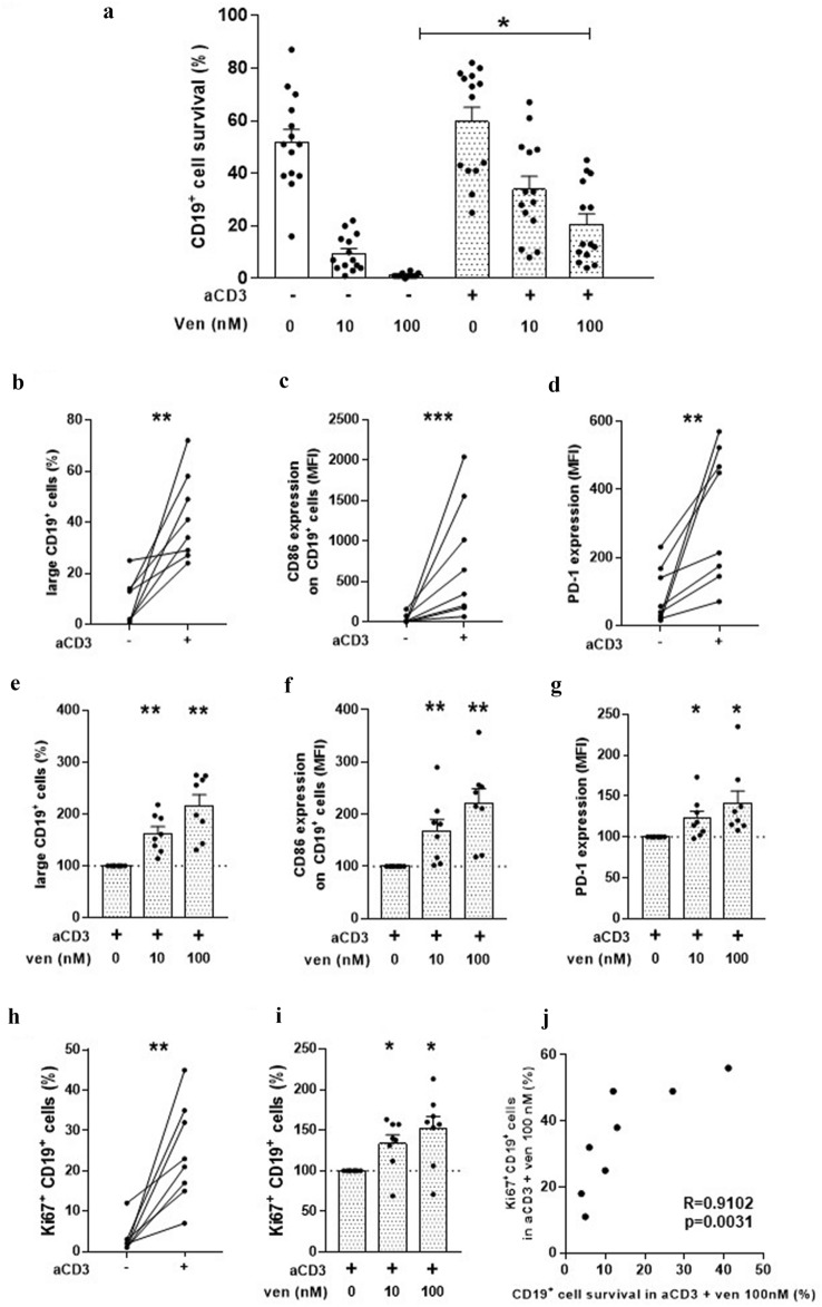
Venetoclax treatment has demonstrated efficacy and a safety profile in chronic lymphocytic leukemia (CLL) patients, however the emergence of resistant cells is a current complication. We and others, previously reported that the activation of CLL cells by signals that mimic microenvironment stimuli favors the upregulation of anti-apoptotic proteins from B cell lymphoma-2 (BCL-2) family that are not targeted by venetoclax, reducing malignant cell sensitivity to the drug. We here studied venetoclax-resistant CLL cells generated in vitro by autologous activated T lymphocytes, and found that they showed an aggressive phenotype characterized by increased expression of activation and proliferation markers. Moreover, surviving cells expressed high levels of B cell lymphoma-extra-large (BCL-XL) and/or myeloid cell leukemia-1 (MCL-1), and a sustained resistance to a second treatment with the drug. Interestingly, the spleen tyrosine kinase (SYK) inhibitor entospletinib, and the phosphoinositide 3-kinase delta (PI3Kδ) inhibitor idelalisib, reduced T cell activation, impaired the generation of leukemic cells with this aggressive phenotype, and were able to restore CLL sensitivity to venetoclax. Our data highlight a novel combination to overcome resistance to venetoclax in CLL.

Keywords: CLL; Entospletinib-Idelalisib; Ibrutinib-Acalabrutinib; Venetoclax resistance.


Conflict of interest statement

GC: Astrazeneca honoraria for advisory board. RFB: AbbVie and Astrazeneca honoraria for lectures and Microsules honoraria for advisory board. MRC: AbbVie honoraria for advisory board. GG: Janssen, Astrazaneca, Sanofi, Varifarma, Teva, Roche and Novartis honoraria for advisory board. MAP: Janssen, Astrazeneca, Raffo and AbbVie honoraria for lectures and Advisory boards. RG: Astrazeneca and AbbVie honoraria for lectures. The remaining authors declare no competing financial interests.

Figures

Fig. 1
Fig. 1
Venetoclax resistant CLL cells show an aggressive phenotype. Peripheral blood mononuclear cells (PBMC) from CLL patients (4 × 106 cells/ml) were cultured in complete medium with immobilized anti-CD3 antibodies (aCD3) or the isotype control for 48 h. Then, venetoclax or DMSO were added to the cultures for another 120 h and survival, enlargement of viable cells, and the expression of CD86, PD-1 and Ki-67 on viable CD19+ cells were evaluated by flow cytometry. a The figure shows the mean ± SEM of CD19+ cell survival in each condition. Statistical analysis was performed using Friedman test followed by Dunn post-test, * p < 0.05 (n = 14). b The graph shows the percentage of large CD19+ cells in control and aCD3 cultures 120 h post DMSO. Statistical analysis was performed using Wilcoxon matched pairs signed rank test, ** p < 0.01, (n = 8). c The graph shows the mean fluorescence intensity (MFI) of CD86 expression on CD19+ cells in control and aCD3 cultures 120 h post DMSO. Statistical analysis was performed using Wilcoxon matched pairs signed rank test, *** p < 0.001, (n = 8). d The graph shows the MFI of PD-1 expression on CD19+ cells in control and aCD3 cultures 120 h post DMSO. Statistical analysis was performed using Wilcoxon matched pairs signed rank test, ** p < 0.01, (n = 8). e The bars show the percentage of large CD19+ cells relative to control. Statistical analysis was performed using Wilcoxon Signed Rank Test, ** p < 0.001 (n = 8). f The figure shows the mean ± SEM of the expression of CD86 on CD19+ cells relative to control. Statistical analysis was performed using Wilcoxon signed rank test, ** p < 0.01, (n = 8). g The bars show the expression of PD-1 on CD19+ cells relative to control. Statistical analysis was performed using Wilcoxon signed rank test, * p < 0.05 (n = 8). h The graph shows the percentage of viable Ki67+ CD19+ cells in control and aCD3 cultures after 120 h post DMSO. Statistical analysis was performed using Wilcoxon matched pairs signed rank test, ** p < 0.01, (n = 8). i The figure shows the mean ± SEM of the percentage of Ki67+ CD19+ cells relative to control. Statistical analysis was performed using Wilcoxon signed rank test, * p < 0.05, (n = 8). j The graph shows the positive correlation between CD19+ cell survival and Ki-67 expression in CD19+ cells in aCD3 plus venetoclax 100 nM cultures. Statistical analysis was performed using Spearman rank correlation (p = 0.0031, R = 0.9102)
Fig. 2
Fig. 2
Venetoclax resistant CLL cells show high expression of MCL-1 and BCL-XL. PBMC from CLL patients (4 × 106 cells/ml) were cultured in complete medium with aCD3 or the isotype control. After 48 h, venetoclax 100 nM or DMSO were added to the cultures. After 24 h, non-viable cells were excluded by employing a dead cell removal kit, and then CLL cells were purified using a CLL purification kit as detailed in the supplementary materials and methods section. Then, whole cell lysates were prepared with purified viable CLL cells and proteins were separated on a standard 15% SDS-PAGE and transferred to a PVDF membrane. Membranes were probed with primary antibodies for MCL-1, BCL-XL, BCL-2 and β-Actin, followed by the corresponding secondary antibody. Then, specific bands were quantified by employing ImageJ and quantitative densitometry protein expression relative to β-actin as loading control was obtained for each culture condition. a The figure shows the expression of MCL-1, BCL-XL and BCL-2 relative to the expression obtained in control cultures. Statistical analysis was performed using Wilcoxon test, * p < 0.05 (n = 5). b The figure shows the results obtained with one representative CLL sample
Fig. 3
Fig. 3
Venetoclax resistant CLL cells are more resistant to a second treatment with the drug. PBMC from CLL patients (4 × 106 cells/ml) were cultured in complete medium with aCD3 or the isotype control. After 48 h, venetoclax 100 nM or DMSO were added to the cultures for another 120 h (First DMSO/venetoclax treatment). a The figure shows a schematic representation of the protocol. PBMC from control cultures with DMSO, and PBMC from aCD3 cultures with and without venetoclax, were extensively washed and cultured with venetoclax 100 nM or DMSO for 24 h (second DMSO/venetoclax treatment). The values of CD19 cell survival in each culture condition are also shown. b The figure shows the mean ± SEM of the percentage of viable CD19+ cells in venetoclax cultures relative to DMSO cultures. Statistical analysis was performed using Wilcoxon signed rank test and Friedman test followed by Dunn post-test, * p < 0.05 (n = 9)
Fig. 4
Fig. 4
Entospletinib and idelalisib overcome venetoclax resistance in CLL cells. PBMC from CLL patients (4 × 106 cells/ml) were cultured in complete medium with aCD3 or the isotype control in presence or absence of entospletinib 1 µM (En), idelalisib 1 µM (Id), ibrutinib 0,1 µM (Ib) and acalabrutinib 0,6 µM (Ac) for 48 h. Then, venetoclax 100 nM or DMSO were added to the cultures for additional 24 h. a The figure shows the mean ± SEM of the percentage of CD86+ CD19+ cells in aCD3 cultures with BCR-KI relative to aCD3 cultures without BCR-KI at 48 h. To compare BCR-KI versus control cultures we employed Wilcoxon Signed Rank Test, * p < 0.05. The comparison amongst BCR-KI were performed using Fridman test following Dunn's multiple comparisons test, # p < 0.05 (n = 7). b The figure shows the mean ± SEM of the percentage of Ki-67+ CD19+ cells in aCD3 cultures with BCR-KI relative to aCD3 cultures without BCR-KI at 48 h. To compare BCR-KI versus control cultures, we employed Wilcoxon Signed Rank Test, * p < 0.05. The comparison amongst BCR-KI were performed using Fridman test following Dunn's multiple comparisons test, # p < 0.05 (n = 7). c-e Purified viable CLL cells from control cultures and from aCD3 cultures with and without BCR-KI for 48 h were analyzed by western blot as detailed in Fig. 2. The expression of MCL-1 c, BCL-XL d and BCL-2 e is shown relative to the expression obtained in control cultures. Statistical analysis was performed using Fridman test following Dunn's multiple comparisons test, * p < 0.05 (n = 6). f The figure shows the results obtained with one representative CLL sample. g The figure shows the mean of CD19+ cell survival in each condition. Statistical analysis was performed using Friedman test followed by Dunn´s test, *p < 0.05 (n = 7)

Comment in

References

    1. Roberts AW, Davids MS, Pagel JM, Kahl BS, Puvvada SD, Gerecitano JF, Kipps TJ, Anderson MA, Brown JR, Gressick L, Wong S, Dunbar M, Zhu M, Desai MB, Cerri E, Heitner Enschede S, Humerickhouse RA, Wierda WG, Seymour JF. Targeting BCL2 with venetoclax in relapsed chronic lymphocytic Leukemia. N Engl J Med. 2016;374(4):311–322. doi: 10.1056/NEJMoa1513257. - DOI - PMC - PubMed
    1. Seymour JF, Mobasher M, Kater AP. Venetoclax-rituximab in chronic lymphocytic Leukemia. N Engl J Med. 2018;378(22):2143–2144. doi: 10.1056/NEJMc1805135. - DOI - PubMed
    1. Fischer K, Al-Sawaf O, Bahlo J, Fink AM, Tandon M, Dixon M, Robrecht S, Warburton S, Humphrey K, Samoylova O, Liberati AM, Pinilla-Ibarz J, Opat S, Sivcheva L, Le Du K, Fogliatto LM, Niemann CU, Weinkove R, Robinson S, Kipps TJ, Boettcher S, Tausch E, Humerickhouse R, Eichhorst B, Wendtner CM, Langerak AW, Kreuzer KA, Ritgen M, Goede V, Stilgenbauer S, Mobasher M, Hallek M. Venetoclax and obinutuzumab in patients with CLL and coexisting conditions. N Engl J Med. 2019;380(23):2225–2236. doi: 10.1056/NEJMoa1815281. - DOI - PubMed
    1. Mato AR, Thompson M, Allan JN, Brander DM, Pagel JM, Ujjani CS, Hill BT, Lamanna N, Lansigan F, Jacobs R, Shadman M, Skarbnik AP, Pu JJ, Barr PM, Sehgal AR, Cheson BD, Zent CS, Tuncer HH, Schuster SJ, Pickens PV, Shah NN, Goy A, Winter AM, Garcia C, Kennard K, Isaac K, Dorsey C, Gashonia LM, Singavi AK, Roeker LE, Zelenetz A, Williams A, Howlett C, Weissbrot H, Ali N, Khajavian S, Sitlinger A, Tranchito E, Rhodes J, Felsenfeld J, Bailey N, Patel B, Burns TF, Yacur M, Malhotra M, Svoboda J, Furman RR, Nabhan C. Real-world outcomes and management strategies for venetoclax-treated chronic lymphocytic leukemia patients in the United States. Haematologica. 2018;103(9):1511–1517. doi: 10.3324/haematol.2018.193615. - DOI - PMC - PubMed
    1. Elias EE, Almejun MB, Colado A, Cordini G, Vergara-Rubio M, Podaza E, Risnik D, Cabrejo M, Fernandez-Grecco H, Bezares RF, Custidiano MDR, Sanchez-Avalos JC, Vicente A, Garate GM, Borge M, Giordano M, Gamberale R. Autologous T-cell activation fosters ABT-199 resistance in chronic lymphocytic leukemia: rationale for a combined therapy with SYK inhibitors and anti-CD20 monoclonal antibodies. Haematologica. 2018;103(10):e458–e461. doi: 10.3324/haematol.2018.188680. - DOI - PMC - PubMed

MeSH terms