. 2021 Oct 27;95(22):e0090421.
doi: 10.1128/JVI.00904-21. Epub 2021 Sep 1.

Skeletal Muscle Is an Early Site of Zika Virus Replication and Injury, Which Impairs Myogenesis

Affiliations

Skeletal Muscle Is an Early Site of Zika Virus Replication and Injury, Which Impairs Myogenesis

Daniel Gavino-Leopoldino et al. J Virol. .

Abstract

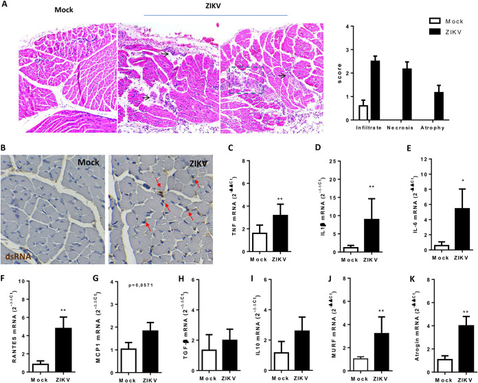
Zika virus (ZIKV) infection became a worldwide concern due to its correlation with the development of microcephaly and other neurological disorders. ZIKV neurotropism is well characterized, but the role of peripheral viral amplification to brain infection remains unknown. Here, we found that ZIKV replicates in human primary skeletal muscle myoblasts, impairing its differentiation into myotubes but not interfering with the integrity of the already-formed muscle fibers. Using mouse models, we showed ZIKV tropism to muscle tissue either during embryogenesis after maternal transmission or when infection occurred after birth. Interestingly, ZIKV replication in the mouse skeletal muscle started immediately after ZIKV inoculation, preceding viral RNA detection in the brain and causing no disruption to the integrity of the blood brain barrier, and remained active for more than 2 weeks, whereas replication in the spleen and liver were not sustained over time. In addition, ZIKV infection of the skeletal muscle induces necrotic lesions, inflammation, and fiber atrophy. We also found a reduction in the expression of regulatory myogenic factors that are essential for muscle repair after injury. Taken together, our results indicate that the skeletal muscle is an early site of viral amplification and lesion that may result in late consequences in muscle development after ZIKV infection. IMPORTANCE Zika Virus (ZIKV) neurotropism and its deleterious effects on central nervous system have been well characterized. However, investigations of the initial replication sites for the establishment of infection and viral spread to neural tissues remain underexplored. A complete description of the range of ZIKV-induced lesions and others factors that can influence the severity of the disease is necessary to prevent ZIKV's deleterious effects. ZIKV has been shown to access the central nervous system without significantly affecting blood-brain barrier permeability. Here, we demonstrated that skeletal muscle is an earlier site of ZIKV replication, contributing to the increase of peripheral ZIKV load. ZIKV replication in muscle promotes necrotic lesions and inflammation and also impairs myogenesis. Overall, our findings showed that skeletal muscle is involved in pathogenesis and opens new fields in the investigation of the long-term consequences of early infection.

Keywords: Zika virus replication; muscle inflammation; myogenesis; pathogenesis; skeletal muscle; viral dissemination.


Figures

FIG 1
FIG 1
ZIKV replicates in human skeletal muscle cells. Human primary skeletal myoblast and differentiated myotube cultures were infected with ZIKV at an MOI of 5 and temporally assessed. (A) ZIKV released by myoblast (white bars) and myotubes (black bars) at different times postinfection was quantified in culture supernatants by using a plaque assay (n = 4 to 5 for each point). (B) Immunofluorescence analysis for the detection of ZIKV-positive myoblasts and myotubes at 36 hpi using anti-flavivirus E protein 4G2 and anti-NS2B of ZIKV. White arrows indicate some positive cells. (C) Quantitative analysis of 4G2-positive cells expressed as the percentage of the total (nuclear staining with DAPI) was performed using at least 10 fields of two independent experiments with ImageJ software. (D) Cell viability was determined by MTT reduction at 48 h after ZIKV infection relative to mock-treated cells for three independent experiments performed in triplicate. (E) Detection and quantification of fibers for mock- and ZIKV-infected differentiated myotube cultures were performed using staining with anti-myosin heavy chain (MF20) at 48 hpi. For quantification of the fiber area, the fluorescence for at least 10 fields of three independent experiments was determined and normalized by the total number of cells (nuclear staining with DAPI) using ImageJ software. Data were analyzed using two-way ANOVA (A and C) or one-way ANOVA (D), followed Sidak and Turkey multiple comparisons, respectively (*, P < 0.05; **, P < 0.01; ***, P < 0.001; ###, P < 0.001 [compared to ZIKV-infected groups]).
FIG 2
FIG 2
ZIKV infection inhibits myogenesis. Skeletal muscle progenitor cells were subjected to differentiation into myotubes. At day 1 of differentiation, cells were mock or ZIKV infected at an MOI of 1 and then cultured until day 5 of differentiation. (A) Cells were fixed at days 1 and 5 of differentiation, and formed fibers were detected by immunofluorescence using MF20 (red); the cell nucleus was stained with DAPI (blue). Fluorescence images were quantified using at least 10 fields of three independent experiments with ImageJ software to obtain the fiber area (B), the fusion index (%) (C), and the cell number per field (D) for mock (white bars)- and ZIKV (black bars)-infected cells. Positive nuclei were used for counting on each field and to normalize the values per field. (E) ZIKV released at culture supernatant at different times postinfection was quantified by plaque assay (n = 3, each point). Statistical analysis was performed by two-way ANOVA (****, P < 0.0001).
FIG 3
FIG 3
ZIKV shows muscle tropism during embryogenesis. (A) Schematic representation of the period of ZIKV infection with pregnant SVA129 mice (red arrow). For this model (model 1), SVA129 females were mock or ZIKV infected (105 PFU) at different pregnancy periods (12.5, 14.5, and 18.5 days of gestation, at least n = 3 each), and tissues were analyzed at birth. (B) Numbers of pups per littermate after maternal exposure to mock or ZIKV infections at different pregnancy periods of infection (PPI). (C) Different tissues from female infected at day 12.5 of pregnancy were collected after delivery, and ZIKV RNA was quantified by qPCR (n = 4 to 6, each point). (D and E) Skeletal muscle from the hind legs (D) and brains (E) of pups were collected at birth after ZIKV inoculation at different PPI. ZIKV RNA in tissue was quantified by qPCR in samples from at least three independent experiments. (F) Muscle samples from pups either mock infected or at 12.5 PPI were fixed for histological analysis. The muscle tissues were embedded in paraffin after dehydration, and tissue sections of 5 μm were prepared, stained with H&E, and scored. Black arrows indicate areas of inflammation, red arrows indicate atrophy areas, and dashed lines indicate areas of lesion. (G) The ZIKV replication intermediary double-stranded RNA was stained in pup muscle sections at birth in mock- and ZIKV-infected groups with an anti-dsRNA J2 antibody. Slides were imaged on Sight DS-5M-L1 digital camera (Nikon, Melville, NY) connected to an Eclipse 50i light microscope (Nikon). Red arrows indicate positive staining areas. Values are plotted as means ± the standard errors of mean (SEM). Statistical analysis was performed by one-way ANOVA, followed of Tukey’s multiple-comparison test (*, P < 0.05; **, P < 0.01; ***, P < 0.001).
FIG 4
FIG 4
Muscle is a main site of peripheral ZIKV replication in vivo, preceding brain infection. (A) Three-day-old WT SV129 mice were ZIKV infected (106 PFU), and the replication kinetics in skeletal muscle from the hind legs, livers, spleens, brains, spinal cords (SC), and dorsal root ganglia (DRG) were determined by qPCR (n = 4 to 6, each point). N, nondetectable. (B) Blood-brain barrier permeability was analyzed in WT mice before infection (3 day old) and in mock- or ZIKV-infected mice after 2 and 6 days. These mice were subcutaneously inoculated with 0.1% Evans Blue solution or PBS. After 1 h, the animals were perfused with PBS, and tissues were removed for visualization. EB, Evans Blue solution; B, brain; S, spleen; L, liver; M, muscle. (C) Evans’s Blue incorporated in brain tissue at 6 dpi was recovered in formamide solution, and the amount was determined by spectrophotometry. (D) Negative strand amplification was detected by qPCR in neural structures at 6 dpi, and the CT (cycle threshold) values were plotted. (E) The ZIKV load was detected at late times postinfection in muscle and brain by qPCR. Values are plotted as means ± the SEM.
FIG 5
FIG 5
ZIKV induces skeletal muscle inflammatory lesions and atrophy in neonate infection. Three-day-old WT SV129 mice were ZIKV infected (106 PFU), and skeletal muscle tissues from the hind legs were collected at 6 dpi. (A) Muscle samples were embedded in paraffin after dehydration, and 5-μm tissue sections were prepared, stained with H&E, and scored. Black arrows indicate areas of inflammation; dashed lines indicate areas of lesion. (B) The ZIKV replication intermediary double-stranded RNA (dsRNA) was stained in muscle sections at 6 dpi using anti-dsRNA J2 antibody in mock- and ZIKV-infected groups. Slides were imaged on a Sight DS-5M-L1 digital camera (Nikon, Melville, NY) connected to an Eclipse 50i light microscope (Nikon). Red arrows indicate positive staining areas. (C to K) Skeletal muscle from hind legs of neonates was bilaterally collected, and the levels of TNF-α, IL-1β, IL-6, RANTES, MCP-1, TGF-β, IL-10, MURF, and Atrogin expression were determined relative to the mock-infected group by qPCR using β-actin expression as an endogenous control. Values are plotted as means ± the SEM. Statistical analysis was performed using a two-sided Mann-Whitney test (*, P < 0.05; **, P < 0.01).
FIG 6
FIG 6
ZIKV impairs activation of myogenic regulatory factors after injury and promoted a replication-dependent reduction in myogenesis in mouse cells. (A to D) Three-day-old WT SV129 mice were ZIKV infected (106 PFU), and skeletal muscle tissues from hind legs were bilaterally collected at 6 dpi. The levels of PAX-7, Myf5, MyoD, and MyoG expression were determined relative to the mock-infected group by qPCR using β-actin expression as an endogenous control. (E) Myoblast C2C12 cells were subjected to differentiation into myotubes. At day 1 of differentiation, the cells were mock treated, inoculated with UV light-inactivated ZIKV (iZIKV), or infected with ZIKV and then cultured until day 5 of differentiation. The cells were fixed at days 1 and 5 of differentiation, and formed fibers were detected by immunofluorescence using MF20 (red); the cell nucleus was stained with DAPI (blue). (F to H) Fluorescence images were quantified using at least 10 fields of three independent experiments with ImageJ software to obtain the cell number per field (F), the fiber area (G), and the fusion index (%) (H) of mock-infected cells (white bars) and ZIKV-infected cells (black bars). Nuclei labeled positive were counted in each field and used to normalize the values per field. (I) ZIKV released in the culture supernatant at different times postinfection was quantified by using a plaque assay (n = 5, each point). (J) An immunofluorescence assay was performed to detect NS2B ZIKV protein-positive cells at 5 days of differentiation. Arrows indicate mononucleated (white) and multinucleated (red) positive cells. Values are plotted as means ± the SEM. Statistical analysis was performed using the Mann-Whitney test (A) and one-way ANOVA, followed by Tukey’s multiple-comparison test (F to I) (**, P < 0.01; ***, P < 0.001 [compared to mock infection]; ##, P < 0.01 [compared to iZIKV]).

References

    1. Musso D, Gubler DJ. 2016. Zika virus. Clin Microbiol Rev 29:487–524. 10.1128/CMR.00072-15. - DOI - PMC - PubMed
    1. Krauer F, Riesen M, Reveiz L, Oladapo OT, Martinez-Vega R, Porgo TV, Haefliger A, Broutet NJ, Low N, Group W, WHO Zika Causality Working Group. 2017. Zika virus infection as a cause of congenital brain abnormalities and Guillain-Barre syndrome: systematic review. PLoS Med 14:e1002203. 10.1371/journal.pmed.1002203. - DOI - PMC - PubMed
    1. Carteaux G, Maquart M, Bedet A, Contou D, Brugieres P, Fourati S, Cleret de Langavant L, de Broucker T, Brun-Buisson C, Leparc-Goffart I, Mekontso Dessap A. 2016. Zika virus associated with meningoencephalitis. N Engl J Med 374:1595–1596. 10.1056/NEJMc1602964. - DOI - PubMed
    1. Nascimento OJM, da Silva IRF. 2017. Guillain-Barre syndrome and Zika virus outbreaks. Curr Opin Neurol 30:500–507. 10.1097/WCO.0000000000000471. - DOI - PubMed
    1. Garcez PP, Loiola EC, Madeiro da Costa R, Higa LM, Trindade P, Delvecchio R, Nascimento JM, Brindeiro R, Tanuri A, Rehen SK. 2016. Zika virus impairs growth in human neurospheres and brain organoids. Science 352:816–818. 10.1126/science.aaf6116. - DOI - PubMed

Publication types