Review
. 2021 Sep;16(14):1135-1151.
doi: 10.2217/fmb-2021-0044. Epub 2021 Sep 1.

Coronaviruses construct an interconnection way with ERAD and autophagy

Affiliations
Review

Coronaviruses construct an interconnection way with ERAD and autophagy

Aref Movaqar et al. Future Microbiol. 2021 Sep.

Abstract

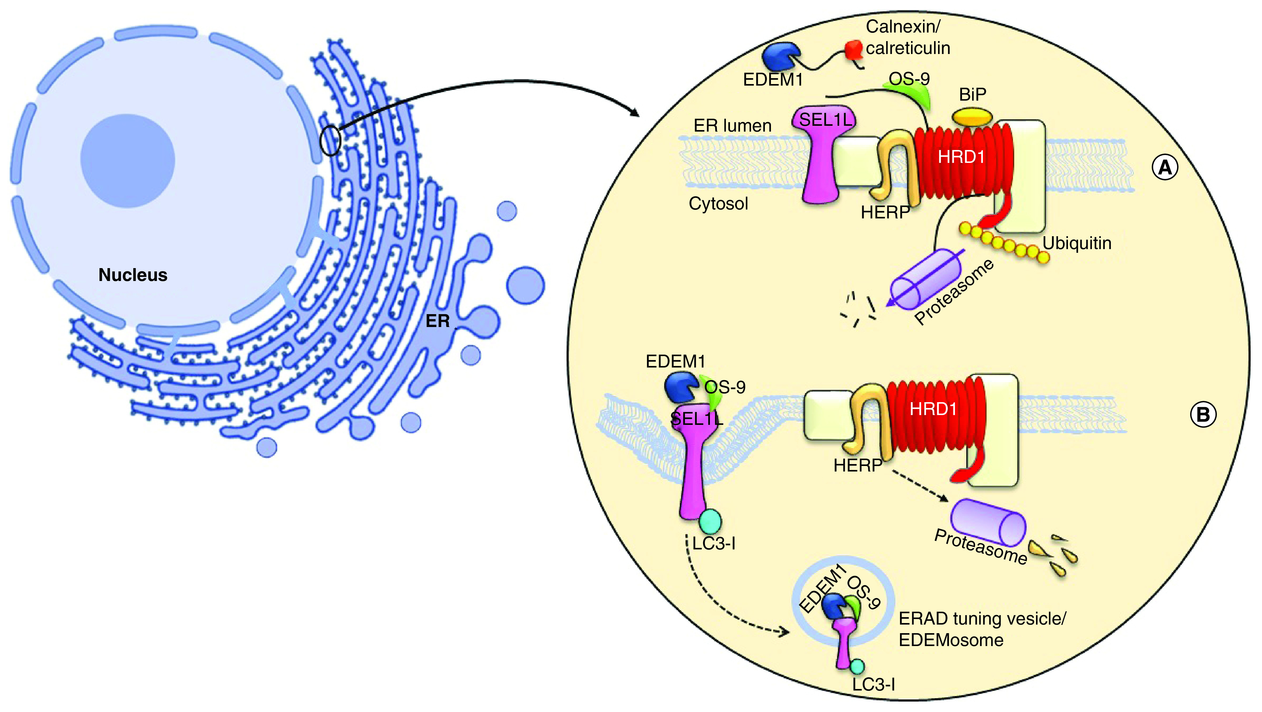
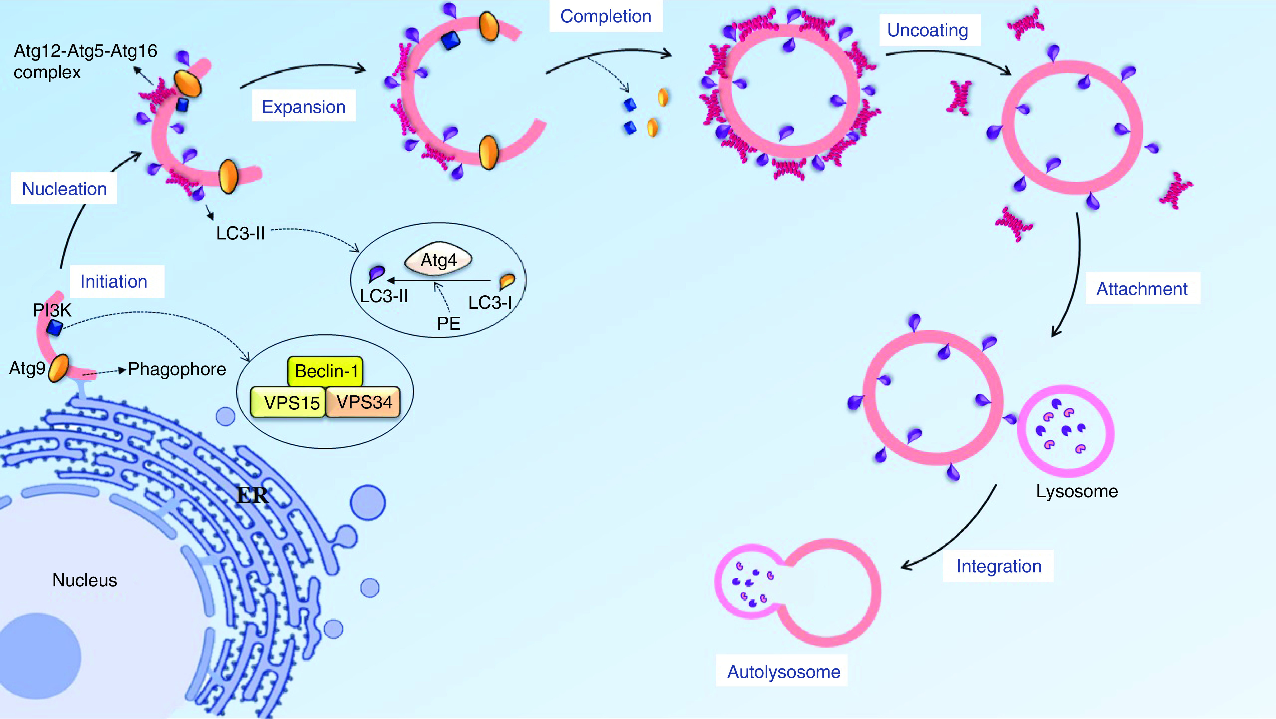
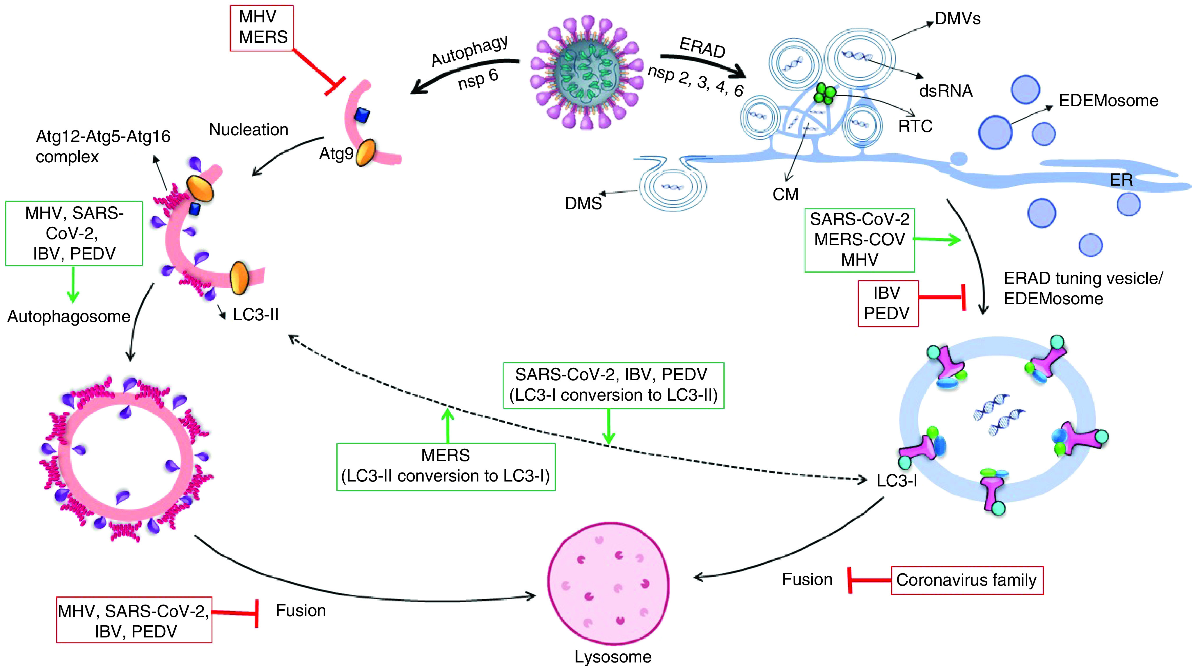
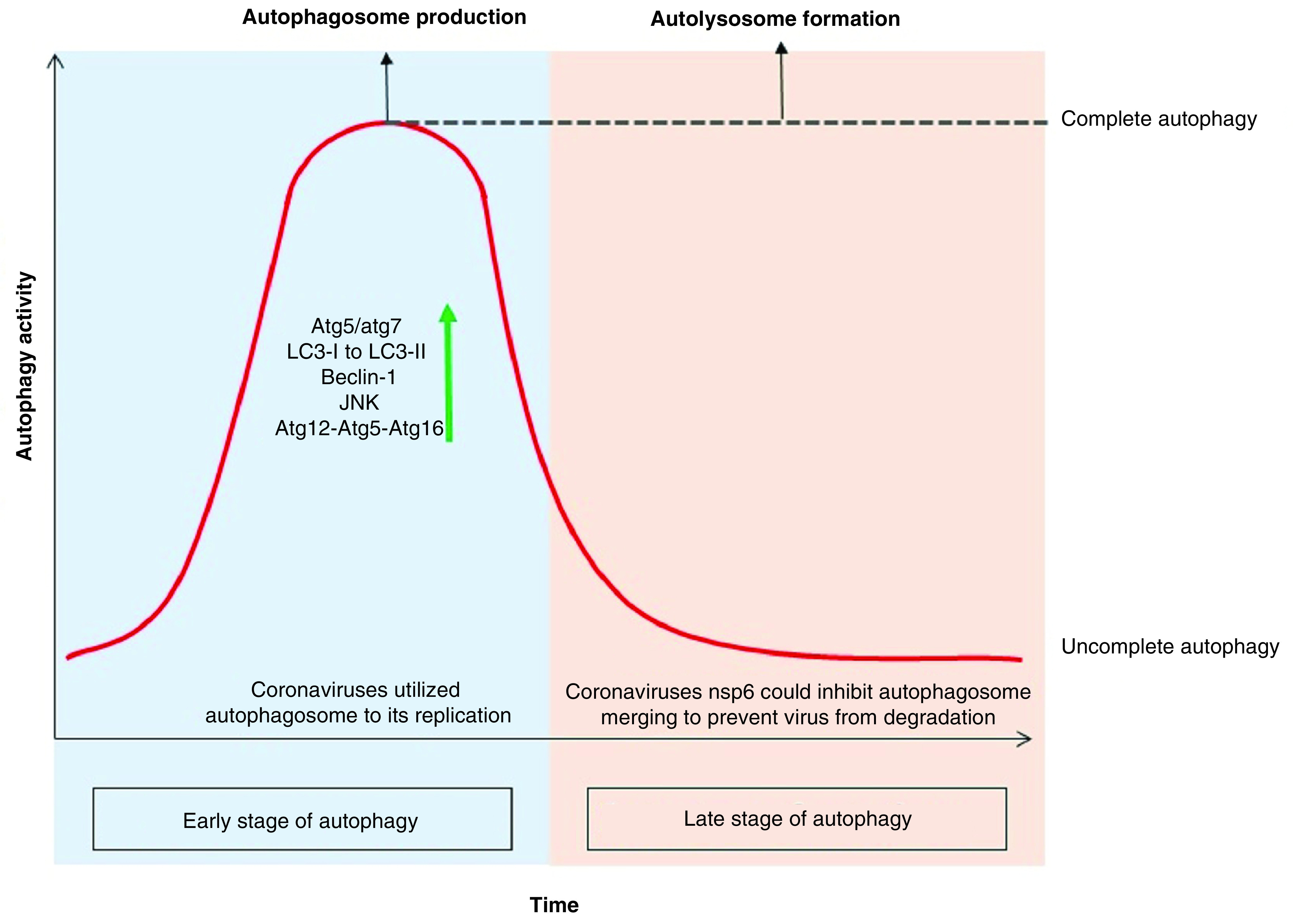
Coronaviruses quickly became a pandemic or epidemic, affecting large numbers of humans, due to their structural features and also because of their impacts on intracellular communications. The knowledge of the intracellular mechanism of virus distribution could help understand the coronavirus's proper effects on different pathways that lead to the infections. They protect themselves from recognition and damage the infected cell by using an enclosed membrane through hijacking the autophagy and endoplasmic reticulum-associated protein degradation pathways. The present study is a comprehensive review of the coronavirus strategy in upregulating the communication network of autophagy and endoplasmic reticulum-associated protein degradation.

Keywords: COVID-19; ERAD; SARS-CoV-2; autophagy; coronaviruses; viral infection.


Conflict of interest statement

Financial & competing interests disclosure

The authors have no relevant affiliations or financial involvement with any organization or entity with a financial interest in or financial conflict with the subject matter or materials discussed in the manuscript. This includes employment, consultancies, honoraria, stock ownership or options, expert testimony, grants or patents received or pending, or royalties.

No writing assistance was utilized in the production of this manuscript.

Figures

Figure 1.
Figure 1.. Endoplasmic reticulum-associated protein degradation and endoplasmic reticulum-associated protein degradation-tuning vesicles process.
(A) The ERAD process contains three major stages: in recognition, calnexin and calreticulin were binding to misfolded protein along with EDEM1, which responsible to remove mannose residue from polypeptide, then its targeting for OS-9 and forwarding whole complexes to the near of lumen; in retrotranslocation stage SEL1L, Hrd1 and HERB create the transportation complexes with misfolded protein. Then, the polypeptide exits from ER membrane and it undergoes ubiquitination by P97 simultaneously. In final stage, the ubiquitinated protein degrade via proteasome. (B) A short half-living ERAD regulators or low amount of misfolded protein causes to segregate a transport vesicle from ER, which has coating with LC3-I. The OS-9 and EDEM-1 is budding while LC3-I attaching noncovalently to SEL1-L and construct EDEMosome, the vesicle to be formed deliver to lysosome. ER: Endoplasmic reticulum; ERAD: Endoplasmic reticulum-associated protein degradation.
Figure 2.
Figure 2.. The progression of the autophagy process.
Autophagy initiates by sequestration of the lipid membrane from intracellular organelle like endoplasmic reticulum, Beclin-1 and atg9 could knock together a lipid collection machine to extend the lipid bilayer. In nucleation stage, atg12-atg5-atg16 complex is located near the phagophore surface to protected lipid bilayer from integration with other vesicles and also have the E3 conjugating system for LC3-II to progress LC3-I conversion, the lipid bilayer turns into phagosome and then another autophagy factor separates from the vesicle, with the exception of LC3-II. In the final stage, the autophagosome and lysosome could migrate on a microtubule and attach together via the SNARE protein and LC3-II marker.
Figure 3.
Figure 3.. In replication of coronaviruses: after genome uncoating and produce mediate dsRNA, it needs an enclosed region for transcription as well as completion of its proteins.
Coronaviruses can construct three types of enclosed chambers with the recruits of ER membranes by the way of RTC making near of ER. DMV is a circular bilayer membrane made by most members of the coronavirus family, including SARS-CoV-2. CM is an irregularly enclosed area that forms a grid and it is formation by most member of coronaviruses. DMS is an omega-shaped membrane caused by the protrusion of a bilayer membrane that only IBV can make. LC3-I:LC3-II concentration ratio is linkage between autophagosome and EDEMosome fabrication level in cells, LC3-I conversion to LC3-II to get ability to putting into lipid membrane, also LC3-II delipidation to LC3-I via atg4 for another autophagosome creation. MERS-CoV is the only member of the coronavirus family that tends to produce more LC3-I. In addition to limiting EDEMosome production, IBV increases autophagosome production and LC3-II construction. SARS-CoV-2 induces ERAD and autophagy by its nonstructural proteins: nsp3, nsp4 with nsp6 cause to promote ERAD-tuning vesicle production and nsp6 lonely could induce autophagy activity. SARS-CoV-2 makes DMV and CM by the way of extending ER membrane or EDEMosome hijacking to translate dsRNA-mediated. Furthermore, SARS-CoV-2 increases autophagosome formation and prevents its fusion with lysosome. CM: Convoluted membrane; DMS: Double-membrane spherule; DMV: Double-membrane vesicle; ER: Endoplasmic reticulum; ERAD: Endoplasmic reticulum-associated protein degradation; IBV: Infectious bronchitis virus; MERS: Middle East respiratory syndrome; nsp: Nonstructural protein; PEDV: Porcine epidemic diarrhea virus; RTC: Replication and transcription complex; SARS-CoV: Severe acute respiratory syndrome coronavirus.
Figure 4.
Figure 4.. Autophagy activity in time course of coronavirus infection.
Since the cell infection with the coronavirus, the autophagy activity has been elevating sharply, including an increase in the production of PI3K components like atg12-atg5-atg16 and beclin-1. It also induces the JNK pathway leading to autophagy, which can be induced by promulgate the xeroderma pigmentosum type B splicing of the inositol-requiring enzyme 1 pathway or by a direct effect on the MAPK. According to the studies, deletion of atg5 and atg7 genes reduces coronavirus replication. Coronavirus-infected cells experience increased LC3-I modification due to the addition of glycine to the C-terminal as well as the covalently binding of phosphatidylethanolamine to its amino terminus. This modification results in the production of LC3-II that bind to the outer membrane of the autophagosome and increase the autophagosome marker inner cell. In the late stage of autophagy, coronavirus with the nsp6 infectious protein could constrain autolysosome constructs and turns the cell toward ‘incomplete autophagy’ to keep the virus away from the lysis enzyme of autolysosome.

References

    1. Lin L-Y, Tran TH. Coronaviruses pandemics: can neutralizing antibodies help? Life Sci. 255, 117836 (2020). - PMC - PubMed
    1. Escalera-Zamudio M, Gutiérrez B, Thézé J, Pybus OG. A60 revealing the evolution of virulence in RNA viruses. Virus Evol. 5(Suppl. 1), 11–23 (2019).
    1. Soleimanpour S, Yaghoubi A. COVID-19 vaccine: where are we now and where should we go? Expert Rev. Vaccines 20(1), 23–44 (2021). - PMC - PubMed
    1. Yang N, Shen H-M. Targeting the endocytic pathway and autophagy process as a novel therapeutic strategy in COVID-19. Int. J. Biol. Sci. 16(10), 1724 (2020). - PMC - PubMed
    2. • Describes the necessity of the SARS-CoV-2 vaccines as an essential part of the long-term solution to COVID-19.

    1. Smith M, Wilkinson S. ER homeostasis and autophagy. Essays Biochem. 61(6), 625–635 (2017). - PMC - PubMed

MeSH terms

Substances

LinkOut - more resources