Review
. 2022 Apr;289(8):2362-2381.
doi: 10.1111/febs.16176. Epub 2021 Sep 13.

AgRP neurons: Regulators of feeding, energy expenditure, and behavior

Affiliations
Review

AgRP neurons: Regulators of feeding, energy expenditure, and behavior

Jennifer D Deem et al. FEBS J. 2022 Apr.

Abstract

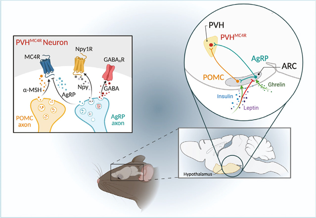
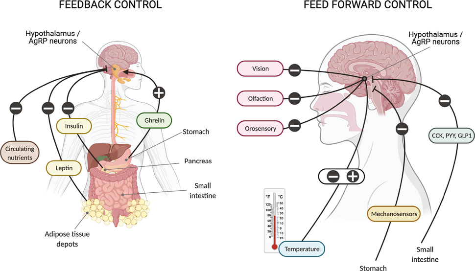
Neurons in the hypothalamic arcuate nucleus (ARC) that express agouti-related peptide (AgRP) govern a critical aspect of survival: the drive to eat. Equally important to survival is the timing at which food is consumed-seeking or eating food to alleviate hunger in the face of a more pressing threat, like the risk of predation, is clearly maladaptive. To ensure optimal prioritization of behaviors within a given environment, therefore, AgRP neurons must integrate signals of internal need states with contextual environmental cues. In this state-of-the-art review, we highlight recent advances that extend our understanding of AgRP neurons, including the neural circuits they engage to regulate feeding, energy expenditure, and behavior. We also discuss key findings that illustrate how both classical feedback and anticipatory feedforward signals regulate this neuronal population and how the integration of these signals may be disrupted in states of energy excess. Finally, we examine both technical and conceptual challenges facing the field moving forward.

Keywords: agouti-related peptide (AgRP); arcuate nucleus; behavior; energy expenditure; energy homeostasis; food intake; hunger; neurocircuits.


Conflict of interest statement

Conflict of Interest

The authors declare no conflict of interest.

Figures

Fig. 1.
Fig. 1.
Central Melanocortin System. The central melanocortin system includes two distinct neuronal populations located in the arcuate nucleus of the hypothalamus (ARC) that express either agouti-related peptide (AgRP) or pro-opiomelanocortin (POMC). Neurons expressing POMC are inhibited by insulin and leptin and release alpha-melanin-stimulating hormone (α-MSH), an agonist that binds to downstream melanocortin-4 receptor (MC4R)-expressing neurons in the paraventricular nucleus of the hypothalamus (PVHMC4R) to inhibit food intake and induce weight loss. In contrast, AgRP neurons are inhibited by insulin and leptin and are activated by the stomach-derived hormone, ghrelin, and stimulate food intake and reduce energy expenditure when activated. These neurons also express neuropeptide Y (Npy), and the inhibitory neurotransmitter, gamma aminobutyric acid (GABA). AgRP neurons mediate their effects through the release of AgRP, which antagonizes the binding of α-MSH to PVHMC4R neurons, Npy acting on downstream Y1 and Y5 receptors, and by inhibiting POMC neurons via synaptic release of GABA.
Fig. 2.
Fig. 2.
Mapped AgRP neuron projections. (A) AgRP neurons project to a broad array of brain areas implicated in a wide range of behaviors. (B) Circuits related to feeding include AgRP projections to the LHA, PVH, BNST, PVT, MeA, and MPOA, which stimulate food intake, while projections to the PBN suppress discomfort and malaise. (C) Downstream projection sites in the PVT have also been found to be involved in smell, the amygdala in fear and aggression, the LHA in taste, Kisspeptin-expressing neurons of the ARC in reproduction, MPOA in maternal behaviors, and the PBN in pain and satiety. AgRP, agouti-related peptide; ARC, arcuate nucleus; BNST, bed nucleus of the stria terminalis; CeA, central amygdala; DMH, dorsomedial hypothalamus; KISS, Kisspeptin neurons; LHA, lateral hypothalamic area; LS, lateral septum; MPOA, medial preoptic area; MeA, medial amygdala; PAG, periaqueductal gray; PBN, parabrachial nucleus; PVH, paraventricular hypothalamus; PVT, paraventricular thalamic nucleus; SCN, suprachiasmatic nucleus; VMH, ventromedial hypothalamus.
Fig. 3.
Fig. 3.
Feedback and Feedforward Regulation of AgRP neurons. Feedback control of AgRP neuron activity is primarily regulated by hormonal (i.e., insulin, leptin, and ghrelin)- and nutrient-related input (i.e., glucose and free fatty acids). Visual and olfactory input related to food presentation and orosensory information related to taste inhibit AgRP neuron activity early in a meal, while mechanosensation and GIderived hormone input inhibit AgRP neuron activity as nutrients enter the gut but before nutrient absorption. Ambient temperature can modulate AgRP neuron activity in either direction, with cold temperatures increasing AgRP neuron activity and warmer temperatures reducing activity.
Fig. 4.
Fig. 4.
Functionally mapped AgRP neuron afferents. AgRP neurons receive afferent input from several different brain areas. Of these, the PVH sends strong, excitatory, glutamatergic input to AgRP neurons in the ARC that stimulate feeding. In contrast, AgRP neurons receive GABAergic input from leptin-receptor-expressing neurons in the DMH that, when activated, inhibit feeding. AgRP, agouti-related peptide; ARC, arcuate nucleus; BNST, bed nucleus of the stria terminalis; DMH, dorsomedial hypothalamus; GABA, gamma aminobutyric acid; LepR, leptin receptor; LS, lateral septum; LHA, lateral hypothalamic area; MPOA, medial preoptic area; NI, nucleus incertus; PVH, paraventricular hypothalamus; PACAP, pituitary adenylate cyclase-activating peptide; PAG, periaqueductal gray; PBN, parabrachial nucleus; SON, supraoptic nucleus; VMH, ventromedial hypothalamus; VTG, ventral tegmental area.

References

    1. Chen WG, Schloesser D, Arensdorf AM, Simmons JM, Cui C, Valentino R, Gnadt JW, Nielsen L, Hillaire-Clarke CS, Spruance V et al. (2021) The emerging science of interoception: sensing, integrating, interpreting, and regulating signals within the self. Trends in Neurosci 44,3–16. - PMC - PubMed
    1. Sternson SM & Eiselt A-K (2015) Three pillars for the neural control of appetite. Annu Rev Physiol 79, 401–423. - PubMed
    1. Padilla SL, Qiu J, Soden ME, Sanz E, Nestor CC, Barker FD, Quintana A, Zweifel LS, Ronnekleiv OK, Kelly MJ et al. (2016) Agouti-related peptide neural circuits mediate adaptive behaviors in the starved state. Nat Neuro 19, 734–741. - PMC - PubMed
    1. Sutton AK & Krashes MJ (2020) Integrating hunger with rival motivations. Trends Endocrinol Metab 31, 495–507. - PubMed
    1. Sternson SM (2020) ScienceDirect Exploring internal state-coding across the rodent brain. Curr Opin Neurobiol 65,20–26. - PubMed

Publication types

MeSH terms

Substances