. 2021 Oct 15;81(20):5161-5175.
doi: 10.1158/0008-5472.CAN-21-1254. Epub 2021 Sep 1.

Novel Mouse Models of Bladder Cancer Identify a Prognostic Signature Associated with Risk of Disease Progression

Affiliations

Novel Mouse Models of Bladder Cancer Identify a Prognostic Signature Associated with Risk of Disease Progression

Soonbum Park et al. Cancer Res. .

Abstract

To study the progression of bladder cancer from non-muscle-invasive to muscle-invasive disease, we have developed a novel toolkit that uses complementary approaches to achieve gene recombination in specific cell populations in the bladder urothelium in vivo, thereby allowing us to generate a new series of genetically engineered mouse models (GEMM) of bladder cancer. One method is based on the delivery of adenoviruses that express Cre recombinase in selected cell types in the urothelium, and a second uses transgenic drivers in which activation of inducible Cre alleles can be limited to the bladder urothelium by intravesicular delivery of tamoxifen. Using both approaches, targeted deletion of the Pten and p53 tumor suppressor genes specifically in basal urothelial cells gave rise to muscle-invasive bladder tumors. Furthermore, preinvasive lesions arising in basal cells displayed upregulation of molecular pathways related to bladder tumorigenesis, including proinflammatory pathways. Cross-species analyses comparing a mouse gene signature of early bladder cancer with a human signature of bladder cancer progression identified a conserved 28-gene signature of early bladder cancer that is associated with poor prognosis for human bladder cancer and that outperforms comparable gene signatures. These findings demonstrate the relevance of these GEMMs for studying the biology of human bladder cancer and introduce a prognostic gene signature that may help to stratify patients at risk for progression to potentially lethal muscle-invasive disease. SIGNIFICANCE: Analyses of bladder cancer progression in a new series of genetically engineered mouse models has identified a gene signature of poor prognosis in human bladder cancer.


Conflict of interest statement

Conflict of interest statement: H. Al-Ahmadie is a Consultant for Bristol-Myers-Squibb, AstraZeneca, Janssen Biotech and Paige.ai. The other authors have no conflicts to report.

Figures

Figure 1.
Figure 1.. Strategies for cell-type specific gene recombination in bladder urothelium
A. Strategy for tumor induction using Adenoviruses expressing Cre recombinase (AdenoCre). AdenoCre was delivered to bladder lumen of mice of the indicated genotypes via surgical or transurethral delivery. B. Strategy for tumor induction of tamoxifen-inducible CreERT2 transgenic alleles. Induction of Cre in the bladder was achieved by systemic administration of tamoxifen or by intravesical delivery of 4-hydroxy-tamoxifen directly into the bladder lumen. C. Schematic showing the cell types in the urothelium and their relationship to the bladder lumen and lamina propria. Subtypes of urothelial cells are distinguished by high, medium (med), or low expression levels of Cytokeratin 5 (Ck5) or Cytokeratin 8 (Ck8), as indicated. D, E. Lineage marking to evaluate the cell-type specificity of gene recombination in bladder urothelium using AdenoCk5-Cre or AdenoCk8-Cre (D) or Ck5-CreERT2 or Ck8-CreERT2 transgenic alleles (E). In panel D, AdenoCk5-Cre or AdenoCk8-Cre were delivered via the surgical method into the bladder lumen of mice of the indicated genotypes at 2 months of age, and analyzed 7 days later. In panel E, tamoxifen was delivered systemically on 4 consecutive days to mice of the indicated genotypes at 2 months of age, and analyzed 10 days later. Panels D and E show representative images of bladder co-stained for YFP (green) and Ck5 (red) or Ck8 (red), as indicated; nuclei were visualized with DAPI staining. Scale bars represent 25 μm. Data represent the summary of 5 or more sections from 3–7 mice/group; quantification of cell counting is shown in Table S1. See also Figures S1 and S2, Supplementary Videos 1–4, and Supplementary procedures.
Figure 2.
Figure 2.
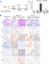
Targeted inactivation of Pten and p53 in basal cells is sufficient for muscle invasive bladder cancer A-D. Tumor induction with AdenoCre. A. Experimental design. AdenoCre was delivered into the bladder lumen of mice of the indicated genotypes at 2 months of age via the surgical method. Mice were monitored for tumor growth by ultrasound imaging for up to 14 months of age. B. Phenotypic analyses. C. Kaplan-Meier survival analysis. D. Immunohistochemistry. E-H. Tumor induction based on transgenic Cre. E. Experimental design. 4-hydroxy-tamoxifen was delivered into the bladder lumen of mice of the indicated genotypes at 2 months of age using the ultrasound guidance method. Mice were monitored for tumor growth by ultrasound imaging for up to 14 months of age. F. Phenotypic analyses. G. Kaplan-Meier survival analysis. H. Immunohistochemistry. Panels B and F show representative bright field images of whole bladder, ultrasound images of tumor monitoring, H&E images at low and high power, and confocal images of YFP lineage-marked cells (green) co-stained with cytokeratin 5 (red) or cytokeratin 8 (red). Scale bar for H&E images represent 100 μm (low-magnification) and 20 μm (high-magnification), and for confocal images scale bars represent 50 μm. Panels C and G show Kaplan-Meier survival analysis based on detection of bladder tumors by ultrasound imaging. P-values were calculated using a log-rank (Mantel-Cox) test. Panels D and H show immunostaining for Ki67, cytokeratin 14 (Ck14), and Vimentin. Scale bars represent 20 μm. See also Figure S3 and S4, and Table 1 and Table S2.
Figure 3.
Figure 3.. Progression from pre-invasive to invasive bladder cancer
A. Experimental design. Mice of the indicated genotypes were induced with tamoxifen at 2 months of age, and analyzed at 5 months (early) or 12 months (late) B. Phenotypic analyses. Representative images showing histological staining (H&E) and immunostaining for Ki67, phosphorylated S6 (pS6), phosphorylated ERK (pERK), cytokeratin 14 (Ck14), and Cd45. Scale bars represent 50 μm for Cd45, and 20 μm for all other images. C. Quantification of Ki67 positive cells. The percentages shown were calculated by counting the number of cells stained by Ki67 divided by number of cells counterstained by hematoxylin. Data represent the summary of 5 sections from 4–5 mice/group. The P-value was calculated using a non-parametric unpaired t test (Mann-Whitney). See also Figure S5.
Figure 4.
Figure 4.. Mechanisms of progression from pre-invasive to invasive bladder cancer
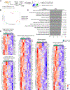
A. Experimental design. Mice of the indicated genotypes were induced with tamoxifen at 2 months of age and sacrificed 3 months later. YFP-marked urothelial cells were isolated by FACS sorting and used to generate organoids or allograft models that were phenotypically analyzed in culture and in vivo, respectively (Figure S6), or were subjected to RNA-sequencing analyses (panels B-D). B. Principal Component Analysis based on gene expression profiles from urothelial cells isolated from mice of the indicated genotypes. C., D. Pathway-based Gene Set Enrichment Analyses (GSEA) based on expression profiles of Ck5-CreERT2; DKO versus Ck8-CreERT2; DKO urothelial cells. Panel C shows a bar graph showing the NES scores and P-values for the positively enriched pathways. Panel D shows heatmaps of the leading edge genes from selected pathways as indicated. See also Figure S6, and Supplementary Datasets 1 and 2.
Figure 5.
Figure 5.. A conserved gene signature of early bladder cancer associated with poor prognosis
A, B. Identification of a conserved gene signature of early bladder cancer. A. Step 1: First, we identified a mouse gene signature of early bladder cancer by comparing the pre-invasive signature from the Ck5-CreERT2; DKO versus Ck8-CreERT2; DKO urothelial cells with a signature of lethal bladder cancer based on comparing AdCMV-Cre induced Ptenflox/flox; p53flox/flox tumors versus non-induced urothelium as described previously (46). The Venn diagram shows the number of genes that are upregulated when comparing the pre-invasive and lethal gene signatures (log2FoldChange >0 and adjusted P < 0.001). This identified 63 genes that comprise the mouse gene signature of early bladder cancer. B. Step 2: To identify a conserved gene signature of early bladder cancer, we performed GSEA using the mouse gene signature (63 genes) to query a human gene signature comparing MIBC versus NMIBS based on the Kim cohort (43). This analysis identified 28 leading edge genes that comprise the conserved gene signature of early bladder cancer. C. Heatmap representation of individual expression levels of the 28 genes from the conserved gene signature of early bladder cancer signature based on the Hedegaard cohort, which is comprised of patient samples with tumor stages from Ta to T4b (n = 476) (42). D,E. Kaplan-Meier survival analyses comparing patients with low (low 25%) and high (high 25%) combined expression of 28 gene signatures in Lund cohort (panel D) (44) and TCGA cohort (panel E) (45). P-values were estimated using a log-rank test. See also Table 2, Tables S4 and S5, Figure S7–11, and Supplementary Dataset 3.

References

    1. Siegel RL, Miller KD, Fuchs HE, Jemal A. Cancer Statistics, 2021. CA Cancer J Clin 2021;71:7–33 - PubMed
    1. Knowles MA, Hurst CD. Molecular biology of bladder cancer: new insights into pathogenesis and clinical diversity. Nat Rev Cancer 2015;15:25–41 - PubMed
    1. Kamat AM, Hahn NM, Efstathiou JA, Lerner SP, Malmstrom PU, Choi W, et al. Bladder cancer. Lancet 2016 - PubMed
    1. Patel VG, Oh WK, Galsky MD. Treatment of muscle-invasive and advanced bladder cancer in 2020. CA Cancer J Clin 2020;70:404–23 - PubMed
    1. Tran L, Xiao JF, Agarwal N, Duex JE, Theodorescu D. Advances in bladder cancer biology and therapy. Nat Rev Cancer 2021;21:104–21 - PMC - PubMed

Publication types

MeSH terms