

Prednisone prevents particle induced bone loss in the calvaria mouse model
- PMID: 34471710
- PMCID: PMC8387912
- DOI: 10.1016/j.heliyon.2021.e07828
Prednisone prevents particle induced bone loss in the calvaria mouse model
Abstract
Introduction: Glucocorticoids are essential in the treatment of many chronic inflammatory and malignant diseases but are known to have detrimental effects on bone. This study aimed to investigate the effects of prednisone on osteoclast functioning in vivo in the calvaria particle-induced bone loss mouse model.
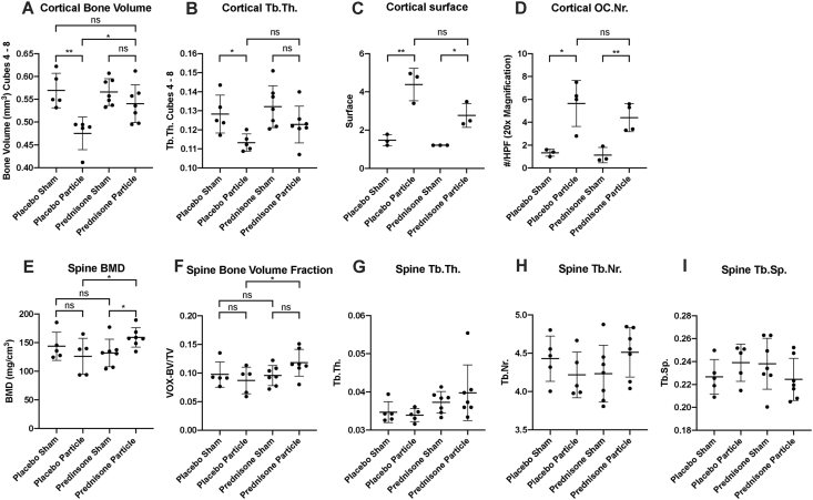
Methods: 12-week-old male C57BL6/J mice received subcutaneously implanted prednisone (2.5 mg/d, 60 day release (n = 14)) or placebo pellets (n = 10). Osteolysis of the calvaria bone was induced two weeks later by application of ultra-high-molecular-weight polyethylene- (UHMWPE) particles to the dome (vs sham operation). The extent of osteolysis was determined histologically and by micro-computer tomography.
Results: Prednisone significantly inhibited particle-induced osteolysis in the skull. No significant difference in osteoclast numbers was seen in mice with prednisone vs placebo treatment. Prednisone treatment alone without particle application did not reduce bone mineral density or deterioration in bone microarchitecture parameters.
Conclusions: The calvaria particle-induced bone loss mouse model can be adapted to investigate osteoclast activity in vivo and the effect of prednisone on osteoclasts. In this preventive experimental design, the application of short-term low-dose prednisone has osteoprotective effects without measurable systemic side effects on bone parameters.
Keywords: Bone; Glucocorticoids; In-vivo; Inflammation; Osteoclasts.
© 2021 The Authors.
Conflict of interest statement
The authors declare no conflict of interest.
Figures


Similar articles
-
Antioxidant impregnated ultra-high molecular weight polyethylene wear debris particles display increased bone remodeling and a superior osteogenic:osteolytic profile vs. conventional UHMWPE particles in a murine calvaria model.J Orthop Res. 2016 May;34(5):845-51. doi: 10.1002/jor.23080. Epub 2015 Nov 23. J Orthop Res. 2016. PMID: 26495749 Free PMC article.
-
Brief Report: Methotrexate Prevents Wear Particle-Induced Inflammatory Osteolysis in Mice Via Activation of Adenosine A2A Receptor.Arthritis Rheumatol. 2015 Mar;67(3):849-55. doi: 10.1002/art.38971. Arthritis Rheumatol. 2015. PMID: 25533750 Free PMC article.
-
[A comparison of the antiresorptive effects of bisphosphonates and statins on polyethylene particle-induced osteolysis].Biomed Tech (Berl). 2005 Jun;50(6):195-200. doi: 10.1515/BMT.2005.027. Biomed Tech (Berl). 2005. PMID: 16003921 German.
-
Metformin suppresses UHMWPE particle-induced osteolysis in the mouse calvaria by promoting polarization of macrophages to an anti-inflammatory phenotype.Mol Med. 2018 May 9;24(1):20. doi: 10.1186/s10020-018-0013-x. Mol Med. 2018. PMID: 30134793 Free PMC article.
-
Anti-oxidation treatment of ultra high molecular weight polyethylene components to decrease periprosthetic osteolysis: evaluation of osteolytic and osteogenic properties of wear debris particles in a murine calvaria model.Curr Rheumatol Rep. 2013 May;15(5):325. doi: 10.1007/s11926-013-0325-3. Curr Rheumatol Rep. 2013. PMID: 23532463 Free PMC article. Review.
Cited by
-
A Review on the Molecular Mechanisms of Action of Natural Products in Preventing Bone Diseases.Int J Mol Sci. 2022 Jul 30;23(15):8468. doi: 10.3390/ijms23158468. Int J Mol Sci. 2022. PMID: 35955603 Free PMC article. Review.
-
The osteocytic actions of glucocorticoids on bone mass, mechanical properties, or perilacunar remodeling outcomes are not rescued by PTH(1-34).Front Endocrinol (Lausanne). 2024 Jul 18;15:1342938. doi: 10.3389/fendo.2024.1342938. eCollection 2024. Front Endocrinol (Lausanne). 2024. PMID: 39092287 Free PMC article.
References
-
- Okano T., Inui K., Tada M., Sugioka Y., Mamoto K., Wakitani S., Koike T., Nakamura H. High frequency of vertebral fracture and low bone quality in patients with rheumatoid arthritis—results from TOMORROW study. Mod. Rheumatol. 2017;27:398–404. - PubMed
-
- Briot K., Geusens P., Em Bultink I., Lems W.F., Roux C. Inflammatory diseases and bone fragility. Osteoporos. Int. 2017;28:3301–3314. - PubMed
-
- Clowes J.A., Riggs B.L., Khosla S. The role of the immune system in the pathophysiology of osteoporosis. Immunol. Rev. 2005;208:207–227. - PubMed
-
- Zerbini C.A.F., Clark P., Mendez-Sanchez L., Pereira R.M.R., Messina O.D., Uña C.R., Adachi J.D., Lems W.F., Cooper C., Lane N.E. On behalf of the IOF Chronic Inflammation, Biologic therapies and bone loss in rheumatoid arthritis. Osteoporos. Int. 2017;28:429–446. - PubMed
LinkOut - more resources
Full Text Sources
