Review
. 2020 Jun 1;2(2):101-119.
doi: 10.1089/bioe.2020.0010. Epub 2020 Jun 17.

Polypyrrole-Incorporated Conducting Constructs for Tissue Engineering Applications: A Review

Affiliations
Review

Polypyrrole-Incorporated Conducting Constructs for Tissue Engineering Applications: A Review

Yeshi Liang et al. Bioelectricity. .

Abstract

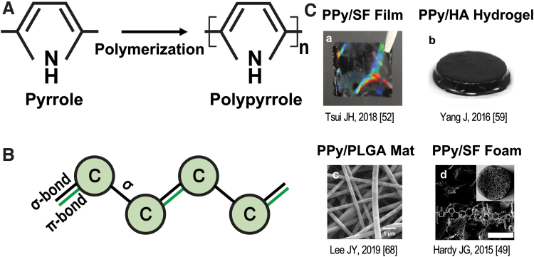
Conductive polymers have recently attracted interest in biomedical applications because of their excellent intrinsic electrical conductivity and satisfactory biocompatibility. Polypyrrole (PPy) is one of the most popular among these conductive polymers due to its high conductivity under physiological conditions, and it can be chemically modified to allow biomolecules conjugation. PPy has been used in fabricating biocompatible stimulus-responsive scaffolds for tissue engineering applications, especially for repair and regeneration of electroactive tissues, such as the bone, neuron, and heart. This review provides a comprehensive overview of the basic properties and synthesis methods of PPy, as well as a summary of the materials that have been integrated with PPy. These composite scaffolds are comparatively evaluated with regard to their mechanical properties, biocompatibility, and usage in tissue engineering.

Keywords: conductive scaffold; conductive tissue engineering; polypyrrole.


Conflict of interest statement

No competing financial interests exist.

Figures

FIG. 1.
FIG. 1.
(A) Polymerization of PPy from pyrrole monomers using FeCl3 as oxidant. Oxidation of pyrrole using FeCl3: nC4H4NH + FeCl3→(C4H2NH)n + FeCl2 + HCl; oxidation (p-doping) of the PPy with dopant in the system to maintain PPy conductivity: (C4H2NH)n + xFeCl3→(C4H2NH)nCl + xFeCl2. (B) Illustration of the conjugated backbone of conductive polymer; alternating pattern of double and single bonds in the backbone. Black bond represents Sigma-bond, which strengthens the electrons; green bond represents the Pi-bond, which exists in the double bond, with lower strength. (C) Incorporation of PPy with different materials as scaffolds. (a) PPy/SF film; (b) PPy/HA hydrogel; (c) PPy/PLGA mat; (d) PPy/SF foam. FeCl3, ferric chloride; PPy, polypyrrole; SF, silk fibroin; HA, hyaluronic acid; PLGA, poly(lactic-co-glycolic acid).
FIG. 2.
FIG. 2.
(A) Process of CHI/PPy hydrogel generation. (B) CHI and CHI/PPy hydrogel coated on Petri dish. Reproduced from Cui et al. with permission from Theranostics. (C) Fabrication of SA/CM-CHI/PPy hydrogel. (D) Cyclic voltammograms for the SA/CM-CHI/PPy hydrogel. Reproduced from Bu et al. with permission from The Royal Society of Chemistry. CM-CHI, carboxymethyl chitosan; SA, sodium alginate.
FIG. 3.
FIG. 3.
(A) Generation of ALG and PPy/ALG hydrogel. (B) Various PPy/ALG hydrogels synthesized with different pyrrole monomer and oxidant concentrations. (C) ATR-IR spectra of PPy/ALG samples. Reproduced from Yang et al. with permission from Marcomolecular Bioscience. ALG, alginate.
FIG. 4.
FIG. 4.
(A) Diazonium coupling reaction used to synthesize AMSF. (B) SF and AMSF films exposed to 50 mM pyrrole and 7.5 mM FeCl3 in water for the times given. (C) ATR-FTIR spectra of SF and AMSF after Ppy deposition for 2 h. Reproduced from Romero et al. with permission from ACS Applied Materials and Interfaces. (D) Fabrication of AMSF and AMSF/PPy. (E) I–V curves of AMSF and AMSF/PPy substrates. Reproduced from Tsui et al. with permission from The Royal Society of Chemistry. AMSF, acid-modified silk fibroin; FTIR, Fourier Transform Infrared; SF, silk fibroin.
FIG. 5.
FIG. 5.
(A) Fluorescence images of SCs cultured on different nanofiber membranes without ES (a–d) and with ES (e–h); SEM images of SCs cultured on different nanofiber membranes without ES (i–l) and with ES (m–p). PLCL/SF-PPy-1, -2, -3 represent different Py concentrations, from 25, 37.5, 50 μL, respectively. Scale bars are 100 mm. Reproduced from Sun et al. with permission from The Royal Society of Chemistry. (B) Fluorescence images of electrically stimulated PC12 cells on PPy/PLGA scaffolds with random fibers at 0 mV/cm (a); at 10 mV/cm (b); with aligned fibers at 0 mV/cm (c); at 10 mV/cm (d). Scale bars are 50 μm. (C) Median neurite lengths (a) and percentages of neurite-bearing PC12 cells (b) with or without electrically stimulated. Reproduced from Lee et al. with permission from Biomaterials. PLCL, poly(l-lactic acid-co-ɛ-caprolactone); PLGA, poly(lactic-co-glycolic acid); SC, Schwann cell; SEM, scanning electron microscopy.
FIG. 6.
FIG. 6.
Calcium transients and normalized fluorescence intensity against time for CMs on (A) CHI-coated Petri dish, (B) PPy/CHI-coated Petri dish. Reproduced from Cui et al. with permission from Theranostics. CMs, cardiomyocytes.

References

    1. Zhao YH, Niu CM, Shi JQ, et al. . Novel conductive polypyrrole/silk fibroin scaffold for neural tissue repair. Neural Regen Res 2018;13:1455–1464. DOI: 10.4103/1673-5374.235303 - DOI - PMC - PubMed
    1. Gajendiran M, Choi J, Kim SJ, et al. . Conductive biomaterials for tissue engineering applications. J Ind Eng Chem 2017;51:12–26. DOI: 10.1016/j.jiec.2017.02.031 - DOI
    1. Tayalia P, Mooney DJ. Controlled growth factor delivery for tissue engineering. Adv Mater 2009;21:3269–3285. DOI: 10.1002/adma.200900241 - DOI - PubMed
    1. Tian L, Prabhakaran MP, Hu J, et al. . Synergistic effect of topography, surface chemistry and conductivity of the electrospun nanofibrous scaffold on cellular response of PC12 cells. Colloids Surfaces B Biointerfaces 2016;145:420–429. DOI: 10.1016/j.colsurfb.2016.05.032 - DOI - PubMed
    1. Hardy JG, Lee JY, Schmidt CE. Biomimetic conducting polymer-based tissue scaffolds. Curr Opin Biotechnol 2013;24:847–854. DOI: 10.1016/J.COPBIO.2013.03.011 - DOI - PubMed

LinkOut - more resources