Review
. 2020 Jun 1;2(2):120-149.
doi: 10.1089/bioe.2020.0021. Epub 2020 Jun 17.

Electroconductive Nanobiomaterials for Tissue Engineering and Regenerative Medicine

Affiliations
Review

Electroconductive Nanobiomaterials for Tissue Engineering and Regenerative Medicine

Ebrahim Mostafavi et al. Bioelectricity. .

Abstract

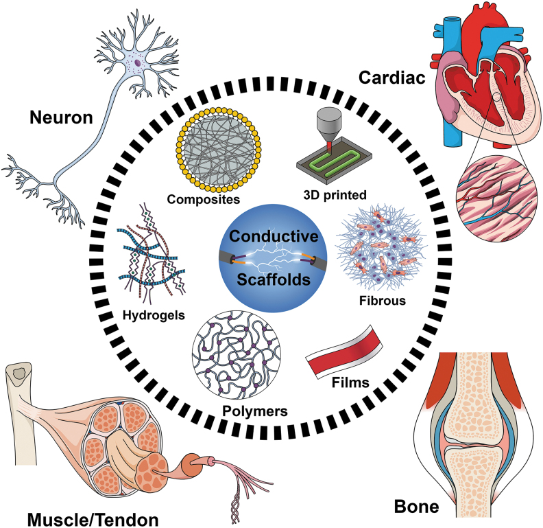
Regenerative medicine aims to engineer tissue constructs that can recapitulate the functional and structural properties of native organs. Most novel regenerative therapies are based on the recreation of a three-dimensional environment that can provide essential guidance for cell organization, survival, and function, which leads to adequate tissue growth. The primary motivation in the use of conductive nanomaterials in tissue engineering has been to develop biomimetic scaffolds to recapitulate the electrical properties of the natural extracellular matrix, something often overlooked in numerous tissue engineering materials to date. In this review article, we focus on the use of electroconductive nanobiomaterials for different biomedical applications, particularly, very recent advancements for cardiovascular, neural, bone, and muscle tissue regeneration. Moreover, this review highlights how electroconductive nanobiomaterials can facilitate cell to cell crosstalk (i.e., for cell growth, migration, proliferation, and differentiation) in different tissues. Thoughts on what the field needs for future growth are also provided.

Keywords: biomaterials; bone; cardiac; electroconductive; extracellular matrix; nanomaterials; nanomedicine; nerve; regenerative medicine; tendon; tissue engineering.


Conflict of interest statement

No competing financial interests exist.

Figures

FIG. 1.
FIG. 1.
Schematic illustration of different electroconductive scaffolds for biomedical applications discussed in this study.
FIG. 2.
FIG. 2.
Schematic overview of various types of electroconductive scaffolds used in cardiac tissue regeneration. (A) Overview of the concept of using a suture-free technology for the attachment of an engineered cardiac patch to the organ. The process from left to the right of the panel can be explained as: Gold nanorod adsorption; cardiac cell seeding; cardiac tissue assembly; patch location and integration by NIR; and finally, the cardiac patch after integration to a rat heart. (B) A schematic illustrating the fabrication steps to produce 3D biohybrid actuators composed of cardiac tissue on top of a multilayer hydrogel sheet impregnated with aligned CNT microelectrodes. (C) Designing a multilayered hybrid scaffold, including nanofibers and hydrogels, that can suitably mimic the native cardiac tissue structure. The first step is to design an interwoven, aligned structure, and scaffold with a network structure from nanofibers of Yarn (NFYs-NET), which possess the benefits for native cardiac tissue. The middle row demonstrates the native myocardium showing a gradual transition of aligned cell layers from the endocardium to the epicardium and shows schematics of multiple layers of NFYs-NETs assembled with a gradual orientation transition. The bottom row shows the fabrication process of one-layer 3D NFYs-NET/GelMA hybrid scaffolds and subsequent CMs cultivation. A single-layer hybrid 3D scaffold is formed through encapsulating a single NFYs-NET layer within a GelMA hydrogel shell after photocrosslinking with UV-radiation. (D) Schematic concept of using a cell-laden hydrogel bioink originating from the patient's own cells that are reprogrammed to become pluripotent and then differentiated to CMs and endothelial cells and encapsulation within the hydrogel for 3D bioprinting functional cardiac tissue (so-called personalized tissue regeneration). (E) Schematic concept of the application of an engineered functional and injectable cardiac patch through a shape/memory scaffold. The scaffolds recover their initial shape following injection. An image of the minimally invasive implanted injectable cardiac patch on the porcine heart without open-heart surgery is also shown. 3D, three-dimensional; CMs, cardiomyocytes; CNT, carbon nanotube; GelMA, gelatin methacryloyl; NIR, near infrared; UV, ultraviolet.
FIG. 3.
FIG. 3.
(A, a) Nanoelectrodes as a minimally invasive wireless device for recording neuronal activities in model animals. (b) Left side of the photo shows traditional bulk implants while the right image displays nanoscale implants as an open, stretchable and flexible framework that induce fewer immune reactions. These networks allow access to many more astrocytes and microglial cells and can be activated at the surface of bulk implants. (c) Multiple forms of signal transduction in neuronal synapses. (d) The size-shrink of metal conductors or semiconductors and change of behavior of iron oxide from paramagnetic to superparamagnetic at the nanoscale compared with bulk-size properties. FETs are gated more easily when nanoscale channels are implemented. Also, nanoscale patterns can lead to a neural response that cannot be detected in a planar neural substrate. (e) The synaptic spaces and several other subcellular spaces are crowded and dynamic. Nanoscale signal transduction can record activities with much higher precision in these crowded spaces compared with traditional methods. (f) Recordings show different signal shapes and amplitudes on different sites of a single neuron. Traces 1–4 show extracellular signal recordings whereas traces I–III show intracellular recordings from micropipettes. All of panel A adopted from Taylor and Francis. (B) Polymeric nanoparticles employed for targeted drug delivery. (i) Schematic representation of paclitaxel-loaded Angiopep-PEG-PCL nanoparticles. (ii) Angiopep-conjugation increased the targeting efficiency of brain tumors. (C) A 3D printed implant, 2-mm in thickness, is used as scaffolding to repair spinal cord injuries in rats. The H shape in the center is the location of the spinal cord and the dots surrounding it are hollow spaces through which stem cell neural implants extend axons into the host tissues. PCL, poly(ɛ-caprolactone).
FIG. 4.
FIG. 4.
Representative examples of the use of electroconductive materials for bone tissue regeneration. (A) Novel in situ polymerization/TIPS method to fabricate conductive nanofibrous PLA scaffolds with well-distributed PANI nanostructures for bone tissue regeneration. Mean for n = 4 ± SD. *P < 0.05, **P < 0.01 (B) Schematic representation of the experimental procedure for the fabrication of electroconductive electrospun CNFs to be used as the substrate for bone cell electrical stimulation; and (C) Study of the effect of the addition of Si-NPs in electrospun PCL membranes to improve the mechanical and osteoconductive properties of the layers. **p < 0.01, ***p = 0.0001, ****p < 0.0001. CNFs, carbon nanofibers; PANI, polyaniline; PLA, poly(lactic acid); SiNPs, silica nanoparticles; TIPS, thermal-induced phase separation.
FIG. 5.
FIG. 5.
Overview of bioengineering approaches for skeletal muscle tissue engineering, redrawn from Nakayama et al.

References

    1. Liu Y, Lim J, Teoh S-H. Development of clinically relevant scaffolds for vascularised bone tissue engineering. Biotechnol Adv 2013;31:688–705 - PubMed
    1. Hosoyama K, Ahumada M, Goel K, et al. . Electroconductive materials as biomimetic platforms for tissue regeneration. Biotechnol Adv 2019;37:444–458 - PubMed
    1. Fennelly C, Soker S. Bioelectric properties of myogenic progenitor cells. Bioelectricity 2019;1:35–45 - PMC - PubMed
    1. Saberi A, Jabbari F, Zarrintaj P, et al. . Electrically conductive materials: opportunities and challenges in tissue engineering. Biomolecules 2019;9:448 - PMC - PubMed
    1. Bassett CAL, Becker RO. Generation of electric potentials by bone in response to mechanical stress. Science 1962;137:1063–1064 - PubMed

LinkOut - more resources