

Under Pressure: Post-Transplant Lymphoproliferative Disease: A Case of Pulmonary Artery External Compression
- PMID: 34471885
- PMCID: PMC8387807
- DOI: 10.1016/j.jaccas.2021.05.011
Under Pressure: Post-Transplant Lymphoproliferative Disease: A Case of Pulmonary Artery External Compression
Abstract
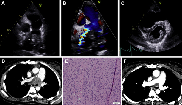
Post-transplant lymphoproliferative disorder (PTLD) is an often fatal complication of cardiac transplantation that occurs in 2% to 6% of transplant recipients. We report a case in which PTLD led to pulmonary artery external compression and multimodality imaging showed key features in the diagnosis, management, and follow-up. (Level of Difficulty: Intermediate.).
Keywords: CT, computed tomography; EBV, Epstein-Barr virus; Ig, immunoglobulin; PTLD, post-transplant lymphoproliferative disorder; TAPSE, tricuspid annular plane systolic excursion; TTE, 2-dimensional transthoracic echocardiogram; biopsy; computed tomography; echocardiography; external compression; post-transplant lymphoproliferative disease; pulmonary artery.
© 2021 The Authors.
Conflict of interest statement
The authors have reported that they have no relationships relevant to the contents of this paper to disclose.
Figures

References
LinkOut - more resources
Full Text Sources
