

Lobar (croupous) pneumonia: old and new data
- PMID: 34472009
- PMCID: PMC8409273
- DOI: 10.1007/s15010-021-01689-4
Lobar (croupous) pneumonia: old and new data
Abstract
Background and aim: Pneumonia remains one of the most frequent death causes worldwide. Among the etiological factors S. pneumoniae-causing lobar pneumonia plays a leading role. According to current textbook knowledge at least three sequential stages of lobar pneumonia are distinguished: congestion, red hepatization and gray hepatization. However, there are no detailed data supporting this stage concept. There are also controversial views on its etiology. In this study, the lung changes in lobar pneumonia were related to the cause and duration of the disease. In addition, the complications of the disease were evaluated. PCR studies verified the etiology of pneumonia.
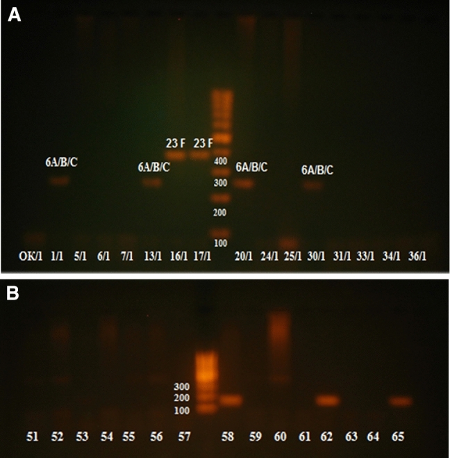
Material and methods: Lobar pneumonia was analyzed in 252 post mortem cases examined in a large hospital in Irkutsk. The pathology, etiology of pneumonia, course of disease and cause of death were recorded and correlated to its clinical course and duration. In the second part of the study, the results in 95 patients were analyzed in detail and related to PCR findings.
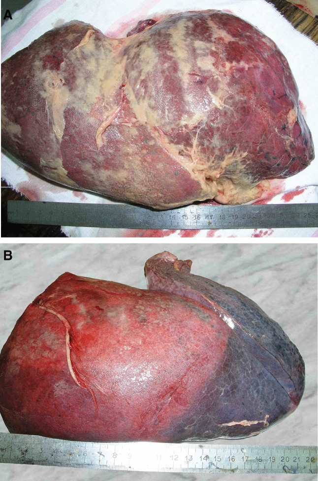
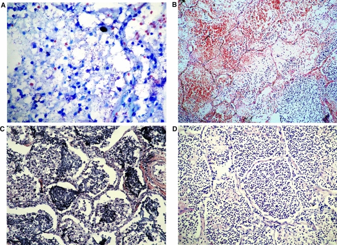
Results: Most patients were adult men of low social status who showed signs of severe alcoholism. Lobar pneumonia was observed in 85% of the patients, while the remaining patients showed sublobar ("lobular", focal) lung involvement. Histologically, three patterns of inflammation were observed, which in most patients occurred concurrently in different parts of the involved lobe: "congestion", characterized by serous exudation with multiple cocci (41% of cases), "red hepatization" (41% of cases) and "gray hepatization" (100% of cases). The latter pattern was subdivided into three subgroups according to the ratio of fibrin-neutrophils and the presence of macrophages. The mean number of different histological patterns observed per patient was 3.8. There was no correlation between the inflammatory patterns and the duration of the disease. In 23% of the patients, the cause of death was of pulmonary origin, while the remaining patients died of extrapulmonary complications (i.e. acute heart failure 26%, acute vascular insufficiency 15% purulent meningitis 11-24.3%. In 29/95 patients (20 with lobar and 9 with focal pneumonia) pneumococcal etiology of pneumonia was established by PCR.
Conclusion: Lobar pneumonia is a distinct clinico-pathological entity caused by S. pneumoniae, demonstrated by PCR testing and/or cytological examinations. Bacteriologic studies frequently give falsenegative results. Lobar pneumonia is characterized by three main histopathological patterns (congestion or microbeous edema, and red and gray hepatization) which usually occur side by side and not in chronological order. Early death is often related to heart failure and septic shock, while meningitis is a frequent complication later in the course.
Keywords: Clinico-pathological correlation; Inflammatory patterns; Lobar pneumonia; PCR; S. pneumoniae.
© 2021. Springer-Verlag GmbH Germany, part of Springer Nature.
Conflict of interest statement
There is no conflict of interest.
Figures
References
-
- Engleberg NC, Di Rita V, Dermodi TS. Schaechter’s mechanisms of microbial disease. Philadelphia: Lippincott Williams & Wilkins; 2007.
-
- Kumar V, Abbas A, Fausto N. Robbins and Cotran pathologic basis of the disease. 9. Philadelphia: Elsevier Saunders; 2020.
-
- Rokitansky C. Handbuch der speziellen pathologischen Anatomie, II. Wien; 1842.
-
- Zinserling A. Pathologische Anatomie der wichtigsten Formen bakteriellen Pneumonien. Zbl Allg Path path Anat. 1990;136:3–13. - PubMed
-
- Zinserling VD. Several questions of pathogenesis of croupous pneumonia in the light of new morphological investigations. Clin Med (Russ) 1939;9–10:3–12.
MeSH terms
LinkOut - more resources
Full Text Sources
Medical
