. 2021 Sep 23;64(18):13373-13393.
doi: 10.1021/acs.jmedchem.1c00633. Epub 2021 Sep 2.

Discovery of Novel Acetamide-Based Heme Oxygenase-1 Inhibitors with Potent In Vitro Antiproliferative Activity

Affiliations

Discovery of Novel Acetamide-Based Heme Oxygenase-1 Inhibitors with Potent In Vitro Antiproliferative Activity

Antonino N Fallica et al. J Med Chem. .

Abstract

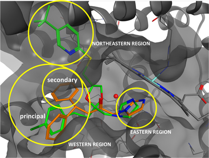
Heme oxygenase-1 (HO-1) promotes heme catabolism exercising cytoprotective roles in normal and cancer cells. Herein, we report the design, synthesis, molecular modeling, and biological evaluation of novel HO-1 inhibitors. Specifically, an amide linker in the central spacer and an imidazole were fixed, and the hydrophobic moiety required by the pharmacophore was largely modified. In many tumors, overexpression of HO-1 correlates with poor prognosis and chemoresistance, suggesting the inhibition of HO-1 as a possible antitumor strategy. Accordingly, compounds 7i and 7l-p emerged for their potency against HO-1 and were investigated for their anticancer activity against prostate (DU145), lung (A549), and glioblastoma (U87MG, A172) cancer cells. The selected compounds showed the best activity toward U87MG cells. Compound 7l was further investigated for its in-cell enzymatic HO-1 activity, expression levels, and effects on cell invasion and vascular endothelial growth factor (VEGF) extracellular release. The obtained data suggest that 7l can reduce cell invasivity acting through modulation of HO-1 expression.


Conflict of interest statement

The authors declare no competing financial interest.

Figures

Figure 1
Figure 1
Chemical structures of azalanstat, 1 (QC-80), 2 (QC-65), 3 (QC-308), and hit compound 4.
Figure 2
Figure 2
Compounds 1 (green) and 3 (orange) inside the HO-1 binding pocket.
Scheme 1
Scheme 1. Reagents and Conditions
(a) α-Bromoacetyl bromide, TEA, dry CH3CN, room temperature, 3 h; and (b) imidazole, K2CO3, dry DMF, room temperature, 2 h or 80% NaH in oil dispersion, dry THF, room temperature, 16 h.
Scheme 2
Scheme 2. Reagents and Conditions
(a) H2NCHO, HCOOH, MW, 150 W, 150 psi, 170 °C, 1.5 h; and (b) LiAlH4 in THF 1 M, dry THF, reflux, 2 h or DIBAL-H in 1 M n-hexane, THF, reflux, 3.5 h, and then room temperature, 16 h.
Scheme 3
Scheme 3. Reagents and Conditions
(a) Appropriate benzyl bromide, K2CO3, KI, acetone, reflux, 3 h.
Scheme 4
Scheme 4. Reagents and Conditions
(a) Benzaldehyde, CH3CO2Na, activated 3 Å molecular sieves, dry CH3OH, room temperature, 12 h, then NaBH4, room temperature, 3 h; (b) acetic anhydride, TEA, 4-dimethylaminopyridine (DMAP), dry CH2Cl2, 0 °C, 10 min, then room temperature, 12 h; and (c) appropriate acyl chloride, TEA, dry CH2Cl2, room temperature, 12 h.
Figure 3
Figure 3
Binding interactions of the four different isomers of 7n (green) compared with the binding pose of 3 in HO-1.
Figure 4
Figure 4
Comparison of compound 3 poses in HO-1 (orange) and HO-2 (light blue). The different residues between HO-2 and HO-1 binding pockets are highlighted in yellow. The highlighted residues belong to HO-2.
Figure 5
Figure 5
Comparison of 3 binding pose (orange) and 7i (light blue), 7l (light pink), 7m (blue), and 7n (green) in HO-1.
Figure 6
Figure 6
Comparison of 3 binding pose (orange) and 7i (light blue), 7l (light pink), 7m (blue), and 7n (green) in HO-2.
Figure 7
Figure 7
Comparison of compound 1 binding pose (orange) and 7o (light blue) in HO-1.
Figure 8
Figure 8
Expression levels of HO-1 in different cancer cell lines. (A) Representative immunoblot of basal HO-1 protein expression detected on cell homogenate of U87Mg, A172, DU145, and A549 cell lines. (B) Bar graphs are representative of results from three independent experiments. Each protein level was expressed as arbitrary units obtained after normalization to actin. (C) Immunolocalization of HO-1 (green fluorescence) in tumor cell lines under basal conditions. Nuclei were stained (blue) with 4′,6-diamidino-2-phenylindole (DAPI). Photomicrographs are representative results of fields taken randomly from slides and scanned by a Zeiss fluorescent microscope.
Figure 9
Figure 9
Effect of 7i and 7l–p treatments on cell viability of (A) U87MG, (B) A172, (C) A549, and (D) DU145 cell lines, assessed by the MTT assay. Results are representative of at least three independent experiments, and the values are expressed as a percentage of control (% of control). Data represent means ± standard error of the mean (SEM). *p < 0.05, **p < 0.01, or ***p < 0.001 vs control.
Figure 10
Figure 10
HO-1 expression after 7i and 7l–p treatments for 48 h. (A–D) Representative immunoblot of HO-1 protein expression detected on cell homogenate of U87Mg, A172, A549, and DU145 cell lines treated with the selected compounds (10 μM). (A′–D′) Bar graphs are representative of results from three independent experiments. Relative band density was quantified by using LI-COR software. Each protein level was expressed as a fold of change after normalization to actin used as a housekeeping protein. Data represent means ± SEM. *p < 0.05 vs control; #p < 0.05 vs control.
Figure 11
Figure 11
(A) HO enzymatic activity in the U87MG cell line untreated and treated with 10 μM compound 7l; the results are representative of at least three independent experiments, and values are expressed as pmol of bilirubin/1 h/mg protein. Data represent means ± SEM. *p < 0.01; vs control. (B) Immunolocalization of HO-1 (green fluorescence) in the U87MG cell line under basal condition or after 48 h of treatment with 7l at 10 μM. Nuclei were stained (blue) with DAPI. Photomicrographs are representative results of fields taken randomly from slides and scanned by Zeiss fluorescent microscope.
Figure 12
Figure 12
Effect of 7l on GBM cell migration. (A) Cell monolayer was scraped by a pipette tip and incubated with 7l compound or vehicle for 48 h. The wounded areas were visualized under a microscope for quantification. Migration was calculated as the average number of cells observed in three random wounded fields/per well in duplicate wells. Scale bar (200 μm). (B) Bar graph shows data expressed as the percentage of control (% of cell migration). Data represent means ± SEM. ***p < 0.0001 vs control 24 h; ###p < 0.0001 vs control 48 h.
Figure 13
Figure 13
Effect of 7l on VEGF expression/release in U87MG human GBM cells and new vessel formation. The expression and release of VEGF were evaluated in U87MG cells treated with vehicle or 10 μM of 7l for 48 h by using Western blot analysis (A, B) and ELISA assay (C). New vessel formation was evaluated by using tube formation assay (D, E). H5V cells were cultured with conditioned medium (CM) derived from U87MG cells treated with vehicle (CM1) or 7l (CM2) for 48 h. In the bar graph, values are expressed as the percentage of control (****p < 0.0001 vs control).

References

    1. Abraham N. G.; Kappas A. Pharmacological and clinical aspects of heme oxygenase. Pharmacol. Rev. 2008, 60, 79–127. 10.1124/pr.107.07104. - DOI - PubMed
    1. Ryter S. W.; Alam J.; Choi A. M. Heme oxygenase-1/carbon monoxide: from basic science to therapeutic applications. Physiol. Rev. 2006, 86, 583–650. 10.1152/physrev.00011.2005. - DOI - PubMed
    1. Chen S.; Wang X.; Nisar M. F.; Lin M.; Zhong J. L. Heme oxygenases: cellular multifunctional and protective molecules against UV-induced oxidative stress. Oxid. Med. Cell. Longevity 2019, 541672810.1155/2019/5416728. - DOI - PMC - PubMed
    1. Sorrenti V.; Raffaele M.; Vanella L.; Acquaviva R.; Salerno L.; Pittala V.; Intagliata S.; Di Giacomo C. Protective effects of caffeic acid phenethyl ester (CAPE) and novel cape analogue as inducers of heme oxygenase-1 in streptozotocin-induced type 1 diabetic rats. Int. J. Mol. Sci. 2019, 20, 244110.3390/ijms20102441. - DOI - PMC - PubMed
    1. Pittalà V.; Vanella L.; Platania C. B. M.; Salerno L.; Raffaele M.; Amata E.; Marrazzo A.; Floresta G.; Romeo G.; Greish K.; Intagliata S.; Bucolo C.; Sorrenti V. Synthesis, in vitro and in silico studies of HO-1 inducers and lung antifibrotic agents. Future Med. Chem. 2019, 11, 1523–1536. 10.4155/fmc-2018-0448. - DOI - PubMed

Publication types

MeSH terms

LinkOut - more resources