Review
. 2022 Mar 10;80(4):677-698.
doi: 10.1093/nutrit/nuab061.

Vitamin K - sources, physiological role, kinetics, deficiency, detection, therapeutic use, and toxicity

Affiliations
Review

Vitamin K - sources, physiological role, kinetics, deficiency, detection, therapeutic use, and toxicity

Přemysl Mladěnka et al. Nutr Rev. .

Abstract

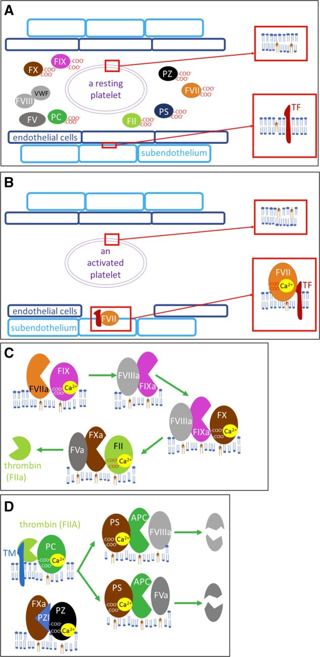
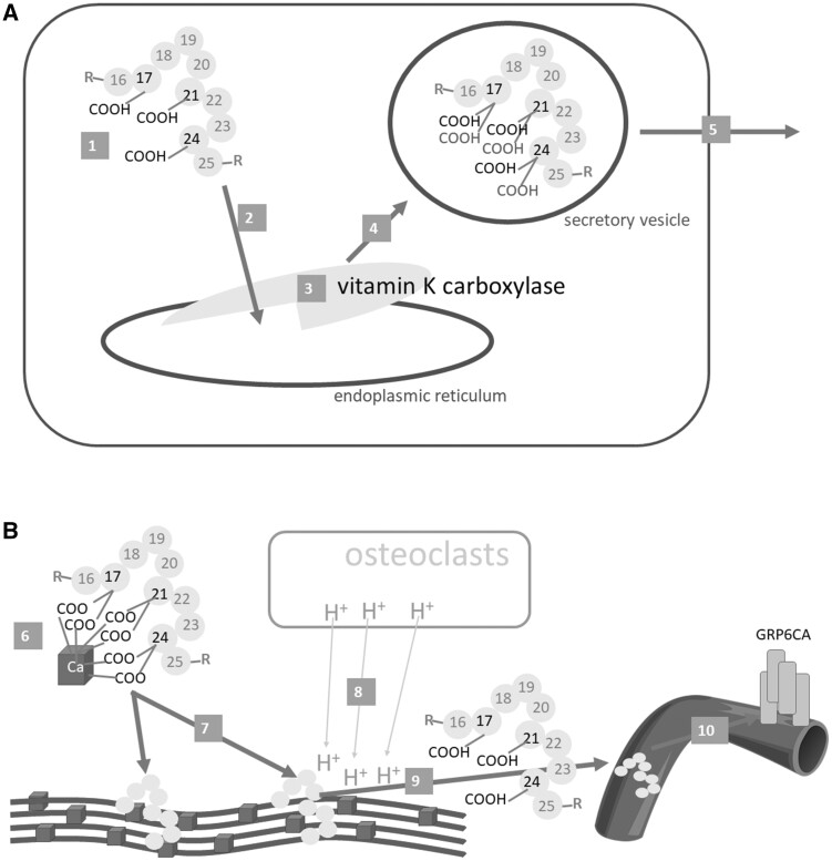
Vitamin K is traditionally connected with blood coagulation, since it is needed for the posttranslational modification of 7 proteins involved in this cascade. However, it is also involved in the maturation of another 11 or 12 proteins that play different roles, encompassing in particular the modulation of the calcification of connective tissues. Since this process is physiologically needed in bones, but is pathological in arteries, a great deal of research has been devoted to finding a possible link between vitamin K and the prevention of osteoporosis and cardiovascular diseases. Unfortunately, the current knowledge does not allow us to make a decisive conclusion about such a link. One possible explanation for this is the diversity of the biological activity of vitamin K, which is not a single compound but a general term covering natural plant and animal forms of vitamin K (K1 and K2) as well as their synthetic congeners (K3 and K4). Vitamin K1 (phylloquinone) is found in several vegetables. Menaquinones (MK4-MK13, a series of compounds known as vitamin K2) are mostly of a bacterial origin and are introduced into the human diet mainly through fermented cheeses. Current knowledge about the kinetics of different forms of vitamin K, their detection, and their toxicity are discussed in this review.

Keywords: bioanalysis; coagulation; diet; menaquinone; phylloquinone.


Figures

Figure 1
Figure 1
Absorption and elimination of vitamin K. A: Oral absorption of vitamin K. 1: Formation of micelles from vitamin K and bile acids. 2: Uptake of vitamin K from a micelle to an enterocyte. 3: Formation and release of a chylomicron containing vitamin K. 4: Through lymphatic circulation, vitamin K in the chylomicron enters systemic circulation (through the vena cava inferior). B: Metabolism of vitamin K in the hepatocyte. A chylomicron loaded with vitamin K binds to low density lipoprotein receptor-related protein 1 (LRP1, 5a) or LDL particles loaded with vitamin K bind to LDL-receptors (LDLRs, 5 b), and this results in the uptake of vitamin K into the hepatocyte. A similar process can also be observed with VLDL in the peripheral tissues (not shown). 6: Vitamin K is ω-hydroxylated by cytochrome 4F2 in the endoplasmic reticulum. 7: This metabolite is subsequently β-oxidized by mitochondrial trifunctional protein (MTP) to 5 C or 7 C or 10 C (not shown), which are subjected to glucuronidation by UDP-glucuronosyltransferase (GT, 8) in the endoplasmic reticulum. 9: Excretion of glucuronides in feces and urine
Figure 2
Figure 2
The general fate of vitamin K–dependent proteins at the site of production. A: Under normal conditions. B: Under vitamin K depletion. 1: Preproprotein is synthesized from mRNA by ribosomes. 2: Preproprotein is targeted to the endoplasmic reticulum (ER), where the ER signaling sequence is removed and the protein is processed by (vitamin K–dependent) γ-glutamyl carboxylase. 3: Proprotein is carboxylated when vitamin K is present. 4: Carboxylated or 4a: uncarboxylated proprotein is transported to the Golgi apparatus, where the PRO-sequence is mostly cleaved (some proteins are also glycosylated there, not shown). 4b: In some cases under vitamin K deficiency, the uncarboxylated protein is cleft by proteasome. 5: Mature protein is extruded via a secretory vesicle into the extracellular space
Figure 3
Figure 3
Probable steps in the carboxylation process mediated by vitamin K in the endoplasmic reticulum. A protein that contains a PRO-sequence is targeted and subsequently bound to the carboxylase in the first step (A). This binding markedly increases the enzymatic function of the carboxylase. The quinone form of vitamin K is reduced to hydroquinone by VKORC1. Hydroquinone is deprotonated by carboxylase in the next step (B). Oxygen reacts with deprotonated vitamin K hydroquinone to produce alkoxide (C). This strong base deprotonates the γ-carbon of glutamyl residue to form a carbanion, which reacts with carbon dioxide (D–E). At the same time, vitamin K epoxide is formed (E). γ-glutamyl carboxylation is accomplished and the formed protein is released from the enzyme and further transported to the Golgi apparatus (not shown), while vitamin K epoxide is converted first to vitamin K quinone and then to vitamin K hydroquinone (F) by VKORC1. Data for this figure were taken from Rishavy et al (2004), Down et al (1995), Ayombil et al (2020) and Berkner (2000)Abbreviations: carboxylase, vitamin K–dependent γ-glutamyl carboxylase; VKORC1, vitamin K epoxide reductase
Figure 4
Figure 4
Vitamin K (pro)coagulatory and anticoagulatory factors. A: Resting state: coagulation factors are found in inactive forms in circulation, phosphatidylserine is not at the surface of the platelets (see the upper magnification), and tissue factor (TF) is not in direct contact with the blood (see the lower magnification). B: Activation of blood coagulation: in case of vascular damage, TF on subendothelial cells is now available for factor VII (FVII), which is activated. Platelet activation leads to modifications in the structure of the plasmatic membrane, with the exposure of phosphatidylserine on its surface. C: Activation of vitamin K–dependent coagulation factor: activated factor VII (FVIIa) cleaves factor IX (FIX) into an active enzyme (factor IXa, FIXa), which needs activated factor VIII (FVIIIa) for its activity. This complex activates factor X (FX) into an active enzyme (FXa), which needs activated factor V (FVa) for its activity. The whole FXa, FVa, calcium and phospholipid complex is also known as prothrombinase, and it activates thrombin (factor II, FII). Factor V (FV) or factor VIII (FVIII) are activated either by FXa or thrombin (not shown). D: Regulatory anticoagulant vitamin K–dependent factors: thrombin with thrombomodulin (TM) cleaves inactive plasma protein C (PC) into the active enzyme APC. For its enzymatic activity, APC also needs protein S (PS). PS does not require proteolysis in order to be active in PC-catalyzed lysis. This complex cleaves both FVa and FVIIIa. Protein Z (PZ) is a cofactor of PZ-dependent protease inhibitor (PZI), which blocks the enzymatic activity of FXa
Figure 5
Figure 5
Osteocalcin. A: Synthesis and release from the osteoblast. B: Effect on bones and systemic release. Osteocalcin (1) is synthesized as preproprotein, then travels to the endoplasmic reticulum (2), where 3 glutamic acid residues at positions 17, 21 and 24 are γ-carboxylated by γ-glutamyl carboxylase (vitamin K carboxylase) (3). The carboxylated osteocalcin is further processed (eg, see Figure 4), transported in the vesicles (4), and released into the bone matrix (5), where it binds calcium ions in hydroxyapatite (6). This is a crucial step in bone formation – the correct alignment of collagen fibers with hydroxyapatite (7). When osteoresorption takes place, osteoclasts decrease the pH level in the bones (8); this can lead to the decarboxylation of osteocalcin to form un(der)carboxylated osteocalcin, which is released into the systemic circulation (9), where it could exert its hormonal effect, in particular by binding to the GRP6CA receptor (10)

References

    1. Raju TNK. The Nobel Chronicles. Lancet .1999;353:761. - PubMed
    1. Shearer M, Newman P.. Recent trends in the metabolism and cell biology of vitamin K with special reference to vitamin K cycling and MK-4 biosynthesis. J Lipid Res .2014;55:345–362. - PMC - PubMed
    1. Tarento TDC, McClure DD, Dehghani F, et al.Pilot-scale production of phylloquinone (vitamin K1) using a bubble column photo-bioreactor. Biochem Eng J. 2019;150:107243.
    1. Tarento TDC, McClure DD, Talbot AM, et al.A potential biotechnological process for the sustainable production of vitamin K1. Crit Rev Biotechnol .2019;39:1–19. - PubMed
    1. Manzotti P, Nisi P, Zocchi G.. Vitamin K in plants. Funct Plant Sci Biotechnol. 2008;2:29–35.

Publication types