Review
. 2021 Aug 31;62(4):399-410.
doi: 10.3325/cmj.2021.62.399.

Cerebrospinal fluid dynamics

Affiliations
Review

Cerebrospinal fluid dynamics

Shinya Yamada. Croat Med J. .

Abstract

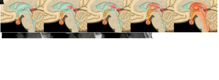
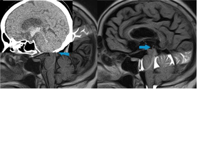
The classical cerebrospinal fluid (CSF) circulation theory has been accepted as an established theory of CSF physiology. It describes bulk CSF flow from production site to absorption site. However, much controversy remains regarding the basic CSF physiology and the mechanisms behind the development of hydrocephalus. In the recent observations made using advanced magnetic resonance imaging (MRI) technique, namely, the time spatial inversion pulse (Time-SLIP) method, CSF was used as internal CSF tracer to trace true CSF movement. Observation of the CSF dynamics using this method reveals aspects of CSF dynamics that are different from those of classical CSF circulation theory. Cerebrospinal fluid shows pulsation but does not show bulk flow from production site to absorption site, a theory that was built upon externally injected tracer studies. Observation of the exogeneous tracer studies were true but misinterpreted. Causes of misinterpretations are the differences between results obtained using the true CSF tracer and exogenous tracers. A better understanding of the real CSF physiology can be significant for the advancement of medical sciences in the future. Revisiting CSF flow physiology is a necessary step toward this goal.


Figures

Figure 1
Figure 1
(A) Contrast-medium ventriculography. (B) Mid-sagittal magnetic resonance imaging (MRI) time spatial inversion pulse image. The contrast medium is directly injected into the human lateral ventricle through a cannula and the outflow of the contrast medium from the lateral ventricle is observed. The contrast medium injected into the ventricles is rapidly cleared through the ventricle system and flowed into the subarachnoid space rapidly (A). No pulsation of the cerebrospinal fluid could be depicted with this method. Cerebrospinal fluid labeled with MRI radiofrequency pulse does not flow out or drain from the ventricle at all. Cerebrospinal fluid only pulsates but does not show unidirectional flow (B).
Figure 2
Figure 2
Coronal views of the lateral and third ventricle at the foramen of Monro. Magnetic resonance imaging time spatial inversion pulse imaging. Cerebrospinal fluid (CSF) turbulent flow is seen in the third ventricle (A). The animation illustrates CSF exchange between the third ventricle and the lateral ventricle. It is a different type of flow from a simple “to and fro” motion. It is more likely replacement of the CSF between the two ventricles (B).
Figure 3
Figure 3
Mid-sagittal images at the third ventricle on magnetic resonance time spatial inversion pulse imaging. Cerebrospinal fluid turbulent flow is seen in the third ventricle (A).The animation illustrates the volume transmission of the ventricular system adjacent to the circumventricular organs (B). Substances, chemicals, or hormones (symbolically showing orange and pink) secreted into the third ventricle are agitated by the vortex of the third ventricle and homogenized in the cerebrospinal fluid.
Figure 4
Figure 4
Magnetic resonance real-time time spatial inversion pulse imaging 72 msec/frame. Cardiac beats are shown as a number in left upper corner of each image. Orange arrow indicates cerebrospinal fluid (CSF) motion through the aqueduct of Sylvius driven by respiration. A large pulsation associated with breathing from the fourth ventricle to the third ventricle is observed in the cerebral aqueduct of Sylvius at the fifth cardiac beat (A). These images are taken immediately after images in Figure 4A. Respiration-driven CSF motion can be seen on the third and fourth cardiac beat (B).
Figure 5
Figure 5
Mid-sagittal magnetic resonance imaging real-time time spatial inversion pulse (SLIP) image. Simultaneous bidirectional flow of cerebrospinal fluid (CSF) is sometimes seen in the aqueduct of Sylvius. (A) Caudo-cephalad CSF flow is seen on the dorsal side of the aqueduct of Sylvius (orange arrow), while cephalo-caudal CSF is seen through the ventral side of the aqueduct of Sylvius (blue arrow) (A). Simultaneous bidirectional CSF flow is captured by real-time time SLIP imaging (B). Blue arrow indicates caudo-cephalad flow and orange arrow indicate cephalo-caudal flow.
Figure 6
Figure 6
Magnetic resonance imaging (MRI) real-time time spatial inversion pulse (SLIP) image. Cerebrospinal fluid in the body of lateral ventricle was stirred by inertia (head shaken). Cerebrospinal fluid agitation become settled 3 and a half minutes after head is shaken. In the middle of head shaking (A), after 1 minute (B), after 2 minutes (C), and after 3 and a half minutes (D).
Figure 7
Figure 7
A 70-year-old woman suddenly developed mild disturbance of consciousness at the onset. Computed tomography scan showed right caudate nucleus hemorrhage and intra-ventricular hemorrhage without acute progressive ventriculomegaly (A). Bilateral foramen Monro, third ventricle, and cerebral aqueduct of Sylvius are occupied by hematomas. No cerebrospinal fluid pulsation through the cerebral aqueduct of Sylvius (complete obstruction, blue arrow) was depicted by magnetic resonance imaging real-time time spatial inversion pulse (B).

References

    1. Cushing H. Studies on the cerebro-spinal fluid: I. Introduction. J Med Res. 1914;31:1–19. - PMC - PubMed
    1. Weed LH.Studies on cerebro-spinal fluid. No. iv: the dual source of cerebro-spinal fluid J Med Res 1914. 31:93-118 11 - PMC - PubMed
    1. Weed LH. Studies on cerebro-spinal fluid. No. III: The pathways of escape from the subarachnoid spaces with particular reference to the arachnoid villi. J Med Res. 1914;31:51–91. - PMC - PubMed
    1. Weed LH. Studies on cerebro-spinal fluid. No. II: The theories of drainage of cerebro-spinal fluid with an analysis of the methods of investigation. J Med Res. 1914;31:21–49. - PMC - PubMed
    1. Sachs EWH, Crawford FS. Studies on cerebrospinal circulation by new method. Arch Neurol Psychiatry. 1929:130–51.