. 2021 Sep 1;10(11):3.
doi: 10.1167/tvst.10.11.3.

Macular Rod Function in Retinitis Pigmentosa Measured With Scotopic Microperimetry

Affiliations

Macular Rod Function in Retinitis Pigmentosa Measured With Scotopic Microperimetry

Arun K Krishnan et al. Transl Vis Sci Technol. .

Abstract

Purpose: To investigate the validity and reliability of macular rod photoreceptor function measurement with a microperimeter.

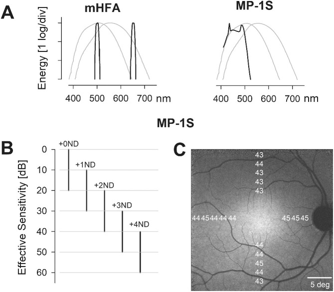
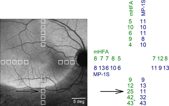
Methods: Macular sensitivity in dark-adapted retinitis pigmentosa (RP) patients (22 eyes; 9-67 years of age) and controls (five eyes; 22-55 years of age) was assessed with a modified Humphrey field analyzer (mHFA), as well as a scotopic microperimeter (Nidek MP-1S). Sensitivity loss (SL) was estimated at rod-mediated locations. All RP eyes were re-evaluated at a second visit 6 months later. The dynamic range of the MP-1S was expanded with a range of neutral-density filters (NDFs).

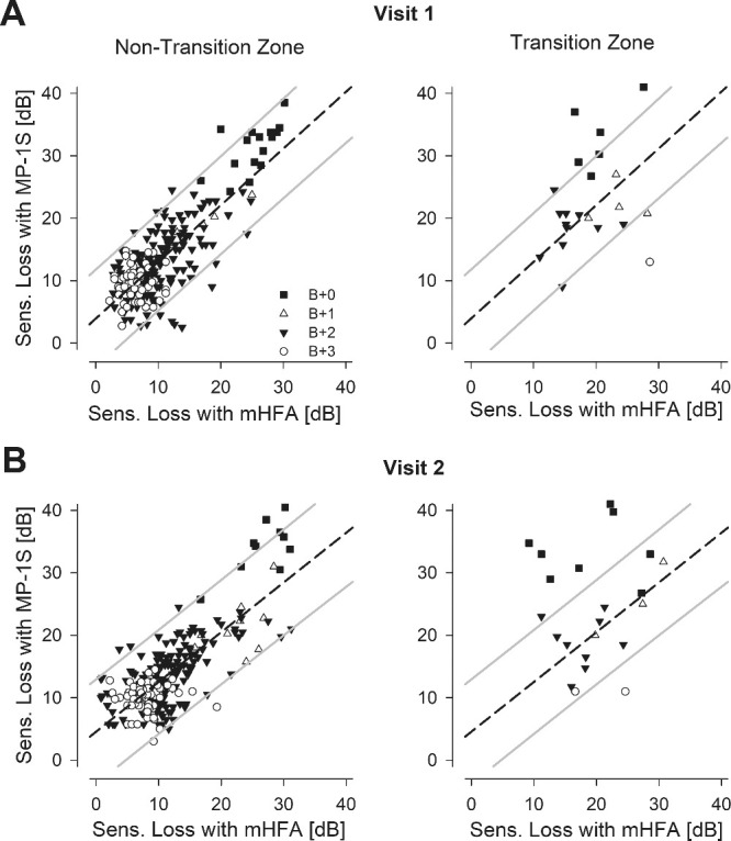
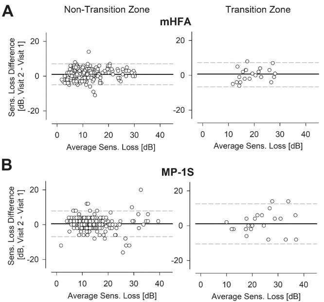
Results: In controls, a 4 NDF was used at all macular locations tested. In patients with RP, 0 to 3 NDFs were used, depending on the local disease severity. At rod-mediated locations (n = 281), SL estimates obtained with the MP-1S were highly correlated (r = 0.80) with those of the mHFA. The inter-perimeter difference of SL averaged less than 3 decibels (dB) with all NDFs, except those with most severe locations evaluated with a 0 NDF, where the difference averaged more than 6 dB. The results were similar on the second visit.

Conclusions: The MP-1S estimates of SL are highly correlated with those of the mHFA over a wide range of disease severity replicated at two visits; however, there was an unexplained bias in the magnitude of SL estimated by the MP-1S especially at loci with severe disease.

Translational relevance: MP-1S scotopic microperimetry can be used to evaluate changes to macular rod function, but evaluation of treatment potential by quantitative comparison of SL to retinal structure will be more challenging.


Conflict of interest statement

Disclosure: A.K. Krishnan, None; A.J. Roman, None; M. Swider, None; S.G. Jacobson, None; A.V. Cideciyan, None

Figures

Figure 1.
Figure 1.
Evaluating macular rod function using the mHFA and MP-1S. (A) Spectral distribution of the monochromatic blue and red colored stimuli used with the mHFA (left) and the broadband blue stimulus used with the MP-1S (right), superimposed over the human scotopic and photopic luminosity functions (thin gray curves). (B) Effective sensitivities obtained with the MP-1S by extending the limited dynamic range of the blue colored stimuli with the use of a range of ND filters. Each log unit of ND filter (+1 to +4 ND) shifts the instrument dynamic range to lower luminances by 10 dB. (C) A total of 18 locations were sampled at 2° intervals, between 6° and 14° eccentricity from the fovea along the horizontal and vertical meridia. The mean effective thresholds for the MP-1S in controls did not vary substantially across the test locations.
Figure 2.
Figure 2.
Residual macular rod function in a representative subject with retinitis pigmentosa. The 18 macular test locations (left, white squares) spanning the central 28° are shown superimposed on the near-infrared excited reduced-illuminance autofluorescence image. Regions with retained retinal pigment epithelium (RPE) melanization appear brighter than regions with RPE demelanization, which are relatively darker (inferior retina). The test location near the transition between these two regions is indicated (black arrow). The sensitivity loss (SL in dB, right), estimated as the difference between average effective threshold of control subjects for each locus and the corresponding threshold in this subject, was similar for the two perimeters for most locations except at the transition zone (black arrow). *Cone-mediated location.
Figure 3.
Figure 3.
Comparison of the SL estimated from the two perimeters in all eyes with retinitis pigmentosa. (A) The estimated MP-1S SL for the majority of the samples not located at a transition zone (left, n = 259) were highly correlated with the corresponding mHFA SL. Different symbols (+0 ND, filled square; +1 ND, unfilled triangle; +2 ND, filled triangle; +3 ND, unfilled circle) represent the range of ND filters used for the MP-1S testing. The relationship among the SL estimates was well fit by a linear regression (dashed line). The 95% prediction intervals are also shown (gray lines). The regression fit and prediction interval from the non–transition-zone data were used to understand the relationship at limited transition zone loci (right, n = 22). The SL estimates at the transition zones (right) tended to show greater mismatches between the two perimeters. (B) Results from a second study visit displayed comparably to those shown in panel A.
Figure 4.
Figure 4.
Test–retest variability of the SL estimated from the two perimeters with blue stimuli in the non–transition-zone and transition-zone loci in subjects with RP. Bland–Altman plots revealed some variability across two consecutive tests 6 months apart for the mHFA (A) and the MP-1S (B), with higher variability for loci in the transition zone (right) as opposed to the loci in the non-transition zones (left).

References

    1. Stone EM, Andorf JL, Whitmore SS, et al. .. Clinically focused molecular investigation of 1000 consecutive families with inherited retinal disease. Ophthalmology. 2017; 124(9): 1314–1331. - PMC - PubMed
    1. Hanany M, Rivolta C, Sharon D.. Worldwide carrier frequency and genetic prevalence of autosomal recessive inherited retinal diseases. Proc Natl Acad Sci USA. 2020; 117(5): 2710–2716. - PMC - PubMed
    1. Kim LS, McAnany JJ, Alexander KR, Fishman GA.. Intersession repeatability of Humphrey perimetry measurements in patients with retinitis pigmentosa. Invest Ophthalmol Vis Sci. 2007; 48(10): 4720–4724. - PubMed
    1. Cideciyan AV, Hufnagel RB, Carroll J, et al. .. Human cone visual pigment deletions spare sufficient photoreceptors to warrant gene therapy. Hum Gene Ther. 2013; 24(12): 993–1006. - PMC - PubMed
    1. Jacobson SG, Cideciyan AV, Peshenko IV, et al. .. Determining consequences of retinal membrane guanylyl cyclase (RetGC1) deficiency in human Leber congenital amaurosis en route to therapy: residual cone-photoreceptor vision correlates with biochemical properties of the mutants. Hum Mol Genet. 2013; 22(1): 168–183. - PMC - PubMed