. 2021 Sep 1;109(17):2691-2706.e5.
doi: 10.1016/j.neuron.2021.06.015. Epub 2021 Jul 19.

IL-23/IL-17A/TRPV1 axis produces mechanical pain via macrophage-sensory neuron crosstalk in female mice

Affiliations

IL-23/IL-17A/TRPV1 axis produces mechanical pain via macrophage-sensory neuron crosstalk in female mice

Xin Luo et al. Neuron. .

Abstract

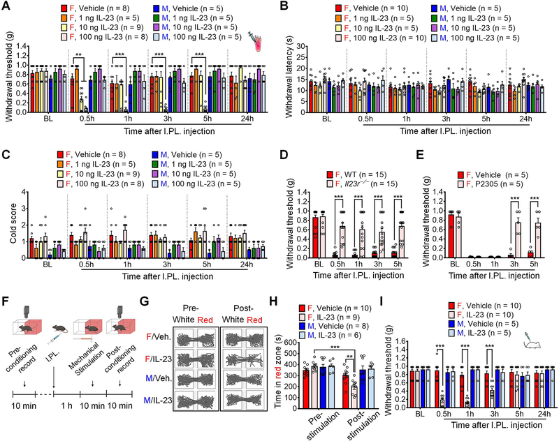
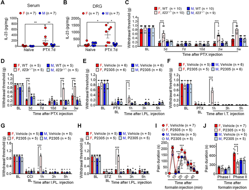
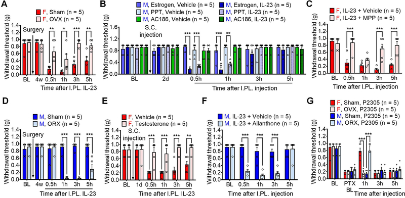
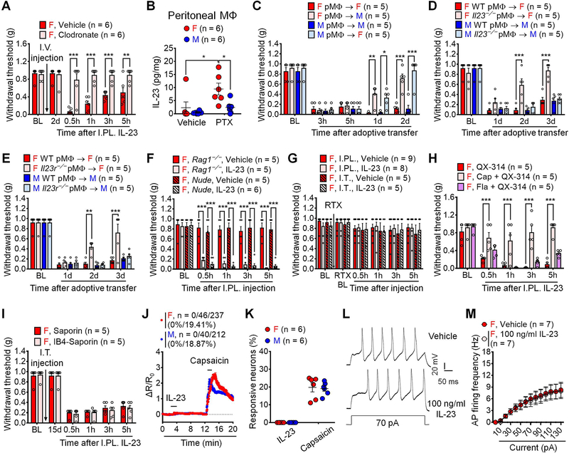
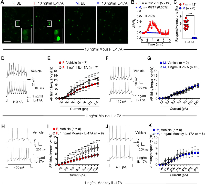
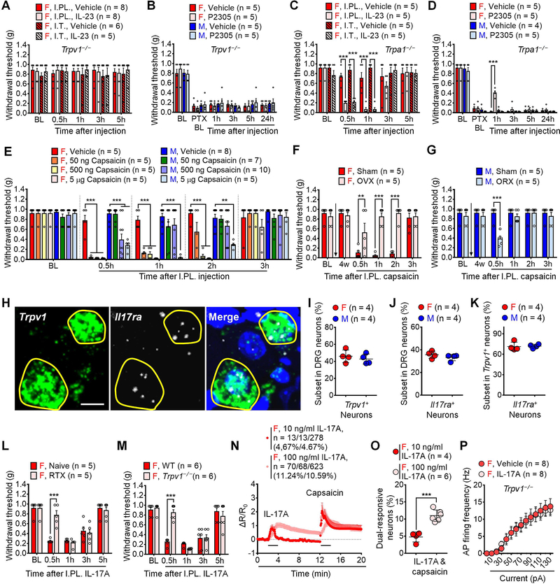
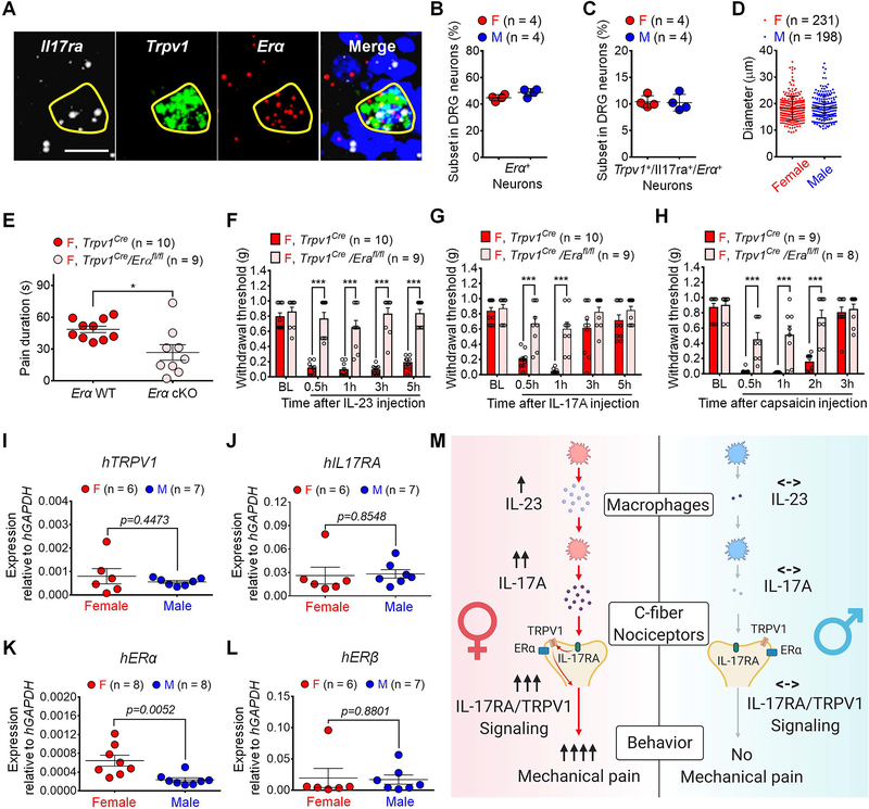
Although sex dimorphism is increasingly recognized as an important factor in pain, female-specific pain signaling is not well studied. Here we report that administration of IL-23 produces mechanical pain (mechanical allodynia) in female but not male mice, and chemotherapy-induced mechanical pain is selectively impaired in female mice lacking Il23 or Il23r. IL-23-induced pain is promoted by estrogen but suppressed by androgen, suggesting an involvement of sex hormones. IL-23 requires C-fiber nociceptors and TRPV1 to produce pain but does not directly activate nociceptor neurons. Notably, IL-23 requires IL-17A release from macrophages to evoke mechanical pain in females. Low-dose IL-17A directly activates nociceptors and induces mechanical pain only in females. Finally, deletion of estrogen receptor subunit α (ERα) in TRPV1+ nociceptors abolishes IL-23- and IL-17-induced pain in females. These findings demonstrate that the IL-23/IL-17A/TRPV1 axis regulates female-specific mechanical pain via neuro-immune interactions. Our study also reveals sex dimorphism at both immune and neuronal levels.

Keywords: IL-17; IL-23; dorsal root ganglion; estrogen receptor α; human; macrophage; mechanical allodynia; nociceptor; nonhuman primate; sex dimorphism.


Conflict of interest statement

Declaration of interests R.-R.J. is a consultant of Boston Scientific and received research grant from the company. These activities are not related to this study. The other authors declare no competing interests.

Figures

Figure 1.
Figure 1.. IL-23 induces mechanical pain in female but not male mice.
(A) I.PL. injection of IL-23 at 1, 10 or 100 ng induces dose-dependent mechanical allodynia in female but not male mice compared to the vehicle. (B-C) I.PL. IL-23 fails to induce thermal hyperalgesia (B, Hargreaves test) or cold allodynia (C, Acetone test) in either sex. (D-E) Mechanical pain induced by IL-23 (I.PL., 100 ng) in females is abolished in Il23r−/− mice (D) and suppressed by IL-23R antagonist P2305 (I.PL., 10 μg, 30 min before IL-23 injection) in WT mice (E). (F-H) Brief protocol of CPA test (F) and representative traces in two chambers following vehicle and IL-23 (I.PL., 100 ng) treatment (G). (H) CPA test indicates that von Frey stimulation (0.04 g) produces aversive behavior in IL-23-treated female mice (I.PL., 100 ng, 1 h before stimulation) but not males. (I) Intrathecal IL-23 (I.T., 100 ng) produces mechanical allodynia in female but not male mice. Data are mean ± SEM. **p < 0.01, ***p < 0.001; Two-Way RM ANOVA with Bonferroni’s post hoc test (A-E and H-I). F (35, 252) = 4.405 (A); F (35, 210) = 0.9888 (B); F (35, 210) = 0.9914 (C); F (4,112) = 11.73 (D); F (4, 32) = 20.56 (E); F (3, 29) = 8.907 (H); and F (15, 130) = 11.19 (I). I.PL.: Intraplantar; I.T.: Intrathecal; CPA: Conditioned preference aversion; BL, baseline.
Figure 2.
Figure 2.. IL-23/IL-23R axis regulates pathological pain in female but not male mice.
(A-B) ELISA showing increased serum (A) and DRG (B) levels of IL-23 in female mice on day 7 after PTX treatment (4 x PTX, 2 mg/kg). (C-D) PTX-evoked mechanical allodynia is reduced in Il23−/− (C) and Il23r−/− (D) female mice in CIPN (1 x PTX, 6 mg/kg). (E-F) P2305 (10 μg), given by I.PL. injection (E) or I.T. injection (F), reduces PTX-induced mechanical allodynia in female but not male mice (4 x PTX, 2 mg/kg). (G-H) P2305 (I.PL., 10 μg) reduces mechanical allodynia in females in the CCI model (G) and STZ model (H) of neuropathic pain. (I-J) P2305 (10 μg, I.T., 30 min before I.PL. 5% formalin) reduces formalin-evoked spontaneous pain in females. (I) Time course. (J) Phase I (0–10 min) and Phase II (10–45 min) responses. Data are mean ± SEM. *p < 0.05, **p < 0.01, ***p < 0.001; Non-parametric Mann-Whitney test (A and B); Two-Way RM ANOVA with Bonferroni’s post hoc test (C-J). F (15, 175) = 7.090 (C); F (15, 80) = 4.781 (D); F (12, 80) = 1.746 (E); F (12, 72) = 5.353 (F); F (12, 64) = 9.924 (G); F (12, 64) = 15.03 (H); F (27, 162) = 2.359 (I); F (3, 18) = 7.333 (J). PTX: Paclitaxel; I.T.: Intrathecal; I.PL.: Intraplantar; CIPN: Chemotherapy-induced peripheral neuropathy; CCI: Chronic constrictive injury; STZ: Streptozotocin; BL: Baseline.
Figure 3.
Figure 3.. Sex hormones regulate IL-23/IL-23R-mediated pain.
(A) Estrogen deficiency by OVX reduces IL-23-induced pain in female mice. (B) S.C. pretreatment (2 mg/kg) of estrogen or ERα agonist PPT, but not ERβ agonist AC186, enables IL-23-induced pain (I.PL., 100 ng) in males. (C) Co-I.PL. injection of ERα receptor antagonist MPP (30 μg) with IL-23 (100 ng) reduces IL-23-induced pain in males. (D) Androgen deficiency by ORX enables IL-23-induced pain in male mice. (E) Testosterone treatment (S.C., 2 mg/kg) suppresses IL-23-induced pain (I.PL., 100 ng) in females. (F) Co-intraplantar injection of androgen receptor antagonist Ailanthone (30 μg) with IL-23 (100 ng) enables IL-23-induced pain in males. (G) Estrogen deficiency abolishes P2305-produced analgesia (I.PL., 10 μg) in females, whereas androgen deficiency enables P2305-produced analgesia (I.PL., 10 μg) in males in CIPN (7d, 1 x PTX, 6 mg/kg). Data are mean ± SEM. *p < 0.05, **p < 0.01, ***p < 0.001; Two-way RM ANOVA with Bonferroni’s post hoc test; F (5, 40) = 6.348 (A); F (25, 120) = 5.896 (B); F (4, 32) = 8.470 (C); F (5, 40) = 26.84 (D); F (5, 40) = 10.74 (E); F (4, 40) = 14.26 (F); and F (12, 64) = 6.988 (G). OVX: Ovariectomy; ORX: Orchiectomy; I.PL.: Intraplantar; S.C.: Subcutaneous; ER: Estrogen receptor; BL: Baseline.
Figure 4.
Figure 4.. IL-23/IL-23R axis-mediated pain requires macrophages and C-fiber nociceptors.
(A) Clodronate (I.V., 1 mg / 200 μl) abolishes IL-23-induced pain (I.PL., 100 ng) in females. (B) ELISA showing PTX (1 μg/ml, 16 h) enhanced IL-23 release from female but not male peritoneal macrophages in vitro. (C) PTX-pretreated macrophages (1 μg/ml, 16 h, 105 cells) produce more potent and sustained pain in same sex vs. opposite sex recipients. (D-E) Loss of IL-23 (Il23−/−, D) or IL-23 receptor (Il23r−/−, E) attenuates mechanical pain by PTX-pretreated macrophages (1 μg/ml, 16 h, 105 cells) in females. (F) IL-23 (I.PL., 100 ng) induced mechanical allodynia is intact in female mice lacking T cells in Rag1−/− mice or nude mice. (G) RTX-induced C-fiber elimination abolishes mechanical allodynia by IL-23-induced (I.PL. or I.T., 100 ng) in females. (H) Blockade of TRPV1+ (peptidergic) C-fibers (6 mM QX314 + 5 μg capsaicin) but not TLR5+ Aβ fibers (6 mM QX314 + 0.3 μg flagellin) reduces IL-23-induced mechanical pain in females (I.PL., 100 ng). (I) Blockade of IB4-binding non-peptidergic C-fibers by IB4-Saporin (I.T. 1.5 μg) fails to affect IL-23-induced pain (I.PL., 100 ng). (J-K) Effects of IL-23 (100 ng/ml, 2 min) and capsaicin (300 nM, 2 min) on Ca2+ influx in dissociated DRG sensory neurons from AdvillinCre/GCamp6f mice. (J) Combined Ca2+ responses from all neurons. Among 237 female neurons, 0 (0%) and 46 (19.41%) show responses to IL-23 and capsaicin, respectively. Among 212 male neurons, 0 (0%) and 40 (18.87%) show responses to IL-23 and capsaicin, respectively. (K) Quantification of % neurons responding to each treatment. n = 6 cultures from 3 mice per sex. (L-M) IL-23 (100 ng/ml, 2 min) does not potentiate action potentials in dissociated small-sized DRG neurons from females. (L) Traces of action potentials. (M) Firing rate of action potentials. Data are mean ± SEM. *p < 0.05, **p < 0.01, ***p < 0.001; Two-way RM ANOVA with Bonferroni’s post hoc test (A, C-I, M); Two-way ordinary ANOVA with Bonferroni’s post hoc test (B); F (5, 50) = 13.30 (A); F (1, 20) = 2.377 (B); F (12, 64) = 9.161 (C); F (9, 48) = 6.432 (D); F (9, 48) = 4.139 (E); F (12, 68) = 7.357 (F); F (15, 115) = 0.2149 (G); F (8, 48) = 5.838 (H); F (5, 40) = 0.2094 (I); F (13, 78) = 14.48 (M). Unpaired two-tailed Student’s t test, t = 0.2307 (K). I.V.: intravenous; I.PL.: Intraplantar; I.T.: Intrathecal; PTX: Paclitaxel; RTX: Resiniferatoxin; MΦ: Macrophage; CAP: Capsaicin; BL: Baseline.
Figure 5.
Figure 5.. Low doses of IL-17A induce female-dominant mechanical pain.
(A) PTX (1 μg/ml, 16h) promotes IL-17 release from both female and male peritoneal macrophages in vitro, which is abolished by Il23r−/− only in females. (B) IL-23 incubation (100 ng/ml, 16h) induces greater IL-17 release from female peritoneal macrophages than male counterparts in vitro. (C) I.PL. IL-17A induces mechanical pain in females at 1–100 ng and males only at 100 ng. (D-E) I.PL. IL-17A at 1–100 ng fails to induce heat hyperalgesia (D) or cold hypersensitivity (D) in either sex. (F) AUC (0.5–3h) of Fig. 5C showing female-dominant mechanical pain by IL-17A (10 and 100 ng). (G) Co-I.PL. injection of IL-17A neutralizing antibody or IL-17RA neutralizing antibody (2 μg) with IL-23 (100 ng) abolishes IL-23-induced pain in females. (H) Clodronate (I.V., 1 mg / 200 μl) does not affect IL-17A-induced pain (I.PL., 10 ng) in females. (I) IL-17A (I.PL., 100 ng) induces mechanical allodynia in female Il23−/− mice. (J) Estrogen deficiency by OVX reduces IL-17A-induced mechanical pain (I.PL., 10 ng) in females. (K) Androgen deficiency by ORX enables IL-17A-induced pain (I.PL., 10 ng) in males. (L-M) PTX increases serum IL-17 in both sexes (L) but enhances DRG IL-17 only in females (M) in CIPN (4 x PTX, 2 mg/kg). Data are mean ± SEM. *p < 0.05, **p < 0.01, ***p < 0.001; Two-way ordinary ANOVA with Bonferroni’s post hoc test (A and B; L and M); Two-way RM ANOVA with Bonferroni’s post hoc test (C-H); F (3, 32) = 2.575 (A); F (1, 24) = 4.336 (B); F (28, 128) = 8.077 (C); F (28, 128) = 0.5004 (D); F (28, 128) = 0.9792 (E); F (3, 32) = 7.285 (F); F (8, 84) = 7.870 (G); F (5, 50) = 0.2779 (H); F (4, 40) = 0.3429 (I); F (5, 40) = 13.05 (J); F (5, 40) = 10.26 (K); F (1, 20) = 0.7352 (L); F (1, 20) = 5.715 (M). AUC: Area under curve; PTX: Paclitaxel; I.PL.: Intraplantar; OVX: Ovariectomy; ORX: orchiectomy; BL: Baseline.
Figure 6.
Figure 6.. IL-17A activates C-fiber nociceptors in a female-dominant manner.
(A-C) Acute perfusion of IL-17A (10 ng/ml, 2 min) evokes Ca2+ influx in DRG sensory neurons cultured from female but not male AdvillinCre/GCamp6f mice. (A) Representative images. Enlarged images are exhibited in the boxes at the bottom. Scale bar: 100 μm. (B) Combined Ca2+ responses of all neurons. 5.71% female neurons (69/1209) and 0% (0/717) male neurons show Ca2+ responses. (C) % quantification of responding neurons. n = 8 or 12 cultures from 4 mice of each sex. (D-G) Bath application of IL-17A (1 ng/ml, 2 min) increases action potential firing in small-sized DRG neurons collected from female but not male mice compared to vehicle. (D, F) Traces of action potentials. (E, G) AP firing rates. (H-K) AP recordings in NHP DRG neurons. Bath application of monkey IL-17A (1 ng/ml, 2 min) increases AP firing in small-sized DRG neurons collected from female but not male NHP compared to vehicle. (H, J) AP traces. (I, K) AP firing rates. Data are mean ± SEM. *p < 0.05, **p < 0.01, ***p < 0.001; Unpaired two-tailed student t test, t = 10.44 (C); Two-way RM ANOVA with Bonferroni’s post hoc test (E, G, I, K); F values: F (13, 78) = 23.08 (E); F (13, 104) = 26.74 (G); F (13, 104) = 25.52 (I); F (13, 91) = 9.548 (K). AP: Action potential; NHP: Non-human primate. See additional details of statistics in Supplemental information.
Figure 7.
Figure 7.. TRPV1 promotes IL-23/IL-17A axis-mediated pain in females.
(A) Trpv1−/− abolishes IL-23-induced pain (I.PL. or I.T., 100 ng) in females. (B) Mechanical pain by PTX (4 × 2 mg/kg) is not affected by IL-23R antagonist P2305 (I.PL., 10 μg) in Trpv1−/− mice of both sexes. (C) IL-23 (I.PL. or I.T., 100 ng) induces comparable mechanical pain in female WT and Trpa1−/− mice. (D) P2305 (I.PL.,10 μg) reduces PTX (4 × 2 mg/kg)-induced mechanical allodynia in female but not male Trpa1−/− mice. (E) I. PL. capsaicin induces mechanical pain in females at 50–5000 ng but males at 500 and 5000 ng. (F) Estrogen deficiency by OVX reduces capsaicin-induced mechanical pain (I.PL., 500 ng) in females. (G) Androgen deficiency by ORX promotes capsaicin-induced mechanical pain (I.PL., 500 ng) in males. (H) In situ hybridization (RNAScope) images show co-localization of Il17ra and Trpv1 mRNA in female DRG neurons. Positive neurons are indicated with yellow circles. Blue DAPI staining labels nuclei. Scale bar, 10 μm. (I-K) Quantification of Il17ra+ (I), Trpv1+ (J) and Il17ra+/Trpv1+ (K) subsets of DRG neurons. n = 4 mice per sex (3 sections per mouse). (L-M) C-fiber elimination by RTX or Trpv1−/− reduces IL-17A-induced pain (I.PL., 100 ng) in females. (N-O) Effects of IL-17A (10 and 100 ng/ml, 2 min) and capsaicin (300 nM, 2 min) on Ca2+ responses in dissociated DRG sensory neurons from female AdvillinCre/GCamp6f mice. (N) Combined responses of all neurons. 4.67% neurons (13/278) respond to IL-17A (10 ng/ml) and capsaicin, and 11.24% neurons (70/623) respond to IL-17A (100 ng/ml), and 10.91% neurons (68/623) respond to both IL-17A (100 ng/ml) and capsaicin. (O) % quantification of responding neurons. n = 4 or 6 cultures from 3 mice per group. (P) Trpv1−/− abolishes IL-17A (1 ng/ml, 2 min) potentiation of action potential firing in small-sized DRG neurons from female mice. Data are mean ± SEM. **p < 0.01, ***p < 0.001; Two-way RM ANOVA with Bonferroni’s post hoc test (A-H, L-M, P); F (12, 92) = 0.2910 (A); F (15, 80) = 0.5561 (B); F (12, 64) = 6.774 (C); F (15, 75) = 5.028 (D); F (28, 168) = 7.304 (E); F (5, 40) = 13.23 (F); F (5, 40) = 4.660 (G); F (4, 32) = 8.850 (L); and F (4, 40) = 13.90 (M); F (13, 91) = 31.63 (P). Unpaired and two-tailed student t test (I-K, O): t = 0.678 (I); t = 1.160 (J); t = 0.1768 (K); t = 6.119 (O). I.PL., Intraplantar; I.T., Intrathecal; PTX, Paclitaxel; RTX, Resiniferatoxin; OVX, Ovariectomy; ORX, Orchiectomy; BL, Baseline.
Figure 8.
Figure 8.. Neuronal estrogen/ERα signaling promotes IL-23/IL-17A-mediated and female-dominant pain.
(A) In situ hybridization (RNAScope) showing co-localization of Il17ra, Trpv1 and Erα mRNA in DRG neurons (females). Positive neurons are indicated with yellow circles. Scale bar, 10 μm. (B-C) Quantification of Erα+ neurons (B) and Il17ra+/Trpv1+/Erα+ neurons (C) in DRGs. n = 4 mice per sex (3 sections per mouse). (D) Diameters of Il17ra+/Trpv1+/Erα+ neurons in DRGs from both sexes. n = 4 mice per sex (3 sections per mouse). (E-H) Selective depletion of Erα in TRPV1+ neurons reduces capsaicin-evoked spontaneous pain (I.PL., 500 ng) in females (E). Erα cKO further abolishes mechanical pain, induced by IP.L. IL-23 (100 ng, F), IL-17A (10 ng, G), or capsaicin (500 ng, H) in females. (I-L) qPCR showing mRNA levels of human TRPV1 (I), IL17RA (J), ERα (K) and ERβ (L) in DRG tissues from human donors using human GAPDH as an internal control. See primer information in Table S2. (M) Working hypothesis underlying the female-specific modulation of mechanical pain by IL-23/IL-17A/TRPV1 axis. Note that both macrophage and nociceptor signaling contribute to the sex dimorphism. Data are mean ± SEM. *p < 0.05, ***p < 0.001. Unpaired two-tailed student t test (B-E, I-L): t = 2.142 (B); t = 0.1789 (C); t = 0.6628 (D); t = 2.851 (E); t = 0.7880 (I); t = 0.1874 (J); t = 3.305 (K); t = 0.1544 (L). Two-way ANOVA with Bonferroni’s post hoc test (F-H): F (4, 68) = 15.67 (F); F (4, 68) = 5.576 (G); F (4, 60) = 9.170 (H). I.PL.: Intraplantar; ER: Estrogen receptor; BL: Baseline.

Comment in

  • Addressing the gender pain gap.
    Dawes JM, Bennett DL. Dawes JM, et al. Neuron. 2021 Sep 1;109(17):2641-2642. doi: 10.1016/j.neuron.2021.08.006. Neuron. 2021. PMID: 34473950

References

    1. Askari A, Naghizadeh MM, Homayounfar R, Shahi A, Afsarian MH, Paknahad A, Kennedy D, and Ataollahi MR (2016). Increased Serum Levels of IL-17A and IL-23 Are Associated with Decreased Vitamin D3 and Increased Pain in Osteoarthritis. PLoS One 11, e0164757. - PMC - PubMed
    1. Avona A, Burgos-Vega C, Burton MD, Akopian AN, Price TJ, and Dussor G (2019). Dural Calcitonin Gene-Related Peptide Produces Female-Specific Responses in Rodent Migraine Models. J Neurosci 39, 4323–4331. - PMC - PubMed
    1. Avraham O, Deng P-Y, Jones S, Kuruvilla R, Semenkovich CF, Klyachko VA, and Cavalli V (2020). Satellite glial cells promote regenerative growth in sensory neurons. Nature Communications 11, 4891. - PMC - PubMed
    1. Bang S, Xie YK, Zhang ZJ, Wang Z, Xu ZZ, and Ji RR (2018). GPR37 regulates macrophage phagocytosis and resolution of inflammatory pain. J Clin Invest 128, 3568–3582. - PMC - PubMed
    1. Basbaum AI, Bautista DM, Scherrer G, and Julius D (2009). Cellular and molecular mechanisms of pain. Cell 139, 267–284. - PMC - PubMed

Publication types

MeSH terms