. 2021 Sep 2;19(1):184.
doi: 10.1186/s12915-021-01109-x.

The ATPase SRCAP is associated with the mitotic apparatus, uncovering novel molecular aspects of Floating-Harbor syndrome

Affiliations

The ATPase SRCAP is associated with the mitotic apparatus, uncovering novel molecular aspects of Floating-Harbor syndrome

Giovanni Messina et al. BMC Biol. .

Abstract

Background: A variety of human genetic diseases is known to be caused by mutations in genes encoding chromatin factors and epigenetic regulators, such as DNA or histone modifying enzymes and members of ATP-dependent chromatin remodeling complexes. Floating-Harbor syndrome is a rare genetic disease affecting human development caused by dominant truncating mutations in the SRCAP gene, which encodes the ATPase SRCAP, the core catalytic subunit of the homonymous chromatin-remodeling complex. The main function of the SRCAP complex is to promote the exchange of histone H2A with the H2A.Z variant. According to the canonical role played by the SRCAP protein in epigenetic regulation, the Floating-Harbor syndrome is thought to be a consequence of chromatin perturbations. However, additional potential physiological functions of SRCAP have not been sufficiently explored.

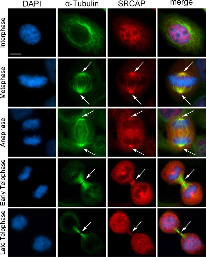
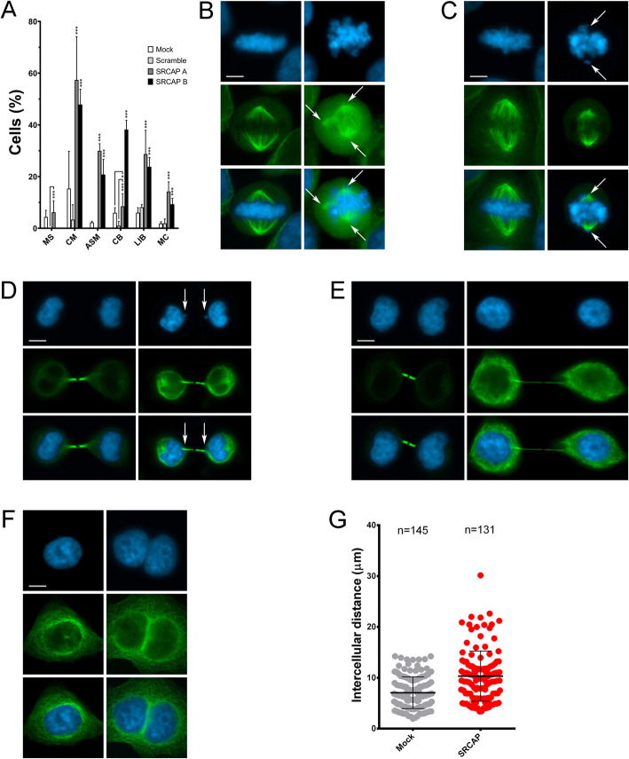
Results: We combined cell biology, reverse genetics, and biochemical approaches to study the subcellular localization of the SRCAP protein and assess its involvement in cell cycle progression in HeLa cells. Surprisingly, we found that SRCAP associates with components of the mitotic apparatus (centrosomes, spindle, midbody), interacts with a plethora of cytokinesis regulators, and positively regulates their recruitment to the midbody. Remarkably, SRCAP depletion perturbs both mitosis and cytokinesis. Similarly, DOM-A, the functional SRCAP orthologue in Drosophila melanogaster, is found at centrosomes and the midbody in Drosophila cells, and its depletion similarly affects both mitosis and cytokinesis.

Conclusions: Our findings provide first evidence suggesting that SRCAP plays previously undetected and evolutionarily conserved roles in cell division, independent of its functions in chromatin regulation. SRCAP may participate in two different steps of cell division: by ensuring proper chromosome segregation during mitosis and midbody function during cytokinesis. Moreover, our findings emphasize a surprising scenario whereby alterations in cell division produced by SRCAP mutations may contribute to the onset of Floating-Harbor syndrome.

Keywords: Cell cycle; Cytokinesis regulators; Floating-Harbor; Midbody; SRCAP.


Conflict of interest statement

The authors declare that they have no competing interests

Figures

Fig. 1
Fig. 1
SRCAP localizes to the centrosomes, spindle, and midbody in HeLa cells. From left to the right: DAPI (blue), anti-α-Tubulin (green), anti-SRCAP (red) and merge. As expected, the SRCAP staining is present in the interphase nuclei. At metaphase, the SRCAP staining is found on spindle poles and spindle fibers, while in later stage decorates centrosomes and central spindle (anaphase) and midbody (telophase). Scale bar = 10 μm
Fig. 2
Fig. 2
Localization of SRCAP on midbodies isolated from HeLa cells. Fixed preparations of midbodies were stained with DAPI (blue), anti-α-Tubulin (green), and anti-SRCAP antibody (red). A Immunolocalization of SRCAP protein to early (left) and late (right) midbodies. No DAPI staining was detected. SRCAP staining clearly decorated the isolated midbodies and overlapped with that of α-Tubulin. Scale bar = 5 μm. B Detection of SRCAP by Western blotting on protein extracts from isolated midbodies. Three high-molecular weight bands were detected (over 270 kD). These bands may be compatible with the three predicted SRCAP isoforms of 343, 337, and 327 kD. In fact, although proteins with minimal size differences should not be separated at high molecular weights, it is well known that the predicted molecular weight of a given protein not always corresponds to that found experimentally by SDS-PAGE. In the case of SRCAP isoforms, post-translational modifications may occur which could affect migration differences. Aurora B was used as a positive control. The ISWI remodeler (negative control) was not detected
Fig. 3
Fig. 3
Depletion of SRCAP affects cell division in HeLa cells. RNAi knockdown was performed by transfecting HeLa cells with specific siRNAs (see the “Methods” section). Cells were stained with DAPI (blue) and anti-α-Tubulin (green). Left panels (mock), right panels (RNAi). Scale bar = 10 μm. Six classes of defects were categorized: A Histograms showing the quantitative analysis of cell division defects; mocks (white histograms), scramble (light gray histograms), SRCAP A (dark gray histograms), and SRCAP B (black histograms). B Multipolar spindles (MS). C Chromosome misalignments (CM) and abnormal spindle morphology (ASM). D Chromatin bridges (CB). E Long intercellular bridges (LIB); no DAPI-stained trapped chromatin was observed. F Multinucleated cells (MC). G Intercellular distance. The quantitative analysis of defects scored in RNAi-treated and control cells (Table 1) is based on the following numbers: at least 100 prometaphases and metaphases for MS, 70 metaphases for CM and ASM, 300 telophases for LIB and CB, and 5500 for MS. Three independent experiments were performed. *P < 0.05; **P < 0.005; and ***P < 0.0005 compared with the controls group (mock and scramble) by Fisher’s exact test
Fig. 4
Fig. 4
Abnormal microtubules re-polymerization in SRCAP depleted HeLa cells. From top to bottom: DAPI (blue), EGFP::α-Tubulin (green) and merge. Hela cells were incubated in ice (1 h) to stimulate microtubules depolymerization (T0). Compared to the non-treated samples (NT), microtubule re-polymerization after 5 min (T5) of rewarming give rise to properly shaped asters in control metaphases (mock), while in SRCAP depleted metaphases (RNAi), aster reformation is clearly aberrant with EGFP::α-Tubulin fluorescence marking only one pole spot, together with sparse and disorganized fibers. The results are based on a total of three experiments; at least 300 cells were scored from both RNAi-treated and control cells. Scale bar = 10 μm
Fig. 5
Fig. 5
SRCAP depletion affects midbody localization of cytokinesis regulators. A Examples of cytokinesis regulators recruitment at midbody in mock and SRCAP depleted HeLa cells (RNAi). From left to the right: DAPI (blue), anti-α-Tubulin (green), cytokinesis regulators (red) and merge. B Histograms showing the quantitative analysis of mis-localizations (see also Table 2); mock (white histograms), SRCAP depleted cells (black histograms). Scale bar = 10 μm. Three independent experiments were performed and at least 300 telophases were scored in both RNAi-treated and control cells. *P < 0.05; **P < 0.005; and ***P < 0.0005 compared with the mock group by Fisher’s exact test
Fig. 6
Fig. 6
SRCAP interacts with cytokinesis regulators in co-IP assays. For immunoprecipitation assays, we used a SRCAP antibody previously validated by Ruhl et al. [9] (Additional file 5: Fig. S4 and Additional file 7: Table S1). A Telophase synchronization in HeLa cells. The scheme summarizes the protocol used for telophase synchronization and subcellular fractionation assay (see the “Methods” section). B Chromatin fractionation of HeLa cells synchronized in telophase. WCE, whole cell extract. P3, nuclear fraction. S2, cytoplasmatic fraction. H3 and α-Tubulin are markers of nuclear and cytoplasmic fraction, respectively. MKLP1 is expressed in late stages of mitosis (telophase synchronization control). C Immunoprecipitation of protein extracts from cytoplasmic fraction of telophase synchronized HeLa cells (S2 fraction). IP sample immunoprecipitated with SRCAP antibody (+ anti-SRCAP) were compared to negative control (- anti-SRCAP). Three independent IP experiments were performed. IN = input, IP = immunoprecipitation
Fig. 7
Fig. 7
DOM-A localizes to interphase nuclei, centrosomes and midbody in Drosophila S2 cells. From left to the right: DAPI (blue), anti-α-Tubulin (green), anti-DOM-A (red) and merge. In addition to interphase nuclei, the anti-DOM-A staining was found on centrosomes (metaphase) and midbody (telophase) pointed by an arrow. Scale bar = 5 μm. *P < 0.05; **P < 0.005; and ***P < 0.0005 compared with the mock group by Fisher’s exact test
Fig. 8
Fig. 8
RNAi-mediated depletion of DOM-A affects mitosis and cytokinesis in Drosophila S2 cells. DAPI staining is shown in blue, α-Tubulin in green. A Quantitative analysis of defects; mock (white histograms), DOM-A depleted cells (black histograms). B From left to right: normal metaphase (mock), multipolar spindle (RNAi), and chromosome misalignments (RNAi). C Left panel: normal telophase (mock); right panel: chromatin bridge (RNAi). D left panel: normal telophase (mock); right panel: long intercellular bridge (RNAi). E Left panel: mononucleated cell (mock); right panel: binucleated cell (RNAi). Scale bar = 5 μm. The quantitative analysis of defects scored in RNAi-treated and mock treated cells is based on about 300 metaphases, telophases, or interphases, scored in at least three independent experiments (Table 3). *P < 0.05; **P < 0.005; and ***P < 0.0005 compared with the mock group by Fisher’s exact test

References

    1. Bickmore WA, van der Maarel SM. Perturbations of chromatin structure in human genetic disease: recent advances. Hum Mol Genet. 2003;12 Spec No 2:R207–R213. doi: 10.1093/hmg/ddg260. - DOI - PubMed
    1. Bouazoune K, Kingston RE. Chromatin remodeling by the CHD7 protein is impaired by mutations that cause human developmental disorders. Proc Natl Acad Sci U S A. 2012;109(47):19238–19243. doi: 10.1073/pnas.1213825109. - DOI - PMC - PubMed
    1. Masliah-Planchon J, Bieche I, Guinebretiere JM, Bourdeaut F, Delattre O. SWI/SNF chromatin remodeling and human malignancies. Annu Rev Pathol. 2015;10(1):145–171. doi: 10.1146/annurev-pathol-012414-040445. - DOI - PubMed
    1. Kumar R, Li DQ, Muller S, Knapp S. Epigenomic regulation of oncogenesis by chromatin remodeling. Oncogene. 2016;35(34):4423–4436. doi: 10.1038/onc.2015.513. - DOI - PubMed
    1. White SM, Morgan A, Da Costa A, Lacombe D, Knight SJ, Houlston R, Whiteford ML, Newbury-Ecob RA, Hurst JA. The phenotype of Floating-Harbor syndrome in 10 patients. Am J Med Genet A. 2010;152A(4):821–829. doi: 10.1002/ajmg.a.33294. - DOI - PubMed

Publication types

Supplementary concepts

LinkOut - more resources