Review
. 2021 Sep 2;7(1):64.
doi: 10.1038/s41572-021-00298-7.

Marfan syndrome

Affiliations
Review

Marfan syndrome

Dianna M Milewicz et al. Nat Rev Dis Primers. .

Erratum in

  • Publisher Correction: Marfan syndrome.
    Milewicz DM, Braverman AC, De Backer J, Morris SA, Boileau C, Maumenee IH, Jondeau G, Evangelista A, Pyeritz RE. Milewicz DM, et al. Nat Rev Dis Primers. 2022 Jan 17;8(1):3. doi: 10.1038/s41572-022-00338-w. Nat Rev Dis Primers. 2022. PMID: 35039531 No abstract available.

Abstract

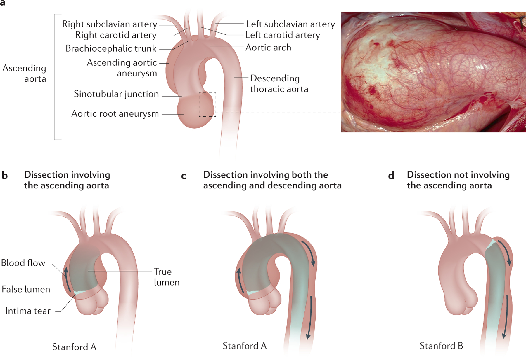
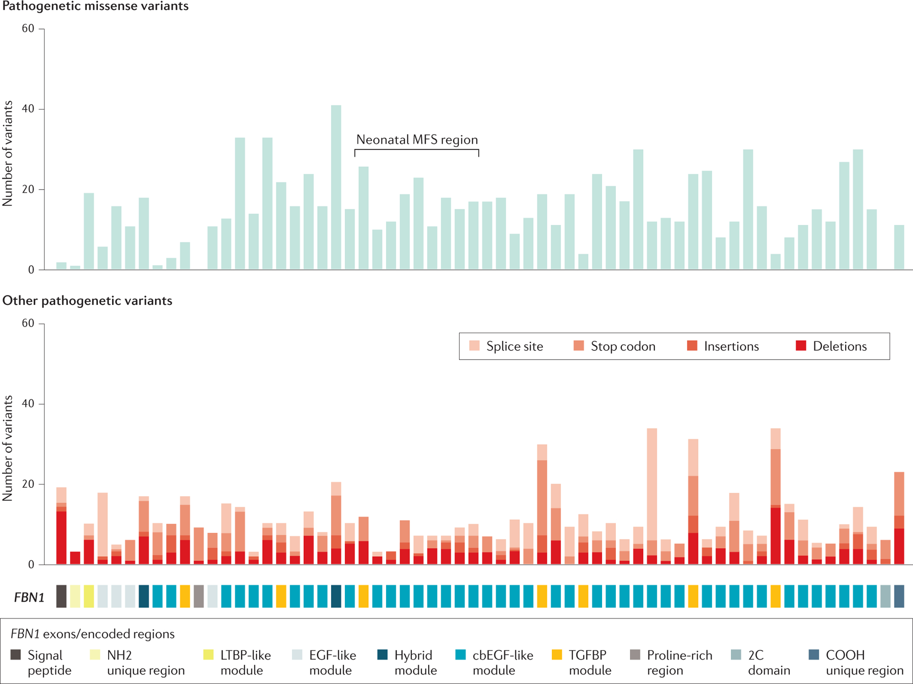
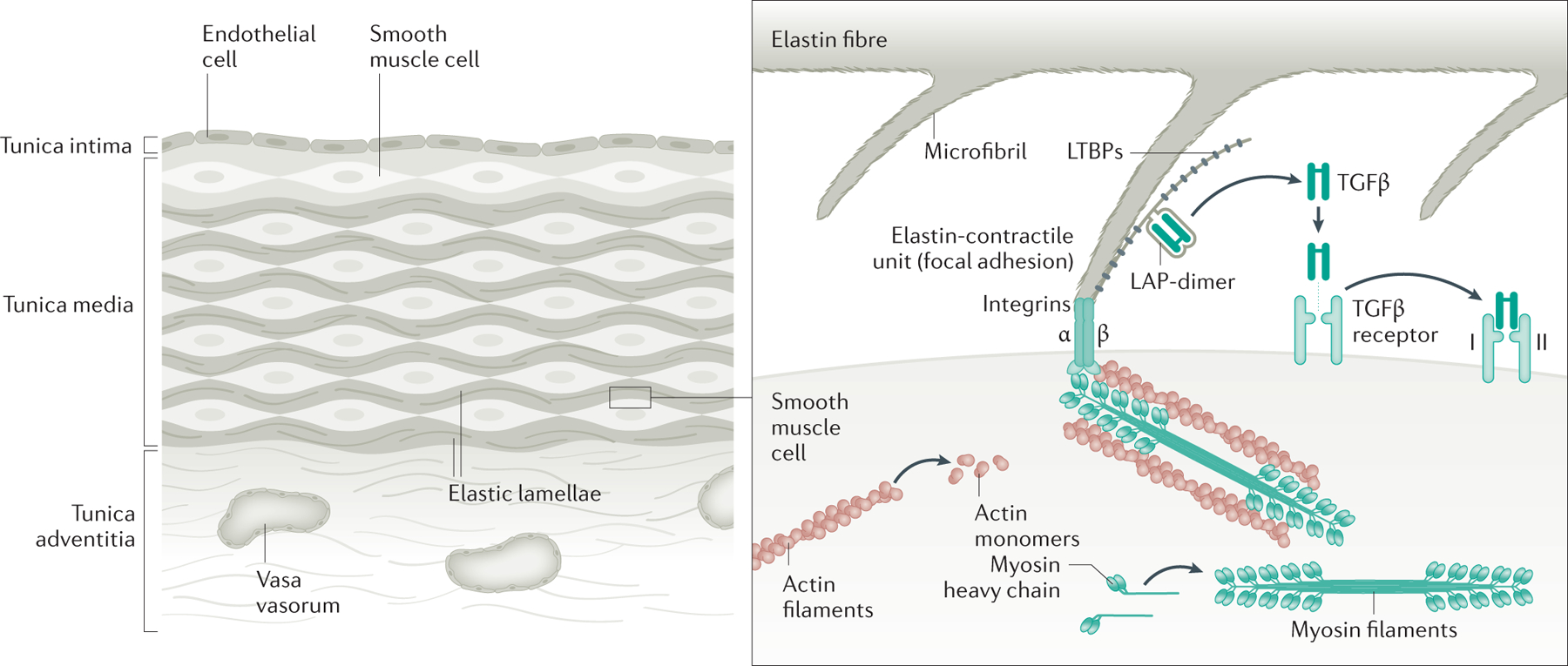
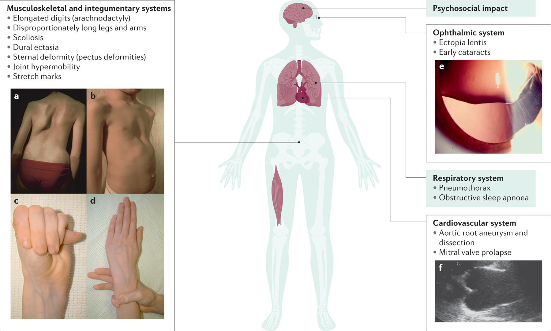
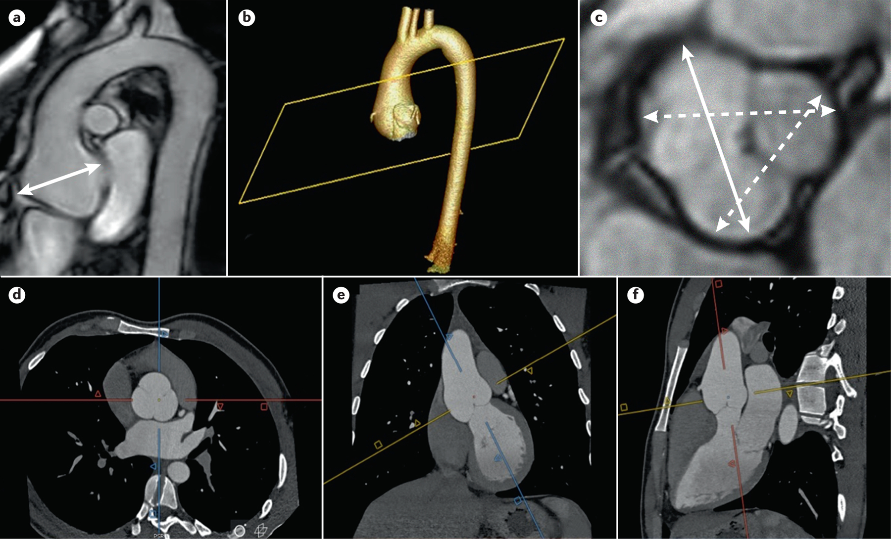
Marfan syndrome (MFS) is an autosomal dominant, age-related but highly penetrant condition with substantial intrafamilial and interfamilial variability. MFS is caused by pathogenetic variants in FBN1, which encodes fibrillin-1, a major structural component of the extracellular matrix that provides support to connective tissues, particularly in arteries, the pericondrium and structures in the eye. Up to 25% of individuals with MFS have de novo variants. The most prominent manifestations of MFS are asymptomatic aortic root aneurysms, aortic dissections, dislocation of the ocular lens (ectopia lentis) and skeletal abnormalities that are characterized by overgrowth of the long bones. MFS is diagnosed based on the Ghent II nosology; genetic testing confirming the presence of a FBN1 pathogenetic variant is not always required for diagnosis but can help distinguish MFS from other heritable thoracic aortic disease syndromes that can present with skeletal features similar to those in MFS. Untreated aortic root aneurysms can progress to life-threatening acute aortic dissections. Management of MFS requires medical therapy to slow the rate of growth of aneurysms and decrease the risk of dissection. Routine surveillance with imaging techniques such as transthoracic echocardiography, CT or MRI is necessary to monitor aneurysm growth and determine when to perform prophylactic repair surgery to prevent an acute aortic dissection.


Conflict of interest statement

Competing interest

The authors declare no competing interests

Figures

Figure 1.
Figure 1.. Aortic root aneurysm and acute aortic dissections in patients with MFS.
A. Illustration of the anatomy of the aorta, with a photo of an aortic root aneurysm. B, C. Patients with MFS present with aortic root aneurysms, which predispose to type A aortic dissections. D. Type B aortic dissection are also part of the disease spectrum. [CE: credit lines needed]
Figure 2.
Figure 2.. Pathogenetic variants in FBN1.
Schematic of the structure of the protein fibrillin-1, with numbers and location of mutations in FBN1, which encodes fibrillin-1, identified in patients with MFS; the location of de novo pathogenetic variants leading to neonatal MFS is also shown. Mutation data were extracted in 2020 from the last update of the UMD-FBN1 database (Ref 289)
Figure 3.
Figure 3.. Role of fibrillin-1 in the SMCs.
The human aorta is composed of over 50 layers of elastic lamellae and SMCs (note that for simplicity only a few layers are shown). Elastin fibers have oblique extensions that have microfibrils at the tips, which connect to focal adhesions on the SMC cell surface, and then to SMC contractile units; these structures are called elastin-contractile units. Fibrillin-1 is the major protein in the connecting microfibrils. Genes that are altered to predispose to heritable thoracic aortic disease disrupt major proteins in the elastin-contractile unit. Research has focused on the role of excessive TGF-β and angiotensin II signalling as drivers of thoracic aortic disease. More recent research has indicated that loss of TGF-β signalling is a primary driver of thoracic aortic disease, but increased signaling may have a role in late stages of the disease process.
Figure 4.
Figure 4.. Clinical manifestations of the Marfan syndrome.
The major manifestations of MFS are illustrated. A. Scoliosis or curvature of the spine. B. Chest wall deformities, such as pectus excavatum. Arachnodactyly as evident by a positive thumb (C) and wrist (D) signs. E. Ectopia lentis. F. Aortic root aneurysm as seen by transthoracic echocardiography in parasternal long-axis view. Panels A and B adapted from https://www.elsevier.com/books/emery-and-rimoin-s-principles-and-practice-of-medical-genetics-and-genomics/pyeritz/978-0-12-812537-3; panels C and D adapted from https://www.nature.com/articles/5201851; [CE: credit lines needed]
Figure 5.
Figure 5.. Imaging for thoracic aortic disease in Individuals with MFS.
Imaging for thoracic aortic disease in MFS patients. A) MRI of the thoracic aorta shows an aortic root aneurysm (double arrow). 3D reconstruction (b) of CTA imaging (d, e, and f) of an aortic root aneurysm. The methodology of acquiring double oblique aortic images using the sagittal and coronal images to achieve perpendicularity to the aortic flow results in a corrected true transversal image of the aortic lumen; C) measurement of aortic root aneurysm by MRI using cusp to cusp diameters at end-diastole. Solid double arrow line shows the maximum aortic root diameter.

References

    1. Hollister DW, Godfrey M, Sakai LY & Pyeritz RE Immunohistologic abnormalities of the microfibrillar-fiber system in the Marfan syndrome. N. Engl. J. Med 323, 152–159 (1990). - PubMed
    1. Sakai LY, Keene DR & Engvall E Fibrillin, a new 350-kD glycoprotein, is a component of extracellular microfibrils. J. Cell Biol 103, 2499–2509 (1986). - PMC - PubMed
    1. Dietz HC et al. Marfan syndrome caused by a recurrent de novo missense mutation in the fibrillin gene. Nature 352, 337–339 (1991 - PubMed
    1. Milewicz DM, Pyeritz RE, Crawford ES & Byers PH Marfan syndrome: defective synthesis, secretion, and extracellular matrix formation of fibrillin by cultured dermal fibroblasts. J. Clin. Invest 89, 79–86 (1992). - PMC - PubMed
    1. Chiu HH, Wu MH, Chen HC, Kao FY & Huang SK Epidemiological profile of Marfan syndrome in a general population: a national database study. Mayo Clin Proc 89, 34–42, doi:10.1016/j.mayocp.2013.08.022 (2014). - DOI - PubMed

Publication types