. 2021 Sep 2;4(1):1028.
doi: 10.1038/s42003-021-02506-2.

Postnatal development in a marsupial model, the fat-tailed dunnart (Sminthopsis crassicaudata; Dasyuromorphia: Dasyuridae)

Affiliations

Postnatal development in a marsupial model, the fat-tailed dunnart (Sminthopsis crassicaudata; Dasyuromorphia: Dasyuridae)

Laura E Cook et al. Commun Biol. .

Abstract

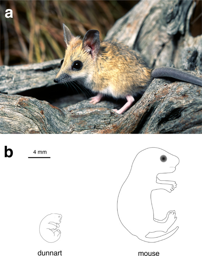
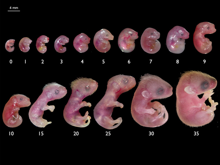
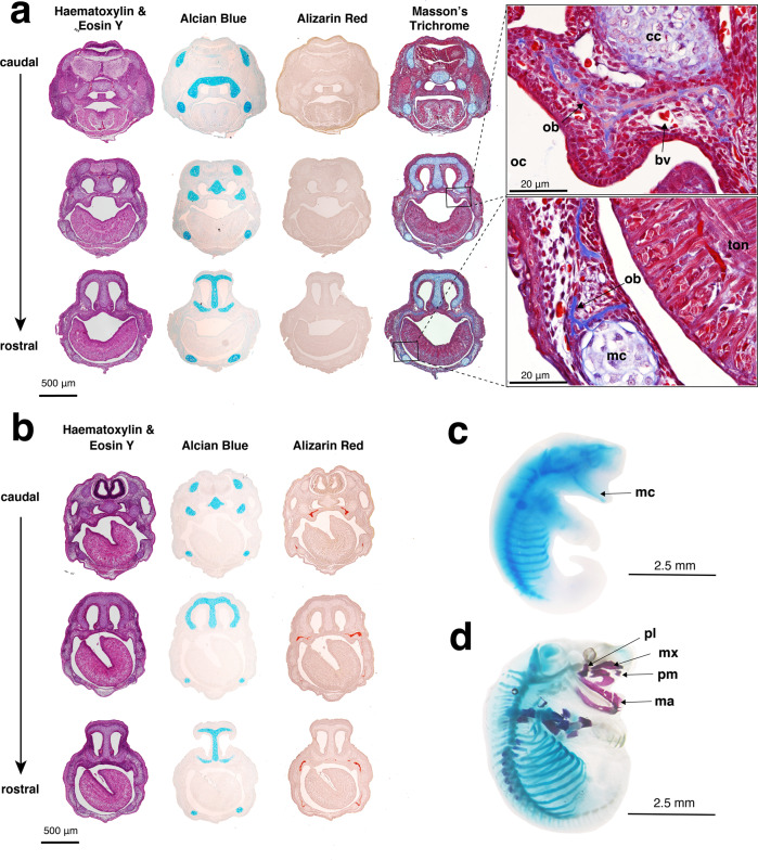
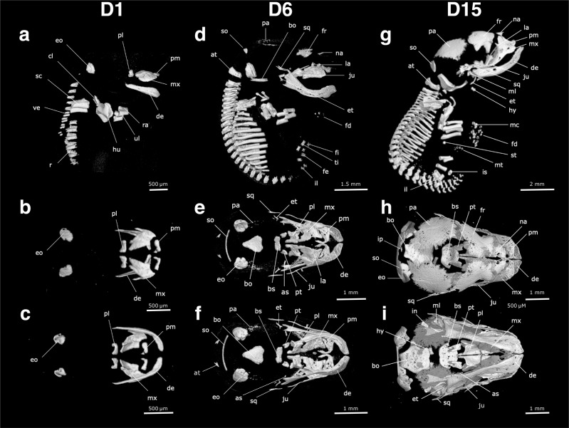
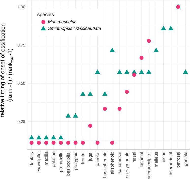
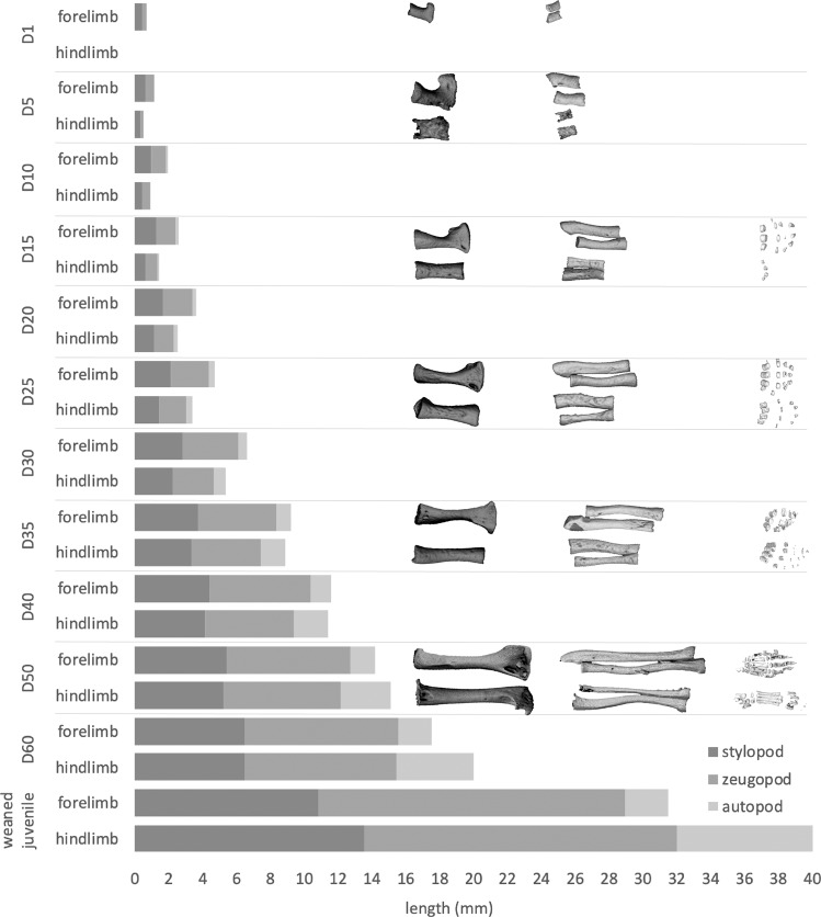
Marsupials exhibit unique biological features that provide fascinating insights into many aspects of mammalian development. These include their distinctive mode of reproduction, altricial stage at birth, and the associated heterochrony that is required for their crawl to the pouch and teat attachment. Marsupials are also an invaluable resource for mammalian comparative biology, forming a distinct lineage from the extant placental and egg-laying monotreme mammals. Despite their unique biology, marsupial resources are lagging behind those available for placentals. The fat-tailed dunnart (Sminthopsis crassicaudata) is a laboratory based marsupial model, with simple and robust husbandry requirements and a short reproductive cycle making it amenable to experimental manipulations. Here we present a detailed staging series for the fat-tailed dunnart, focusing on their accelerated development of the forelimbs and jaws. This study provides the first skeletal developmental series on S. crassicaudata and provides a fundamental resource for future studies exploring mammalian diversification, development and evolution.


Conflict of interest statement

The authors declare no competing interests.

Figures

Fig. 1
Fig. 1. At birth the fat-tailed dunnart represents one of the most altricial mammals in existence.
a Adult fat-tailed dunnart (S. crassicaudata). Image: Alan Henderson—Minibeast Wildlife. b Schematic comparing extent of development of the fat-tailed dunnart and mouse (M. musculus) neonates on the day of birth.
Fig. 2
Fig. 2. Gross morphology of the fat-tailed dunnart throughout postnatal development in the pouch.
Numbers below pouch young refer to the day postpartum.
Fig. 3
Fig. 3. Histological analyses were used to determine ossification onset in D0 and D1 fat-tailed dunnart pouch young.
a D0 head sections stained with haematoxylin and eosin Y (purple = nuclei, pink = cytoplasm), alcian blue (cartilage), alizarin red (mineralised bone) and Masson’s trichrome (blue = collagen, red = muscles, cytoplasm, and keratin). Representative images of D0 at higher magnification (×60) show ossification had begun with collagen present in the maxillary and mandible prominences. b D1 head sections stained with haematoxylin and eosin Y (purple = nuclei, pink = cytoplasm), alcian blue (cartilage), and alizarin red (mineralised bone). Wholemount, c D0 and d D1 fat-tailed dunnart specimens cleared and stained with alcian blue (cartilage) and alizarin red (bone). bv = blood vessel, cc = cartilago cupularis, ma = mandible, mc = Merkel’s cartilage, mx = maxillary, ob = osteoblast, oc = oral cavity, pl = palatine process, ton = tongue.
Fig. 4
Fig. 4. The skeleton of the fat-tailed dunnart (D1, D6 and D15), as revealed in microCT scans of pouch young.
The right lateral side of the skeleton is shown in (a), (d) and (g), the dorsal view of the skull in (b), (e) and (h), and the ventral view of the skull in (c), (f) and (i). Pouch young one day after birth (D1) are shown in (a), (b) and (c). Pouch young six days (D6) after birth are shown in (d), (e) and (f). Pouch young 15 days after birth (D15) are shown in (g), (h) and (i). as = alisphenoid, at = atlas, bo = basioccipital, bs = basisphenoid, cl = clavicle, de = dentary, eo = exooccipital, et = ectotympanic, fd = forelimb digits, fe = femur, fi = fibula, fr = frontal, hu = humerus, hy = hyoid, in = incus, il = ilium, ip = interparietal, is = ischium, ju = jugal, la = acrimal, mc = metacarpals, ml = malleus, mt = metatarsals, mx = maxilla, na = nasal, pa = parietal, pl = palatine process, pm = premaxilla, pt = pterygoid, ra = radius, ri = ribs, sc = scapula, so = supraoccipital, sq = squamosal, st = sternum, ti = tibia, ul = ulna, ve = vertebrae.
Fig. 5
Fig. 5. Relative timing of onset of ossification for the bones of the skull in the fat-tailed dunnart (S. crassicaudata) and mouse (M. musculus).
Specimens were ranked in order of bone onset or bone contact timing and relative ranks were normalised for comparison between species. M. musculus is shown with pink circles and S. crassicaudata is shown with green triangles, highlighting the early onset of ossification of the oral region in both species.
Fig. 6
Fig. 6. The secondary jaw joint as revealed in microCT scans in D20 fat-tailed dunnart pouch young.
a Ventral view of the secondary jaw joint with inset white box showing entire skull. b Posterior to anterior view of the secondary jaw joint. c = condyle (orange), et = ectotympanic ring (purple), gf = glenoid fossa (light green), in = incus (yellow), ml = malleus (blue) and sq = squamosal (dark green). Scale bar = 1 mm.
Fig. 7
Fig. 7. The skeleton of fat-tailed dunnart pouch young (D35, D50 and D70), as revealed in microCT scans.
The right lateral side of the skeleton is shown in (a), (d) and (g), the dorsal view of the skull in (b), (e) and (h), and the ventral view of the skull in (c), (f) and (i). Pouch young on D35 are shown in (a), (b) and (c). Pouch young on D50 are shown in (d), (e) and (f). Pouch young on D70 are shown in (g), (h) and (i).
Fig. 8
Fig. 8. Limb heterochrony in the fat-tailed dunnart.
Relative lengths of forelimb and hindlimb elements throughout development, shown as shaded grey bars (n = 1). Limbs are divided into proximal and distal segments: the stylopod (humerus and femur), zeugopod (ulna/radius and tibia/fibula) and autopod (carpals, metacarpals, tarsal, metatarsals and digits).

References

    1. Bininda-Emonds OR, et al. The delayed rise of present-day mammals. Nature. 2007;446:507–12. doi: 10.1038/nature05634. - DOI - PubMed
    1. Luo Z-X, Yuan C-X, Meng Q-J, Ji Q. A Jurassic eutherian mammal and divergence of marsupials and placentals. Nature. 2011;476:442–445. doi: 10.1038/nature10291. - DOI - PubMed
    1. Renfree MB. Review: Marsupials: placental mammals with a difference. Placenta. 2010;31 Suppl:S21–6. doi: 10.1016/j.placenta.2009.12.023. - DOI - PubMed
    1. Tyndale-Biscoe H, Renfree M. Reproductive physiology of marsupials. Cambridge: Cambridge University Press; 1987.
    1. Tyndale-Biscoe CH. Life of Marsupials. Collingwood, Victoria: CSIRO Publishing; 2005.

Publication types