

Endodontic management of a maxillary second molar with three roots and seven canals using cone-beam computed tomography
- PMID: 34475690
- PMCID: PMC8378491
- DOI: 10.4103/jcd.jcd_652_20
Endodontic management of a maxillary second molar with three roots and seven canals using cone-beam computed tomography
Abstract
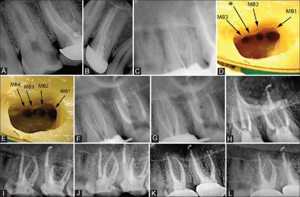
The present case highlights the endodontic management of a maxillary second molar with three roots and seven canals. Root canal treatment was performed for the maxillary second molar diagnosed with symptomatic irreversible pulpitis. During the procedure under magnification, extra canals were detected in the mesiobuccal root. Cone-beam computed tomography (CBCT) evaluation confirmed four canals in the mesiobuccal root with Vertucci's Type XXI (4-1) pattern. The distobuccal root exhibited two canals with Vertucci's Type III (1-2-1) configuration. The palatal canal was single and large. A 4 year follow-up revealed satisfactory clinical and radiographic findings. Magnification and CBCT allow us to explore possible anatomic variations with insights to tackle such situations clinically.
Keywords: Cone beam computed tomography; magnification; maxillary second molar; seven canal system.
Copyright: © 2021 Journal of Conservative Dentistry.
Conflict of interest statement
There are no conflicts of interest.
Figures

References
-
- Kottoor J, Velmurugan N, Surendran S. Endodontic management of a maxillary first molar with eight root canal systems evaluated using cone-beam computed tomography scanning: A case report. J Endod. 2011;37:715–9. - PubMed
-
- Kim JR, Choi SB, Park SH. A maxillary second molar with 6 canals: A case report. Quintessence Int. 2008;39:61–4. - PubMed
-
- Pasternak Júnior B, Teixeira CS, Silva RG, Vansan LP, Sousa Neto MD. Treatment of a second maxillary molar with six canals. Aust Endod J. 2007;33:42–5. - PubMed
-
- Chawla A, Sujlana A, Dixit A. Re-treating a maxillary second molar with 6 root canals assisted by cone beam computed tomography. Gen Dent. 2015;63:e14–6. - PubMed
Publication types
LinkOut - more resources
Full Text Sources