

Etiology of Hydronephrosis in adults and children: Ultrasonographic Assessment in 233 patients
- PMID: 34475906
- PMCID: PMC8377938
- DOI: 10.12669/pjms.37.5.3951
Etiology of Hydronephrosis in adults and children: Ultrasonographic Assessment in 233 patients
Abstract
Objectives: Hydronephrosis (HN) is dilatation of the collecting system of the kidney due to obstruction of urine outflow. This study intended firstly, to investigate the efficacy of ultrasound (US) imaging to determine the cause of HN, and secondly, to list the causes of HN.
Methods: In this retrospective study, 233 patients with HN were scanned to determine the cause of the HN in the period from 1st January 2016 to 31st October 2017. Categorical results were written as frequencies and percentages.
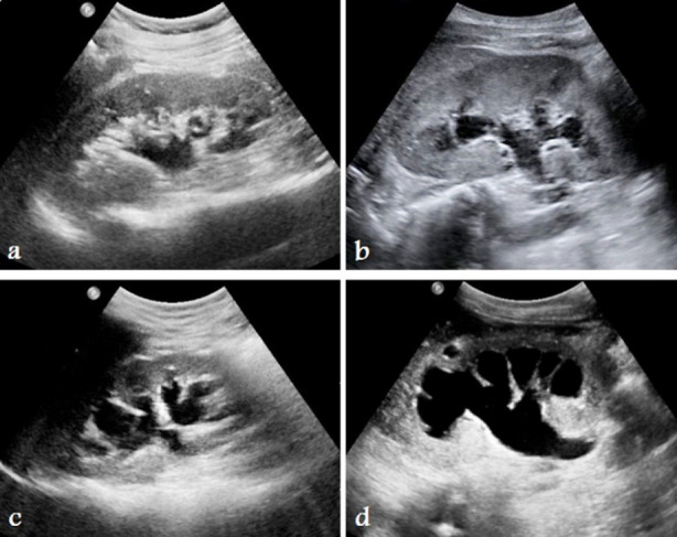
Results: Out of 233, 91.41% were adults and 8.58% were children (P<0.001), 66.10% were male and 33.90% were female (P<0.001). In 55.36%, HN was in the right kidney and 44.64% was in the left (P=0.116). Exactly 58% of patients were suffering from grade-2, 21.5% grade-3, 11.6% grade-1, and 8.2% grade-4 HN. US imaging can determine the cause of HN in 70.4% of patients. Kidney or ureteric calculi were the cause of HN in 54.1% of cases, reflux was in 7.3%, and pelviureteric junction (PUJ) stenosis was in 3.9%.In cases of calculi induced HN, 25.3% of the calculi were in the vesicoureteric (VUJ) junction, 21.5% were in the renal pelvis, 6.4% were in the PUJ or upper ureter, and only 0.9% were in the middle ureter.
Conclusion: Ultrasound imaging can determine the cause of HN in more than two thirds of patients. Calculi are the most common cause of HN even in children and are most common in the VUJ junction.
Keywords: Calculi; Etiology; Hydronephrosis; Pregnancy-induced hydronephrosis; Ultrasound imaging.
Copyright: © Pakistan Journal of Medical Sciences.
Conflict of interest statement
Conflicts of interest: The authors declare no conflicts of interest.
Figures
References
LinkOut - more resources
Full Text Sources