. 2021 Oct;32(19-20):1186-1199.
doi: 10.1089/hum.2021.142.

Investigating the Barrier Activity of Novel, Human Enhancer-Blocking Chromatin Insulators for Hematopoietic Stem Cell Gene Therapy

Affiliations

Investigating the Barrier Activity of Novel, Human Enhancer-Blocking Chromatin Insulators for Hematopoietic Stem Cell Gene Therapy

Penelope-Georgia Papayanni et al. Hum Gene Ther. 2021 Oct.

Abstract

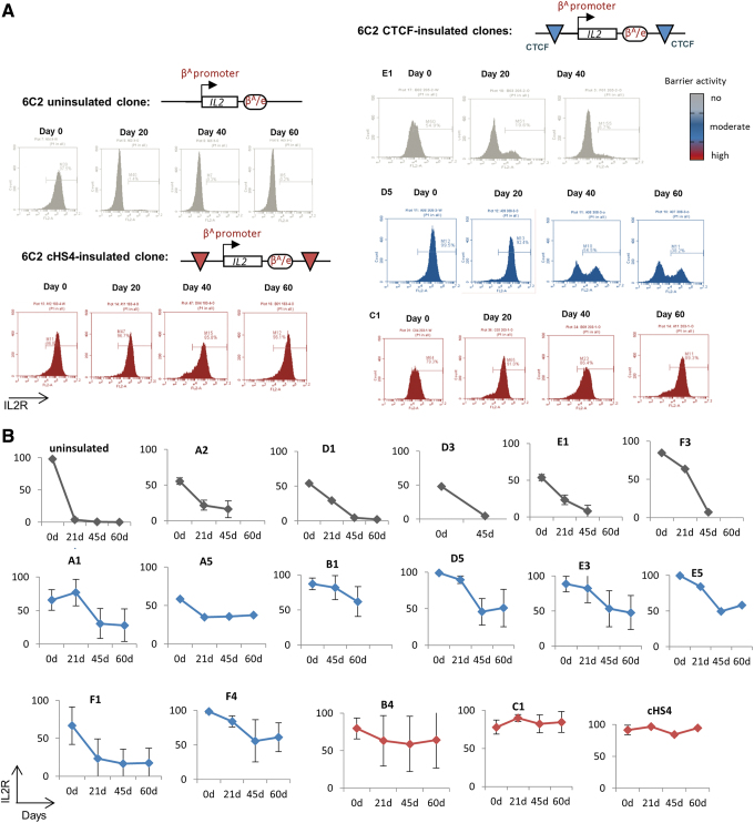
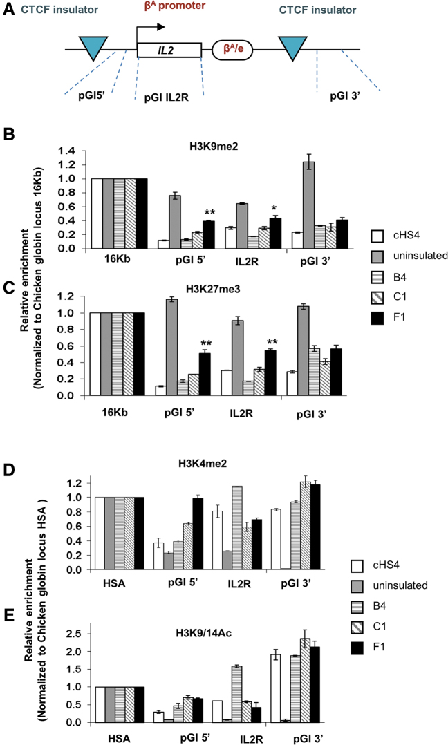
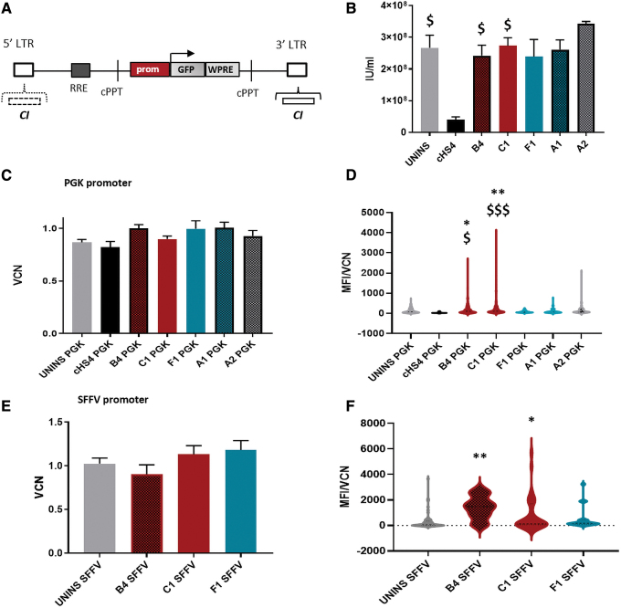
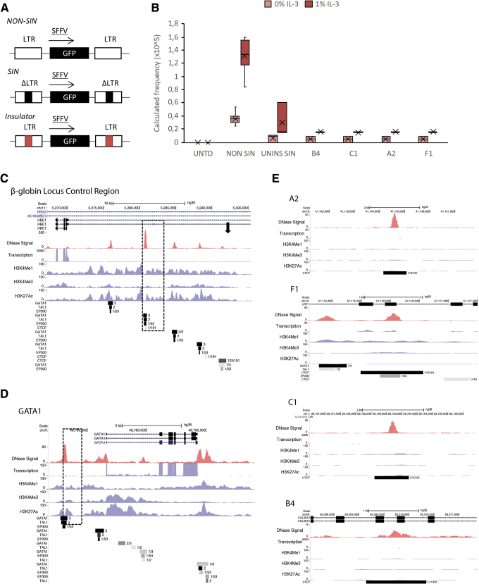
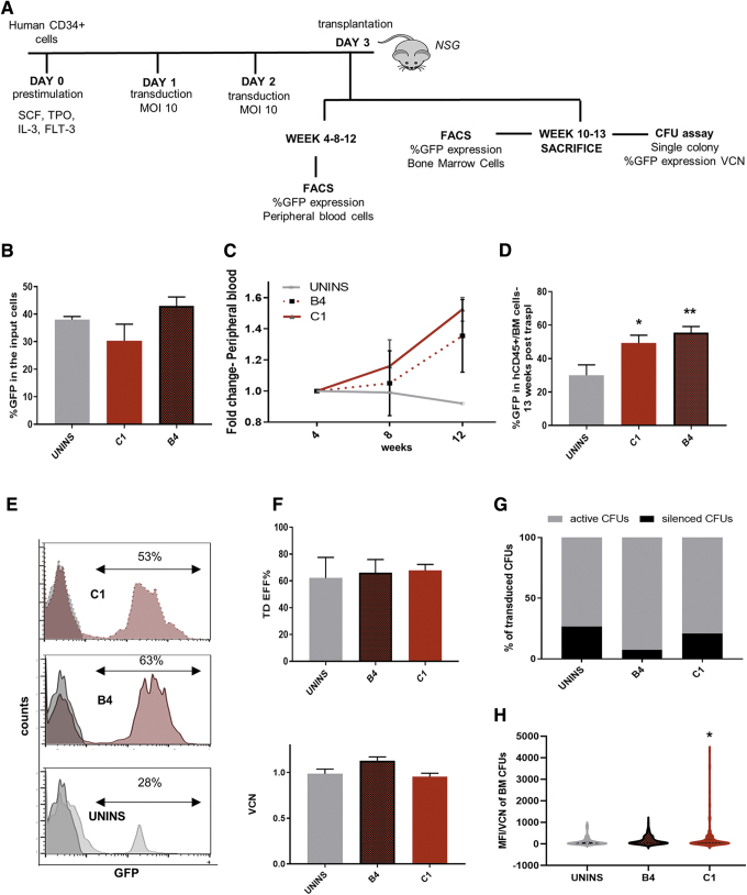
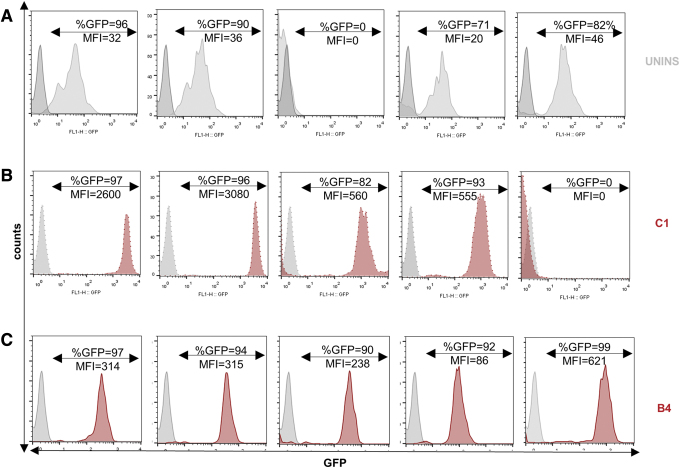
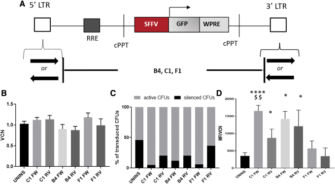
Despite the unequivocal success of hematopoietic stem and progenitor cell gene therapy, limitations still exist including genotoxicity and variegation/silencing of transgene expression. A class of DNA regulatory elements known as chromatin insulators (CIs) can mitigate both vector transcriptional silencing (barrier CIs) and vector-induced genotoxicity (enhancer-blocking CIs) and have been proposed as genetic modulators to minimize unwanted vector/genome interactions. Recently, a number of human, small-sized, and compact CIs bearing strong enhancer-blocking activity were identified. To ultimately uncover an ideal CI with a dual, enhancer-blocking and barrier activity, we interrogated these elements in vitro and in vivo. After initial screening of a series of these enhancer-blocking insulators for potential barrier activity, we identified three distinct categories with no, partial, or full protection against transgene silencing. Subsequently, the two CIs with full barrier activity (B4 and C1) were tested for their ability to protect against position effects in primary cells, after incorporation into lentiviral vectors (LVs) and transduction of human CD34+ cells. B4 and C1 did not adversely affect vector titers due to their small size, while they performed as strong barrier insulators in CD34+ cells, both in vitro and in vivo, shielding transgene's long-term expression, more robustly when placed in the forward orientation. Overall, the incorporation of these dual-functioning elements into therapeutic viral vectors will potentially provide a new generation of safer and more efficient LVs for all hematopoietic stem cell gene therapy applications.

Keywords: CTCF-binding site; barrier activity; chromatin insulator; gene therapy; hematopoietic stem cells.


Conflict of interest statement

The authors declare that they have no competing interests relevant to the subject matter of this article.

Figures

Figure 1.
Figure 1.
In vitro evaluation of barrier activity of enhancer-blocking elements in 6C2 cell line. (A) Vector design and FACS analysis of transgenic IL2R expression. Representative FACS plots of selected clones transduced with uninsulated or cHS4-, E1-, D5-, C1-insulated vectors on days 0, 20, 40, 60. (B) Kinetic expression patterns of IL2R transgene that is uninsulated or insulated by novel CIs. A2, D1, D3, E1, F3, A1, A5, B1, D5, E3, E5, F1, F4, B4, C1, and the cHS4 insulator. CIs, chromatin insulators; FACS, fluorescence activated cell sorting. Color images are available online.
Figure 2.
Figure 2.
The barrier effect in post-translational histone modification. (A) Schematic representation of the insulated IL2R transgene and the positions of PCR primer sets used in ChIP assays. (B–E) ChIP assays of the flanking insulator sites and the integrated IL2R transgenes. The relative enrichment of precipitated DNA fragments using antibodies specific to H3K4me2 (B), H3K27me3 (C), H3K9me2 (D), and H3K9/14ac (E) was normalized to the enrichment of the endogenous HSA or 16 kb condensed chromatin fragment to adjust for immunoprecipitation efficiency. *p < 0.05; **p < 0.01 by Student's t-test. Shown are the mean ± SEM of three experiments. PCR, polymerase chain reaction; SEM, standard error of the mean; HSA, A tissue-specific hypersensitive site. Color images are available online.
Figure 3.
Figure 3.
In vitro evaluation of barrier activity of enhancer-blocking elements, incorporated in lentiviral vectors, after transduction of normal CD34+ cells. (Α) Vector structures in their proviral form. The lentiviral reporter vectors, used for this study, were produced with the SIN vector design, carrying the GFP gene, the WPRE element, an SFFV, or PGK promoter, with or without a candidate CI. (B) Vector titers. The novel CIs do not decrease vector titers (IU/mL), compared with the uninsulated parental vector. (C) VCN in transduced single colonies, using insulated and uninsulated vectors, carrying the PGK promoter. (D) Normalization of MFI per VCN in transduced single colonies, using insulated and uninsulated vectors, carrying the PGK promoter. (E) VCN in transduced single colonies, using insulated and uninsulated vectors, carrying the SFFV promoter. (F) Normalization of MFI per VCN in transduced single colonies, using insulated and uninsulated vectors, carrying the SFFV promoter. Dollars; the comparisons between cHS4-insulated and novel-insulated vectors; asterisks; the comparison between uninsulated parental vector and novel-insulated vectors. *p ≤ 0.05, **p ≤ 0.001, ***p ≤ 0.0001, $p ≤ 0.05, $$$p ≤ 0.0001. cPPT, central polypyrimidine tract; GFP, green fluorescent protein; LTR, long terminal repeat; MFI, mean fluorescence intensity; PGK, phosphoglycerate kinase; prom, promoter; RRE, REV responsive element; SFFV, spleen focus-forming virus; SIN, self-inactivating; VCN, vector copy number; WPRE, woodchuck post-transcriptional regulatory element. Color images are available online.
Figure 4.
Figure 4.
Evaluation of potential enhancer activity of the novel insulators. (A) All insulators were evaluated for a potential enhancer activity by assessing their ability to promote oncogenesis in 32D cells. Vector design: NON SIN positive, SIN negative control, and insulated SIN vectors carrying GFP transgene and SFFV promoter, SIN, GFP, SFFV with LTR. (B) Frequency of IL-3-independent colonies in 32D experiments. (C–E) Screening for enhancer marks in the candidate insulators through the ENCODE database. DNase hypersensitivity, transcription, as well as ChIP-Seq for H3K4Me1, H3K4Me3, H3K27ac, EP300, GATA1, TAL1, and CTCF tracks are shown for (C) the beta globin LCR, (D) the GATA-1 enhancer, (E) four candidate barrier insulators in K562 cells. ChIP-Seq, chromatin immunoprecipitation combined with DNA sequencing; LCR, locus control region. Color images are available online.
Figure 5.
Figure 5.
In vivo evaluation of barrier activity of C1 and B4 enhancer blocker CIs. (Α) Schema of in vivo analyses: normal CD34+ cells were transduced with uninsulated and insulated vectors, with MOI 20. Peripheral blood cells were evaluated, monthly. Post-transduction, %GFP was measured in hCD45+ cells, and CFUs of BM cells were evaluated for %GFP, MFI, VCN. (Β) %GFP of input cells, on day 3. (C) Fold change of hCD45+/GFP+ cells of peripheral blood cells in xenografts 3 months post-transplantation. (D) %GFP expression in human CD45+ cells of BM cells, during the sacrifice of the mice, 10–13 weeks after cell transplantation. (E) Representative flow cytometric plots of transduced BM cells with similar VCNs. (F) TD EFF% and VCN in single colonies derived from the human CD45+ cells of the chimeric BM, after immunomagnetic purification of human cells and in vitro culture in methylcellulose. (G) Silencing in transduced colonies. Silenced CFUs; ≤5% GFP expression, active CFUs; >5% GFP expression in colonies with VCN 0.4–2.2. (H) Normalization of (MFI) per (VCN) in transduced single colonies derived from the human CD45+ cells of the chimeric BM, after immunomagnetic purification of human cells and in vitro culture in methylcellulose. *p ≤ 0.05, **p ≤ 0.001. CFU, colony-forming unit; BM, bone marrow; MOI, multiplicity of infection; TD EFF%, % transduction efficiency. Color images are available online.
Figure 6.
Figure 6.
Flow cytometric analysis of transduced single colonies derived from the human CD45+ cells of the chimeric BM. Human CD45+ cells were culture in methylcellulose medium after immunomagnetic purification of human cells. Single colonies were subsequently analyzed for expression of GFP by flow cytometry. The percentage of GFP+ cells is reported above the indicated gates, and was calculated by subtracting the background from the untransduced controls. (A) CFUs derived from untransduced cells, (B) CFUs derived from C1-transdused cells, (C) CFUs derived from B4-transduced cells. Color images are available online.
Figure 7.
Figure 7.
The effect of CTCF-binding site orientation in barrier activity of CIs. (A) Vector structures in their proviral form. The lentiviral reporter vectors, used for this study, were produced with the SIN vector design, carrying the GFP gene, the WPRE region, the SFFV promoter, with or without CI, either in reverse or in forward orientation. (B) VCN in transduced single colonies, using insulated, with CIs in different orientations (FW or RV), and uninsulated vectors. (C) Silencing in transduced colonies. Silenced CFUs; ≤5% GFP expression, active CFUs; >5% GFP expression in colonies with VCN 0.4–2.2. (D) Normalization of MFI per VCN in transduced single colonies, using insulated, with CIs in different orientations (FW or RV), and uninsulated vectors, asterisks; the comparisons between insulated and uninsulated parental vectors; dollars; the comparison between C1 FW-C1 RV insulated vectors. *p ≤ 0.05, **p ≤ 0.001, ****p ≤ 0.00001, $$p ≤ 0.001. FW, forward; RV, reverse. Color images are available online.

Similar articles

Cited by

References

    1. Cavazzana-Calvo M, Hacein-Bey S, de Saint Basile G, et al. . Gene therapy of human severe combined immunodeficiency (SCID)-X1 disease. Science 2000;288:669–672. - PubMed
    1. Wu X, Li Y, Crise B, et al. . Transcription start regions in the human genome are favored targets for MLV integration. Science 2003;300:1749–1751. - PubMed
    1. Schröder ARW, Shinn P, Chen H, et al. . HIV-1 integration in the human genome favors active genes and local hotspots. Cell 2002;110:521–529. - PubMed
    1. Montini E, Cesana D, Schmidt M, et al. . The genotoxic potential of retroviral vectors is strongly modulated by vector design and integration site selection in a mouse model of HSC gene therapy. J Clin Invest 2009;119:964–975. - PMC - PubMed
    1. Cesana D, Ranzani M, Volpin M, et al. . Uncovering and dissecting the genotoxicity of self-inactivating lentiviral vectors in vivo. Mol Ther 2014;22:774–785. - PMC - PubMed

Publication types

LinkOut - more resources