. 2021 Oct;25(20):9567-9585.
doi: 10.1111/jcmm.16898. Epub 2021 Sep 3.

A novel rat model of vertebral inflammation-induced intervertebral disc degeneration mediated by activating cGAS/STING molecular pathway

Affiliations

A novel rat model of vertebral inflammation-induced intervertebral disc degeneration mediated by activating cGAS/STING molecular pathway

Qihang Su et al. J Cell Mol Med. 2021 Oct.

Abstract

In this study, we describe a new rat model of vertebral inflammation-induced caudal intervertebral disc degeneration (VI-IVDD), in which IVD structure was not damaged and controllable segment and speed degeneration was achieved. VI-IVDD model was obtained by placing lipopolysaccharide (LPS) in the caudal vertebral bodies of rats. Rat experimental groups were set as follows: normal control group, group with a hole drilled in the middle of vertebral body and not filled with LPS (Blank group), group with a hole drilled in the middle of vertebral body and filled with LPS (Mid group), and group with hole drilled in the vertebral body in proximity of IVD and filled with LPS (NIVD group). Radiological results of VI-IVDD rats showed a significant reduction in the intervertebral space height and decrease in MRI T2 signal intensity. Histological stainings also revealed that the more the nucleus pulposus and endplate degenerated, the more the annulus fibrosus structure appeared disorganized. Immunohistochemistry analysis demonstrated that the expression of Aggrecan and collagen-II decreased, whereas that of MMP-3 increased in Mid and NIVD groups. Abundant local production of pro-inflammatory cytokines was detected together with increased infiltration of M1 macrophages in Mid and NIVD groups. Apoptosis ratio remarkably enhanced in Mid and NIVD groups. Interestingly, we found a strong activation of the cyclic GMP-AMP synthase /stimulator of interferon gene signalling pathway, which is strictly related to inflammatory and degenerative diseases. In this study, we generated a new, reliable and reproducible IVDD rat model, in which controllable segment and speed degeneration was achieved.

Keywords: animal model; cGAS/STING signalling pathway; intervertebral disc degeneration; spread of inflammation; vertebral inflammation-induced intervertebral disc degeneration model.


Conflict of interest statement

The authors declared no potential conflicts of interest with respect to the research, authorship and/or publication of this article.

Figures

FIGURE 1
FIGURE 1
Intervertebral disc degenerationmodel (IVDD) establishment. (A) Anatomy of rat tail; (B) experimental groups included the normal control group (Normal group), group with hole drilled in the middle of vertebral body without placing lipopolysaccharide (LPS) (Blank group), group with hole drilled in the middle of vertebral body and treated with LPS (Mid group, also called VI‐IVDD basic model), and group with hole drilled at one side (in proximity of IVD) of the vertebral body and treated with LPS (NIVD group, also called VI‐IVDD acceleration model); (C) experimental procedure included the following steps: (I) localization of vertebral body, IVD and vessel; (II) dissection and localization of subcutaneous tissue and caudal ligament; (III) identification of drilling position, and execution of drilled holes in vertebral bodies with a drill bit (diameter: 1.5 mm), inserted with a 45‐degree inclination; and (IV) in Mid and NIVD groups, LPS was placed and sealed with bone wax, whereas in the Blank group, drilled hole was sealed with bone wax without LPS. After surgical intervention, skin was sutured and disinfected
FIGURE 2
FIGURE 2
Radiological evaluation. (A) Intervertebral space height and the position of drilled holes were evaluated by molybdenum‐target plain X‐ray image at 1, 2 and 4 weeks for each group. The intervertebral space height decreased in Mid and NIVD groups; (B) statistical analysis relative to the quantitative index of intervertebral space height (DHI); (C) MRI images at 1, 2 and 4 weeks for each group. Preliminary evaluation of IVDD revealed that the T2 signal of IVD decreased in Mid and NIVD groups; (D) statistical analysis relative to the total greyscale value of MRI T2 signal of IVD
FIGURE 3
FIGURE 3
Histological stainings (4 weeks) revealed that the NP disappeared, the AF structure was disordered, the fibres were thickened, large numbers of inflammatory cells infiltrated, and the cartilage EP degenerated and disappeared in Mid and NIVD groups. (A) Haematoxylin‐eosin (HE) staining; (B) safranin O/fast green (SOFG) staining. AF, annulus fibrosus; Homolateral EP, homolateral endplate of the vertebral body; NP, nucleus pulposus. Black arrow indicates drilling location in the vertebral body
FIGURE 4
FIGURE 4
Masson's trichrome staining. (A) In Mid and NIVD groups, NP disappeared, AF structure was disordered, fibres were thickened, a large number of fresh collagen fibres were formed (blue), and cartilage EP degenerated and disappeared (4 weeks); (B) many immature fresh collagen fibres (blue) were formed in the degenerative IVD over time, and gradually matured (red) up to 4 weeks. NP: nucleus pulposus, AF, annulus fibrosus, and Homolateral EP, homolateral endplate of the vertebral body
FIGURE 5
FIGURE 5
IHC analysis of the IVD‐related markers Aggrecan, Collagen‐II and MMP‐3. (A) Aggrecan; (B) Collagen‐II; (C) MMP‐3; (D) statistical analysis relative to AOD showed that the expressions of Aggrecan and Collagen‐II significantly decreased in IVD or AF, but increased in EP in Mid and NIVD groups. MMP‐3 was highly expressed in Mid and NIVD groups. AF, annulus fibrosus; Homolateral EP: homolateral endplate of vertebral body; NP, nucleus pulposus. Contralateral EP: contralateral endplate of vertebral body. Black arrow indicates drilling location in the vertebral body
FIGURE 6
FIGURE 6
IHC analysis of IL‐1β, IL‐6 and TNF‐α inflammatory cytokines. (A) IL‐1β; (B) IL‐6; (C) TNF‐α; (D) statistical analysis relative to AOD showed that inflammatory marker expression in Mid and NIVD groups significantly increased. AF, annulus fibrosus; Homolateral EP: homolateral endplate of vertebral body; NP, nucleus pulposus. Contralateral EP: contralateral endplate of vertebral body
FIGURE 7
FIGURE 7
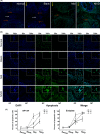
Infiltration of iNOS+and CD68+ M1 macrophages. (A) Double‐IF staining revealed a iNOS and CD68 macrophage infiltrate in Mid and NIVD groups; (B) The co‐location scatter diagram of iNOS (green) and CD68 (red) revealed that iNOS and CD68 co‐localized in Mid and NIVD groups (Rr: Pearson's correlation coefficient; R: overlap coefficient); (C) statistical analysis showed that M1 macrophages were significantly more represented in Mid and NIVD groups than in Normal and Blank groups at the indicated time‐points
FIGURE 8
FIGURE 8
Cell apoptosis was detected by TUNEL assay. (A) The overall pattern of IVD apoptotic cells (green) in each group showed that cell apoptosis was more prominent in Mid and NIVD groups (white arrow: NP, red arrow: cartilage EP, yellow arrow: homolateral to vertebral body); (B) Local apoptosis for each group. The inset panels (Magnified 700x) revealed that cell apoptosis significantly increased in Mid and NIVD groups (NP: nucleus pulposus, AF: annulus fibrosus, EP: endplate); (C) statistical analysis revealed that the number of apoptotic cells was significantly higher in Mid and NIVD groups than that in Normal and Blank groups (^^Normal Blank: p > 0.05, *Mid‐Normal/Blank: p > 0.05; **1–2 weeks (Mid): p > 0.05, ***2–4 weeks (NIVD): p > 0.05. Differences between other unmarked groups at different time‐points were significant, p < 0.05)
FIGURE 9
FIGURE 9
Inflammation signs during development of IVDD in the VI‐IVDD model. (A) Analyses of morphology (SOFG staining), IVD‐related marker (Aggrecan) and inflammatory marker (IL‐6) revealed that inflammation and development of IVDD were time‐dependent. (B) The diagram shows the process of inflammation and degeneration in detail. Early phase: inflammatory factors and cells infiltrate on the side of vertebral body, but no signs of IVDD are present. Mid phase: inflammation reaches IVD, NP degenerates and disappears, AF structure become disordered, and the homolateral EP is damaged. Mid‐late phase: IVDD further worsens and inflammation reaches the contralateral EP. Late phase: severe IVDD and severe damage to the contralateral EP. P‐stage is achieved, and the degeneration shows a stable trend. AF, annulus fibrosus; DEP, degenerative endplate; IF, inflammatory factors/ cells; LPS, lipopolysaccharide; NEP, normal nucleus pulposus; NP, nucleus pulposus
FIGURE 10
FIGURE 10
Activation of the cGAS/STING signalling pathway in the VI‐IVDD model. (A) Double‐IF staining showed high cGAS and STING expression, in degenerative IVD. The two molecules co‐localized; (B) IF of TBK1 and IHC of IRF3 showed high expression of TBK1 and IRF3 in AF (black arrow) and in NP (red arrow); (C) statistical analysis of AOD showed that cGAS, STING, TBK1 and IRF3 expressions were higher in Mid and NIVD groups than in Normal and Blank groups (*NIVD‐Normal/Blank: p < 0.05; **Mid‐Normal/Blank: p < 0.05); (D) hypothetical pattern of mechanisms downstream LPS activation of cGAS/STING signalling pathway, triggering inflammation and IVDD development in the VI‐IVDD model (ECM, extracellular matrix; ER, endoplasmic reticulum; ROS, reactive oxygen species and red P: phosphorylation)

Similar articles

Cited by

References

    1. Vlaeyen JWS, Maher CG, Wiech K, et al. Low back pain. Nat Rev Dis Primers. 2018;4(1):52. 10.1038/s41572-018-0052-1 - DOI - PubMed
    1. Zhao L, Manchikanti L, Kaye AD, et al. Treatment of discogenic low back pain: current treatment strategies and future options‐a literature review. Curr Pain Headache Rep. 2019;23(11):86. 10.1007/s11916-019-0821-x - DOI - PubMed
    1. Wang F, Cai F, Shi R, et al. Aging and age related stresses: a senescence mechanism of intervertebral disc degeneration. Osteoarthritis Cartilage. 2016;24(3):398‐408. 10.1016/j.joca.2015.09.019 - DOI - PubMed
    1. Ruiz‐Fernández C, Francisco V, Pino J, et al. Molecular relationships among obesity, inflammation and intervertebral disc degeneration: are adipokines the common link? Int J Mol Sci. 2019;20(8):2030. 10.3390/ijms20082030 - DOI - PMC - PubMed
    1. Zhang Y, Si M, Li C, et al. Effect of hyperlipidaemia to accelerate intervertebral disc degeneration in the injured rat caudal disc model. J Orthop Sci. 2019;24(1):42‐49. 10.1016/j.jos.2018.08.006 - DOI - PubMed

Publication types

MeSH terms