

Myeloid cells in retinal and brain degeneration
- PMID: 34478598
- PMCID: PMC8891394
- DOI: 10.1111/febs.16177
Myeloid cells in retinal and brain degeneration
Abstract
Retinal inflammation underlies multiple prevalent ocular and neurological diseases. Similar inflammatory processes are observed in glaucomatous optic neuropathy, age-related macular degeneration, retinitis pigmentosa, posterior uveitis, Alzheimer's disease, and Parkinson's disease. In particular, human and animal studies have demonstrated the important role microglia/macrophages play in initiating and maintaining a pro-inflammatory environment in degenerative processes impacting vision. On the other hand, microglia have also been shown to have a protective role in multiple central nervous system diseases. Identifying the mechanisms underlying cell dysfunction and death is the first step toward developing novel therapeutics for these diseases impacting the central nervous system. In addition to reviewing recent key studies defining important mediators of retinal inflammation, with an emphasis on translational studies that bridge this research from bench to bedside, we also highlight a promising therapeutic class of medications, the glucagon-like peptide-1 receptor agonists. Finally, we propose areas where additional research is necessary to identify mechanisms that can be modulated to shift the balance from a neurotoxic to a neuroprotective retinal environment.
Keywords: Alzheimer’s disease; Parkinson’s disease; age-related macular degeneration; glaucoma; inflammation; macrophages; microglia; monocytes; retina; retinitis pigmentosa.
© 2021 Federation of European Biochemical Societies.
Conflict of interest statement
Conflicts of Interest:
Michelle Guo – no disclosures related to this work
Turner D. Schwartz – no disclosures related to this work
Joshua L. Dunaief – no disclosures related to this work
Qi N. Cui – sponsored research agreement (Neuraly, Inc.)
Figures
References
-
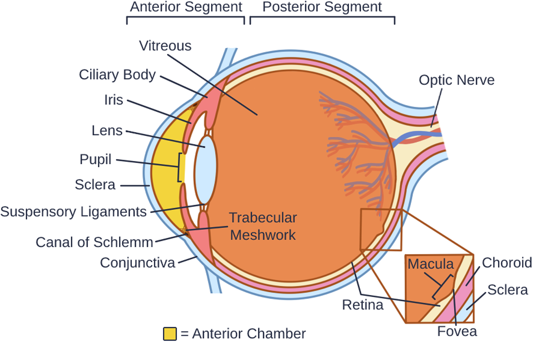
- Tamm ER, Braunger BM & Fuchshofer R (2015) Intraocular Pressure and the Mechanisms Involved in Resistance of the Aqueous Humor Flow in the Trabecular Meshwork Outflow Pathways, Prog Mol Biol Transl Sci. 134, 301–14. - PubMed
-
- Eghrari AO, Riazuddin SA & Gottsch JD (2015) Overview of the Cornea: Structure, Function, and Development, Prog Mol Biol Transl Sci. 134, 7–23. - PubMed
-
- Delaye M & Tardieu A (1983) Short-range order of crystallin proteins accounts for eye lens transparency, Nature. 302, 415–7. - PubMed
-
- Koretz JF & Handelman GH (1988) How the human eye focuses, Sci Am. 259, 92–9. - PubMed
-
- Grus FH, Joachim SC & Pfeiffer N (2007) Proteomics in ocular fluids, Proteomics Clin Appl. 1, 876–88. - PubMed
Publication types
MeSH terms
Grants and funding
LinkOut - more resources
Full Text Sources
Medical
