. 2021 Sep 14;16(9):2351-2363.
doi: 10.1016/j.stemcr.2021.08.002. Epub 2021 Sep 2.

The aryl hydrocarbon receptor promotes differentiation during mouse preimplantational embryo development

Affiliations

The aryl hydrocarbon receptor promotes differentiation during mouse preimplantational embryo development

Ana Nacarino-Palma et al. Stem Cell Reports. .

Abstract

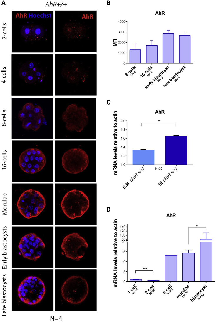
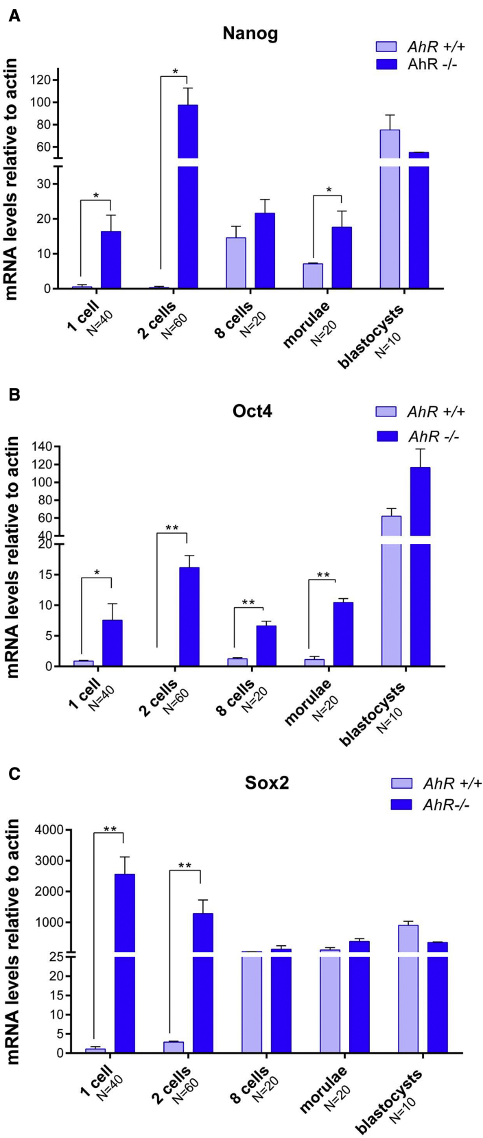
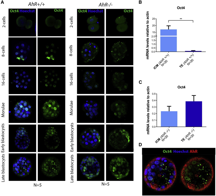
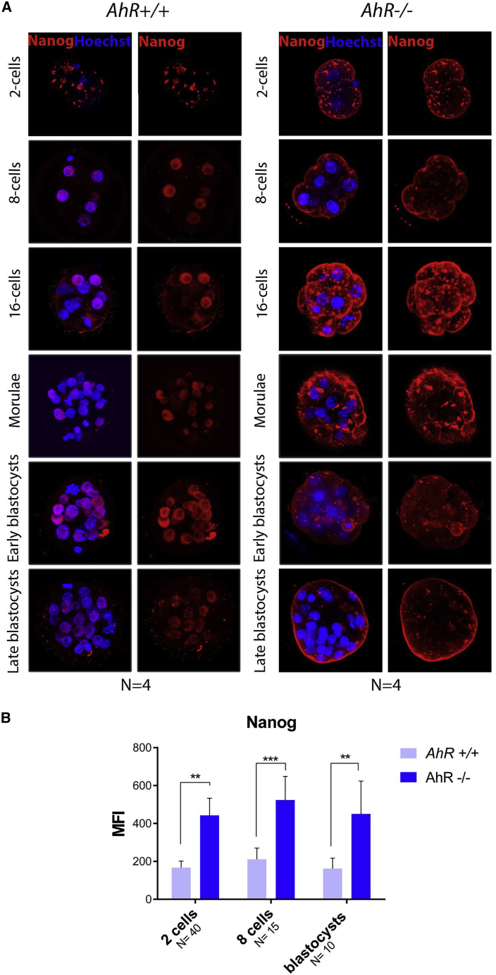
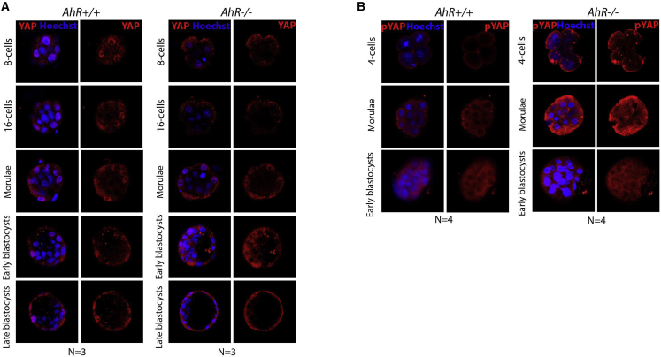
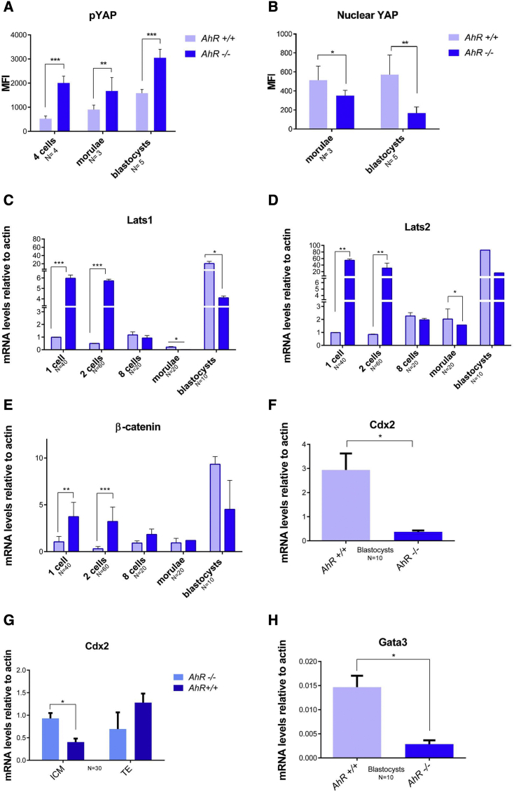
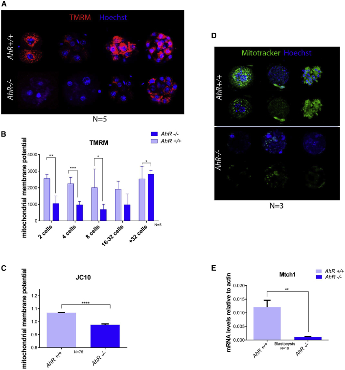
Mammalian embryogenesis is a complex process controlled by transcription factors that regulate the balance between pluripotency and differentiation. Transcription factor aryl hydrocarbon receptor (AhR) regulates OCT4/POU5F1 and NANOG, both essential controllers of pluripotency, stemness and early embryo development. Molecular mechanisms controlling OCT4/POU5F1 and NANOG during embryogenesis remain unidentified. We show that AhR regulates pluripotency factors and maintains the metabolic activity required for proper embryo differentiation. AhR-lacking embryos (AhR-/-) showed a pluripotent phenotype characterized by a delayed expression of trophectoderm differentiation markers. Accordingly, central pluripotency factors OCT4/POU5F1 and NANOG were overexpressed in AhR-/- embryos at initial developmental stages. An altered intracellular localization of these factors was observed in the absence of AhR and, importantly, Oct4 had an opposite expression pattern with respect to AhR from the two-cell stage to blastocyst, suggesting a negative regulation of OCT4/POU5F by AhR. We propose that AhR is a regulator of pluripotency and differentiation in early mouse embryogenesis.

Keywords: Hippo; aryl hydrocarbon receptor; embryo differentiation; pluripotency; preimplantation.


Conflict of interest statement

The authors declare no competing interests.

Figures

Figure 1
Figure 1
AhR expression increases during embryo differentiation (A) Immunofluorescence analysis of AHR at the indicated embryonic stages. Whole AhR+/+ embryos (n = 4) were stained using a specific AHR antibody. Hoechst staining was used to label cell nuclei. Confocal microscopy was used for detection. Scale bar, 20 μm. (B) Immunofluorescence was quantified by calculating the mean fluorescence intensity (MFI) for each developmental stage (the replicates are indicated in the x axis). (C) AhR mRNA expression was quantified by qRT-PCR using RNA purified from TE or ICM fractions previously separated by MACS. (D) AhR mRNA expression was quantified by qRT-PCR in AhR+/+ embryos at the indicated developmental stages using total RNA and the specific primers indicated in Table S1. qRT-PCR was normalized by the expression of β-actin and is represented as 2−ΔΔCt. p < 0.05; ∗∗p < 0.01; ∗∗∗p < 0.001. Data are shown as mean ± SD. The experiments were performed at least four times, and number of embryos analyzed is indicated on the x axis.
Figure 2
Figure 2
Pluripotency factors are upregulated in AhR-null embryos (A–C) AhR+/+ and AhR−/− embryos were obtained at the indicated embryonic stages and used to quantify the mRNA expression of Nanog (A), Oct4 (B), and Sox2 (C) by qRT-PCR. Expression levels were normalized by β-actin and are represented as 2−ΔΔCt. p < 0.05; ∗∗p < 0.01. Data are shown as mean ± SD. The experiments were performed at least four times, and number of embryos analyzed is indicated on the x axis.
Figure 3
Figure 3
AhR depletion alters OCT4 cellular distribution through embryogenesis (A) Immunofluorescence analysis of OCT4 at the indicated embryonic stages. Whole embryos were stained using a specific antibody. Hoechst was used to stain cell nuclei. Scale bar, 20 μm. (B and C) Oct4 mRNA expression was quantified by qRT-PCR using mRNA purified from TE and ICM fractions separated by MACS using the specific primers indicated in Table S1. mRNA expression was normalized by β-actin and is represented as 2−ΔΔCt.. (D) Immunofluorescence analysis of OCT4 (green) and AHR (red) in embryos at the blastocyst stage. Scale bar, 10 μm. ∗∗p < 0.01. Data are shown as mean ± SD. Confocal microscopy was used for detection. The experiments were performed at least three times, and number of embryos analyzed is indicated on the x axis.
Figure 4
Figure 4
NANOG distribution in the embryo is altered in the absence of AhR (A) Immunofluorescence analysis of NANOG at the indicated embryonic stages. Whole embryos were stained using a specific antibody. Hoechst was used for staining cell nuclei. Scale bars, 20 μm. (B) Immunofluorescence was quantified by calculating the mean fluorescence intensity (MFI). ∗∗p < 0.01; ∗∗∗p < 0.001. Data are shown as mean ± SD. The experiments were performed at least four times, and number of embryos analyzed is indicated on the x axis.
Figure 5
Figure 5
AhR deficiency alters localization of the Hippo effector YAP (A and B) Immunofluorescence analysis of YAP (A) and phosphor-YAP (pYAP) (B) at the indicated developmental stages in AhR+/+ and AhR−/− embryos. Whole embryos were stained using specific antibodies for YAP or pYAP. Hoechst was used for staining cell nuclei. Confocal microscopy was used for detection. The experiments were performed at least three (A) and four (B) times. Scale bars, 20 μm.
Figure 6
Figure 6
Hippo effectors and molecular intermediates of pluripotency are altered in the absence of AhR (A) Levels of pYAP were quantified from immunofluorescence and are presented as the mean fluorescence intensity (MFI). (B–H) Levels of nuclear YAP in morulae and blastocysts from AhR+/+ and AhR−/− mice were quantified by immunofluorescence and are presented as the MFI (B). AhR+/+ and AhR−/− embryos at the indicated developmental stages were used to purify embryo mRNA (C–F and H) or mRNA from TE and ICM fractions (G) that were separated by MACS as indicated in experimental procedures. The expression of Lats1 (C), Lats2 (D), β-catenin (E), Cdx2 (F and G), and Gata3 (H) was quantified by qRT-PCR. Expression levels were normalized by β-actin and are represented as 2−ΔΔCt. p < 0.05; ∗∗p < 0.01; ∗∗∗p < 0.001. Data are shown as mean ± SD. The experiments were performed at least three times, and number of embryos analyzed is indicated on the x axis.
Figure 7
Figure 7
AhR-lacking embryos have lower mitochondrial activity (A and B) Mitochondrial membrane potential was measured by TMRM staining at the indicated developmental stages and then analyzed by confocal microscopy. Three embryos per genotype were analyzed. Scale bar, 20 μm. (C) Mitochondrial membrane potential was determined by JC10 staining in pools of 15–20 embryos. (D) Mitochondrial volume was analyzed by mitotracker green staining and visualized by confocal microscopy. Scale bar, 10 μm. (E) Mitochondrial carrier homolog-1 (Mtch1) mRNA expression was measured in blastocysts for each genotype and quantified by RT-qPCR. Expression levels were normalized by β-Actin and represented as 2−ΔΔCt. ∗p < 0.05; ∗∗p < 0.01; ∗∗∗p < 0.001. Data are shown as mean ± SD. The experiments were performed, at least, 3 times and number of embryos analyzed is indicated in the x axis.

References

    1. Abbott B.D., Schmid J.E., Pitt J.A., Buckalew A.R., Wood C.R., Held G.A., Diliberto J.J. Adverse reproductive outcomes in the transgenic Ah receptor-deficient mouse. Toxicol. Appl. Pharmacol. 1999;155:62–70. - PubMed
    1. Bedzhov I., Graham S.J., Leung C.Y., Zernicka-Goetz M. Developmental plasticity, cell fate specification and morphogenesis in the early mouse embryo. Philos. Trans. R. Soc. Lond. B Biol. Sci. 2014;369:20130538. - PMC - PubMed
    1. Boyer L.A., Lee T.I., Cole M.F., Johnstone S.E., Levine S.S., Zucker J.P., Guenther M.G., Kumar R.M., Murray H.L., Jenner R.G., et al. Core transcriptional regulatory circuitry in human embryonic stem cells. Cell. 2005;122:947–956. - PMC - PubMed
    1. Boyer L.A., Mathur D., Jaenisch R. Molecular control of pluripotency. Curr. Opin. Genet. Dev. 2006;16:455–462. - PubMed
    1. Chazaud C., Yamanaka Y. Lineage specification in the mouse preimplantation embryo. Development. 2016;143:1063–1074. - PubMed

Publication types

MeSH terms

Substances