. 2021 Nov 1:175:95-107.
doi: 10.1016/j.freeradbiomed.2021.08.238. Epub 2021 Aug 31.

Hemopexin dosing improves cardiopulmonary dysfunction in murine sickle cell disease

Affiliations

Hemopexin dosing improves cardiopulmonary dysfunction in murine sickle cell disease

Paul W Buehler et al. Free Radic Biol Med. .

Abstract

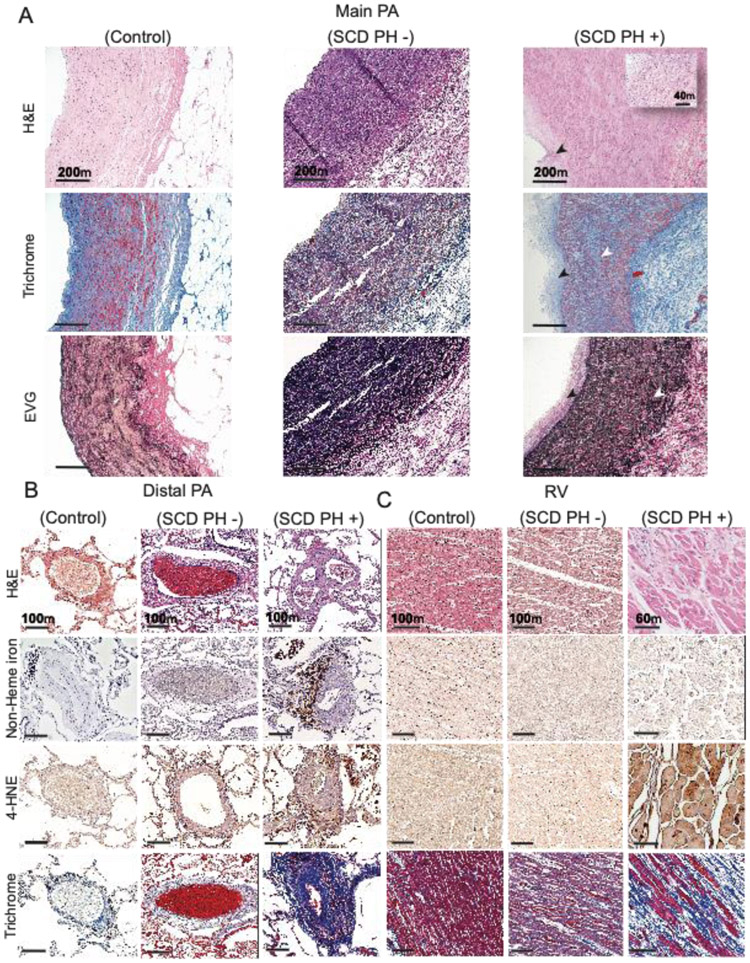
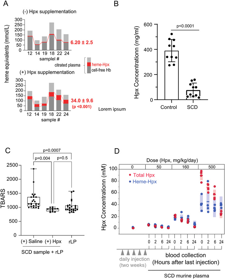
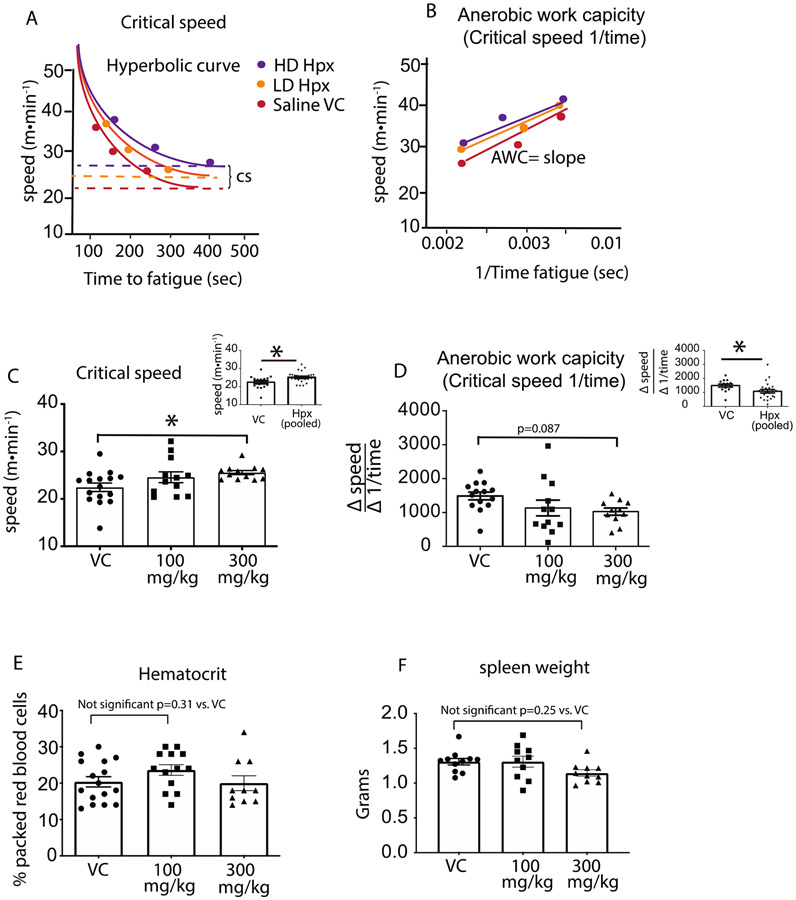
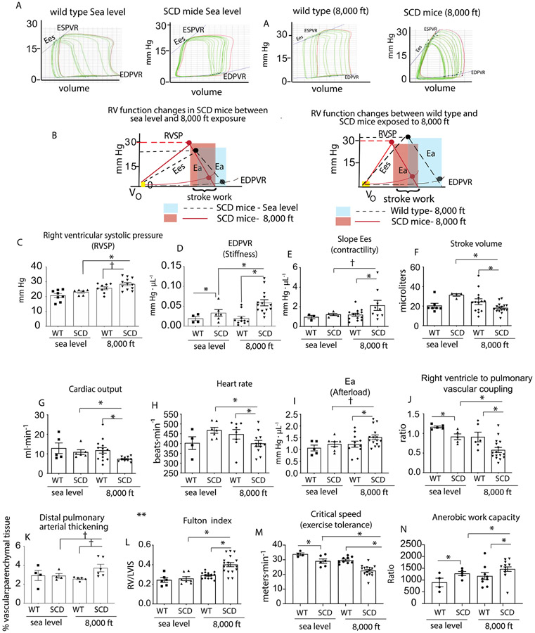
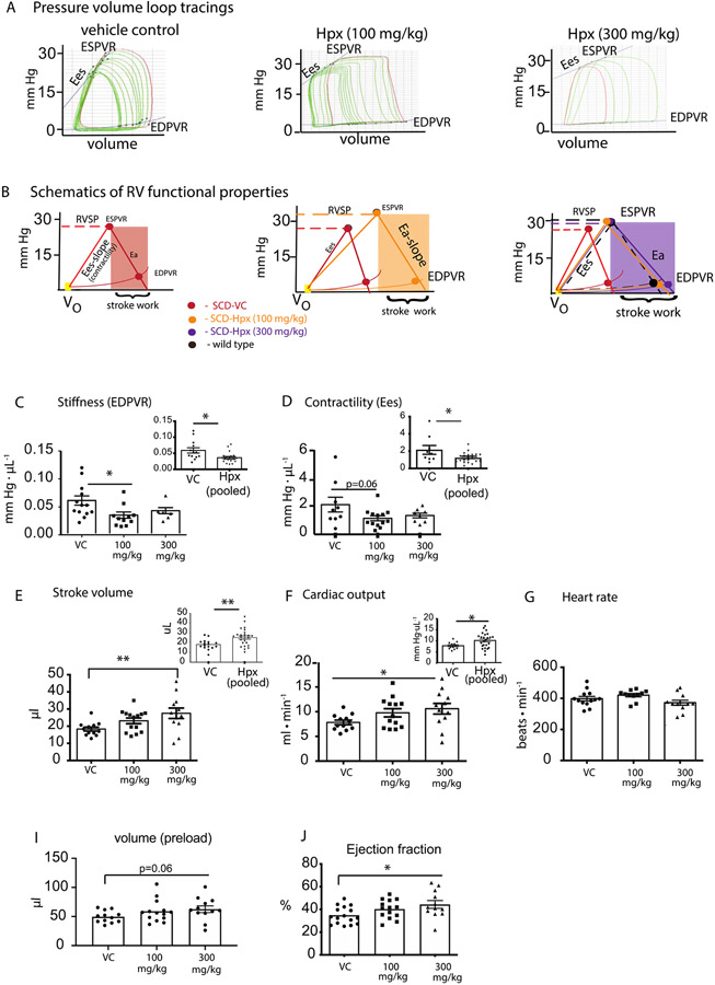
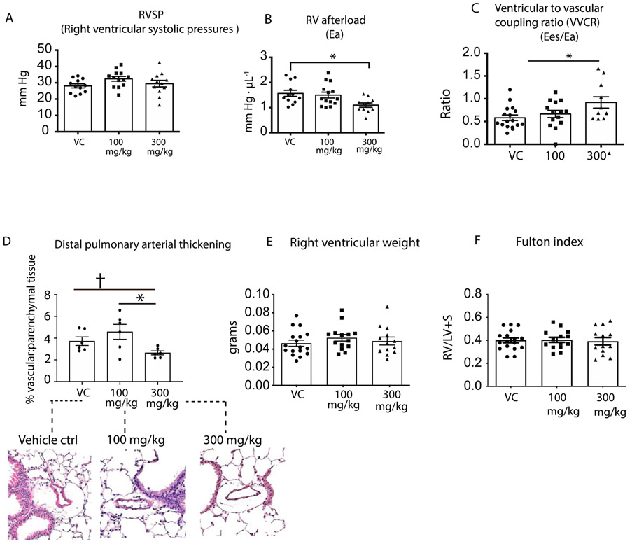
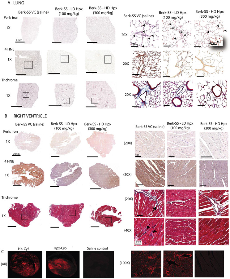
Hemopexin (Hpx) is a crucial defense protein against heme liberated from degraded hemoglobin during hemolysis. High heme stress creates an imbalance in Hpx bioavailability, favoring heme accumulation and downstream pathophysiological responses leading to cardiopulmonary disease progression in sickle cell disease (SCD) patients. Here, we evaluated a model of murine SCD, which was designed to accelerate red blood cell sickling, pulmonary hypertension, right ventricular dysfunction, and exercise intolerance by exposure of the mice to moderate hypobaric hypoxia. The sequence of pathophysiology in this model tracks with circulatory heme accumulation, lipid oxidation, extensive remodeling of the pulmonary vasculature, and fibrosis. We hypothesized that Hpx replacement for an extended period would improve exercise tolerance measured by critical speed as a clinically meaningful therapeutic endpoint. Further, we sought to define the effects of Hpx on upstream cardiopulmonary function, histopathology, and tissue oxidation. Our data shows that tri-weekly administrations of Hpx for three months dose-dependently reduced heme exposure and pulmonary hypertension while improving cardiac pressure-volume relationships and exercise tolerance. Furthermore, Hpx administration dose-dependently attenuated pulmonary fibrosis and oxidative modifications in the lung and myocardium of the right ventricle. Observations in our SCD murine model are consistent with pulmonary vascular and right ventricular pathology at autopsy in SCD patients having suffered from severe pulmonary hypertension, right ventricular dysfunction, and sudden cardiac death. This study provides a translational evaluation supported by a rigorous outcome analysis demonstrating therapeutic proof-of-concept for Hpx replacement in SCD.

Keywords: Critical speed; Exercise tolerance; Pulmonary vascular disease; Right ventricular function.


Figures

Figure 1:
Figure 1:. Pulmonary hypertension in SCD results in lung vascular and right ventricle tissue remodeling
(A) Human main pulmonary artery (MPA) tissue sections from SCD patients with PH (SCD PH (+)) demonstrate distinctive pathology when compared to SCD patients without PH (SCD PH (−)) and controls. H&E staining (a-c), Masson’s trichrome (d-f) and Verhoeff Van Gieson elastic stain, EVG (g-i ). SCD PH + patient H&E stained tissues sections show intimal hyperplasia (black arrows), medial thickening and loss of vascular smooth muscle cell integrity (c, inset). Collagen deposition (f) and elastin fragmentation (i) are observed in the MPA media. (B) Human peripheral lung tissue sections are shown from top to bottom stained for H&E (a-c), Perls iron (non-heme iron) (d-f), 4-hydroxynonenal (4-HNE) (g-i) and Masson’s trichrome (j-l). Staining in SCD PH + patient peripheral lung tissue shows arterial recanalization, vascular plexiform lesions, and intimal thickening (c). Iron positive cellular staining is observed in lung vascular adventitia (f) and immune reactivity to 4-HNE within outer medial and adventitia accumulated cells consistent with iron localization (i). Peripheral arterial lung fibrosis is a prominent pathologic feature of SCD PH and visually observed across all structures of the vessel (l). Limited indicators of pathological processes are visualized in SCD PH - patient tissue sections, except for oxidative stress visually observed as 4-HNE immune reactivity in adventitia accumulated cells (h). No indications of peripheral lung vascular pathology are observed in control tissue sections. (C) Right ventricular pathology is observed in SCD PH (+) as loss of cardiac muscle striations (black arrows) and eosinophilia (back box) (c). Cardiac iron was not observed in any of the tissue sections (e-f). 4-HNE immune reactivity was most intense visually in SCD PH + patient tissue sections (i) and to a lesser extent in SCD PH – tissue sections (h). Interstitial collagen accumulation is visualized in SCD PH + patient tissue sections (l), to a lesser extent in SCD PH – tissue sections (k) and not at all in control in tissue sections. Magnification main PA - 10x; distal PA- 10x; right ventricle- 20x
Figure 2:
Figure 2:. Heme transfer to Hpx and lipid oxidation in heme rich biological samples
(A) Shows heme binding in citrated plasma obtained at the end of exchange transfusions, before and after the addition of Hpx (n=6 samples). Light grey - no Hb or holo-Hpx, red - holo-Hpx and dark grey cell-free Hb. Prior to exogenous Hpx addition, holo-Hpx concentration was 6.20 ± 2.5 mmol/L (Mean ± SD). After Hpx addition, holo-Hpx increased to 34.0 ± 9.6 mmol/L (Mean ± SD) (p <0.001), suggesting a 5-fold increase in heme transfer from non-Hpx proteins and lipids. (B) Shows differences in healthy control donor plasma (390 μg/ml ± 88.7, n=10) and SCD (77.3 ± 55.0, n=10) (p <0.001), data from the same sample cohort shown for quick reference (PMID: 30047285). (C) Shows citrated plasma in the presence of reconstituted lipoprotein (rLP) as a source for heme partitioning and oxidation. Box plots with highest to lowest individual values (n=12-24 per group) show the accumulation of lipid peroxidation products (thiobarbituric acid reactive substances, TBARS). SCD samples + saline + rLP show the greatest generation of TBARS (left), which is significantly reduced (p=0.004) with the addition of Hpx. Alone, rLP does not increase TBARS generation (right). (D) Dose finding for the primary murine study is based on the efficiency of Hpx dosing (50, 160 and 500 mg/kg/day) to bind heme in SCD mice (n=4) after five daily doses followed by blood collections at 0, 2, 6, 24 hours after the final dose. Red circles represent total Hpx, blue circles represent heme bound Hpx, solid blue and red lines indicate mean values and bars indicate SD. Data indicates that doses equal to 50 and 160 mg/kg/day x 5 days consume Hpx within a heme-Hpx complex, while dosing at 500 mg/kg/day exceeds transferable heme availability and suggests a steady state transferable heme concentration of approximately 40 - 80 uM.
Figure 3:
Figure 3:. Hemopexin improves exercise tolerance in SCD mice
(A) Hyperbolic curve fit for critical speed in Hpx treated and untreated SCD mice. (B) Anerobic work capacity demonstrates the liner relationship between exercise intensity vs. 1time. (C) Dose and main effect of Hpx treatment on exercise tolerance as determined from the the quantitative value for critical speed plotted as bar graphs representing animals in each cohort. (D) Dose and main effects of Hpx treatment on the glycolytic state during exercise as quantitatified by the value or slope of the anerobic work capacity. The anerobic work capacity is plotted as a bar graphs representing animals for each cohort. (E) Packed red blood cell volume as measured by the hemotocrit and plotted as bar graphs representing each cohort. (F) High spleen weights associate with red blood hemolysis. Absence any difference in packed red cell volume and spleen weights suggests Hpx therapy is not altering red blood cell oxygen delivery. * p<0.05
Figure 4:
Figure 4:. SCD mice housed for 3 months at moderate hypoxia (8,000 ft) demonstrate progressive pulmonary vascular disease.
(A) Representative tracings of pressure volume (PV) loops during an occlusion for wild type and SCD mice housed at either sea level or moderate hypoxia (8,000 ft) (B) shows the corresponding schematics of the pressure volume relationships for wild type and SCD mice for each cohort. (C-J) Right ventricular functional analysis in wild type and SCD mice showing right ventricular systolic pressure, stiffness, contractility, stroke volume, cardiac output, heart rate, afterload and right ventricle to pulmonary vascular coupling ratio. (K) Pulmonary vascular thickening (L) right ventricular hypertrophy. (M) Critical speed. (N) Anerobic work capacity. Data is represented as means ± standard error of measurement. RVSP- right ventricular pressures; Ea-Afterload; EDPVR-end diastolic pressure volume relationship; ESPVR- end systolic pressure volume relationship. *p<0.05 vs. SCD mice at 8,000 ft; p < 0.05 vs SCD mice t-test
Figure 5:
Figure 5:. Right ventricular functional analysis in hemopexin treated and untreated SCD mice experiencing progressive cardiopulmonary vascular disease.
(A) Representative tracings of pressure volume (PV) loops during an occlusion (B) shows the corresponding schematics of the pressure volume relationships for hemopexin treated and untreated SCD mice. (C-J) Right ventricular stiffness, contractility, stroke volume, cardiac output, heart rate, preload and ejection fraction. Data is represented as means ± standard error of measurement. Inset graphs show low and high dose Hpx treated mice combined into one group. RVSP- right ventricular pressures; Ea-Afterload; EDPVR-end diastolic pressure volume relationship; ESPVR- end systolic pressure volume relationship. *p<0.05, **p<0.005
Figure 6:
Figure 6:. Pulmonary vascular analysis in hemopexin treated and untreated SCD mice experiencing progressive cardiopulmonary vascular disease.
(A) Right ventricular systolic pressures (B) Right ventricular afterload (C) Ventricular to vascular coupling ration (VVCR); (D) Pulmonary vascular thickening (E) Right ventricular weight (F)Fulton index a determinant of RV hypertrophy*p<0.05, †p<0.05 by t test.
Figure 7:
Figure 7:. Microscopic examination of lung and right ventricle tissues in hemopexin treated and untreated SCD mice.
(A) Histopathology of lung tissue . Top row- Perls iron staining; Middle rows show 4HNE staining for lipid peroxidation; Bottom row- Trichrome staining as a marker for fibrosis; (B) Histopathology of right ventricle tissue Top row- Perls iron staining; Middle rows show 4HNE staining for lipid peroxidation (C) Confocal microscopy images of showing the distribution of hemoglobin and hemopexin extravasation into cardiac tissue 30 min after administration.

Similar articles

Cited by

References

    1. McCavit TL. Sickle cell disease. Pediatr Rev. 2012;33(5):195–204; quiz 5-6. - PubMed
    1. Simpson S Sickle cell disease: a new era. Lancet Haematol. 2019;6(8):e393–e4. - PubMed
    1. Quinn CT. Sickle cell disease in childhood: from newborn screening through transition to adult medical care. Pediatr Clin North Am. 2013;60(6):1363–81. - PMC - PubMed
    1. E MJ, and T TA. Sickle Cell Anemia. Medscape. 2020.
    1. Fushimi N, Sawada H, Udaka F, and Kameyama M. [Five-year follow-up studies of laboratory data in the elderly group]. Nihon Ronen Igakkai Zasshi. 1989;26(2):179–80. - PubMed

Publication types